Abstract
Background and objectives. In the evaluation of the growth of head circumference (HC), charts depicting normal growth are of paramount importance. Current Finnish HC growth charts are based on data from only 130 children born 1953–1964. As a secular trend in HC growth has been reported, we updated the HC charts using a large sample of contemporary HC data. Material and methods. Mixed cross-sectional HC data of 19,715 healthy subjects aged 0–7 years were collected from primary health care providers. References for HC for age and HC/height ratio for age were fitted using generalized additive models for location, scale, and shape (GAMLSS).Results. Increased HC for age was seen particularly after 2 years of age in both genders compared to the 1953–1964 reference. The SD for HC was remarkably larger in the 1953–1964 reference. The proportion of 1986–2008 reference subjects exceeding the +2 SD limit of the 1953–1964 reference was much bigger than the proportion below −2 SD. Conclusions. Because of the secular change in HC growth, the HC reference has to be renewed periodically. The new Finnish reference for HC for age should be implemented for monitoring HC growth of children in Finland.
Key words::
| Abbreviations | ||
| BCPE | = | Box-Cox power exponential |
| CDC | = | Centers for Disease Control and Prevention |
| GAMLS | = | Generalized additive models for location, scale, and shape |
| HC | = | head circumference |
| SD | = | standard deviation |
| SDS | = | standard deviation score |
| WHO | = | World Health Organization |
Key messages
Updated HC references (head circumference for age) for Finnish children aged 0–7 years are presented.
Head circumference-to-height ratio for age was constructed for Finnish children aged 0–7 years.
There is a notable positive secular change in HC growth and a reduction in the SD of the new HC reference compared to the one currently in use. Therefore the current HC charts should be replaced by the new HC reference.
Introduction
In the evaluation of the growth of head circumference (HC), charts depicting normal growth are of paramount importance. Head circumference charts currently in use in Finland are based on follow-up data of only 130 children born in years 1953–1964 (Citation1,Citation2). Head circumference is routinely measured at health care visits during infancy and childhood until the age of 7 according to the recommendation by the National Institute for Health and Welfare. The ultimate goal of taking multiple HC measurements is the early detection of underlying pathological processes affecting head growth. Indeed, HC is a good indicator of the growth in brain volume especially in early childhood (Citation3–5), and slow growth of HC may indicate primary pathology in the developing brain (Citation6–8). Excessive growth in turn may indicate a pathological process affecting the circulation of the cerebrospinal fluid leading to hydrocephalus (Citation9). Because of the small number of individuals and positive secular change in HC growth, evident in many countries (Citation10–15), the aim of the current work was to provide an update of the HC charts, for the ages 0–7 years, based on adequate sample size and utilization of recent statistical methods.
Material and methods
Data for the present study were collected from providers of public primary care in the city of Espoo, Finland's second largest city with a population of 241,600 inhabitants. With a significant net migration from all parts of Finland, its population has grown 10.6-fold over the past 60 years. The majority of the population (94.4%) is of Finnish origin, which mirrors the whole of Finland (97.3%). The Finnish social security system provides regular, free-of-charge visits to public primary care child health clinics to permanent residents of Finland regardless of social status or income level. Primary care nurses in Finland are specially trained in child health care and health prevention, and their duties include assessment of health and development at scheduled visits including standardized weight, length/height, and head circumference measurements.
Children in Espoo have regular visits at child health clinics at the ages of 1–2 weeks, 3–6 weeks, and 6–8 weeks; at 2, 3, 4, 5, 6, 8, 10, 12, 18, and 24 months; and then at 3.5, 5, and 6 years of age. Children may have also extra visits if special health concerns are suspected.
Head circumference is measured using a plastic tape measure at every visit to the child health clinic as the maximum occipitofrontal circumference, and the results are rounded to the nearest 0.1 cm. Since 2003, all measurements in the Espoo area have been captured in a networked electronic patient management system named Effica (Tieto Ltd, Finland). The birth measurements, including HC as well as data on premature birth, are recorded to Effica during the first visit to the child health clinic after birth. Permission for the present study was obtained from the Espoo Municipality Institutional Review Board. No contact was made with the study subjects since the data were handled anonymously.
Database cleaning
Finnish growth references for weight and height have been recently renewed (Citation16). The data for these references comprise subjects born 1983–2008. The original sample for the HC reference is the same. Database cleaning for the height standard encompassed three phases: first, the primary care nurses of Espoo municipality excluded the subjects with diseases or medications potentially affecting growth. The nurses had been specifically trained by a pediatric endocrinologist (L.D.). This phase of database cleaning has been reported in detail elsewhere (Citation16). Next, measurements which were obtained outside scheduled visits were excluded. Lastly, all HC outside ± 5 SD were excluded (representing extreme outliers, i.e. physiologically improbable measurements, or being extremely pathological when true measurements). After the cleaning procedure, the data consisted of 146,790 measurements of 19,715 subjects (9,536 girls; 48.4%) born 1986–2008.
Statistical methods
In the construction of growth curves for HC (HC for age and HC/height ratio for age), distribution of response variables and technique in smoothing distribution parameter curves over age were chosen by closely following the guidelines provided by the World Health Organization (WHO) (Citation17). Accordingly, generalized additive models for location, scale, and shape (GAMLSS) were used, choosing the distribution of response variable from the flexible Box-Cox power exponential (BCPE) distribution family and using cubic splines as a smoothing technique. BCPE distribution can be described in terms of four parameters: M for median, S for coefficient of variation, L for Box-Cox transformation power, and T as a parameter related to kurtosis.
R statistical software (GAMLSS package (Citation18)) was used in the analysis. First, an optimal power transformation was calculated for age in relation to the response variable as it was found to improve goodness of fit. Second, optimal degrees of freedom for parameter curves were defined using the optim function and information criteria BIC (which have penalty h of log(n) in the formula −2 * l – hp, where l is the maximized likelihood, p number of parameters in the model, and n number of observations) as it seemed to give optimal smoothness for curves. We started modeling from the normal distribution (BCPE with L = 1 and T = 2), and it turned out to be sufficient for both response variables when comparing fitted percentiles to observed percentiles (Supplementary Figure 1).
Results
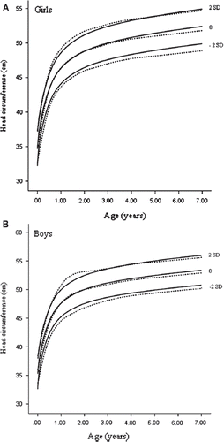
A positive secular change was seen in HC reference 1986–2008 both in girls and boys as compared to the HC reference 1953–1964 (). It was particularly clear after 2 years of age in both genders. The mean HC at birth was 34.8 cm in girls and 35.3 cm in boys in the new reference. The corresponding figures were 34.7 cm and 35.5 cm in the 1953–1964 HC reference, respectively.
Figure 1. The new Finnish HC for age reference (mean ± 2 SD, solid lines). Curves based on 146,790 measurements from 19,715 full-term healthy subjects born between 1986 and 2008 compared to the current Finnish HC reference based on subjects born between 1953 and 1964 (mean ± 2 SD, dashed lines). A: girls aged 0–7 years; B: boys aged 0–7 years.

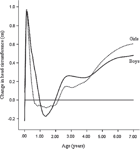
The difference in mean HC between the two references is better illustrated in as an absolute (cm) difference. In girls the greatest difference was at about 0.15 years of age when the mean HC was 1.0 cm greater in the 1986–2008 reference than in the 1953–1964 reference (). After that, the difference between the two references rapidly decreased until it was slightly negative between the ages of 0.60 and 2.15 years. Thereafter, the difference grew continuously to the age of 7 years, being 0.6 cm at the end. In boys the difference in HC between the 1986–2008 reference and the 1953–1964 reference followed quite the same pattern. The maximum difference of 1.0 cm was reached at 0.15 years, and around 1.35 years the mean HC of the 1986–2008 reference was at the lowest point below the 1953–1964 reference (−0.2 cm). After the age of 2 years the mean HC of the 1986–2008 reference became bigger than that of the 1953–1964, and from that point on the difference kept increasing, being 0.5 cm at the age of 7 years.
Figure 2. Age- and sex-specific features of the secular change in HC in Finland. Comparison between HC reference 1986–2008 and HC reference 1953–1964. Curves indicate differences from the HC reference 1953–1964 in mean HC in cm from birth to age 7 years. Dashed line = girls; solid line = boys.

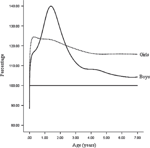
A very identical pattern was seen in the changes in mean HC expressed in standard deviation score (SDS) in both girls and boys. At 7 years, the mean HC of the 1986–2008 reference was 0.42 SDS and 0.35 SDS above the mean of the 1953–1964 reference in girls and boys, respectively. Strikingly the SD was 10%–40% bigger in the HC reference 1953–1964 compared to the new reference () except at birth, when the SD of the HC reference 1986–2008 was larger.
Figure 3. Age- and sex-specific features of the change in head circumference SD. Curves illustrate the ratio between the SD of the HC reference 1953–1964 and HC reference 1986–2008 (horizontal line). Dashed line = girls; solid line = boys.

We assessed how many subjects from the 1986–2008 reference population would fall outside ±2 SD limits of the 1953–1964 HC reference ( and ).
Table I. Number of head circumference measurements of girls in the head circumference (HC) reference 1986–2008 population and percentage of measurements ≤ −2 SD and ≥ 2 SD when compared to the HC reference 1953–1964.
Table II. Number of head circumference measurements of boys in the head circumference (HC) reference 1986–2008 and percentage of measurements ≤ −2 SD and ≥ 2 SD when compared to the HC reference 1953–1964.
In total, 2.3% (range 0.9%–6.0%) of measurements in girls were above +2 SD, which is actually the percentage expected in normally distributed head circumference. However, the proportion of measurements below −2 SD was only 0.5% (range 0.1%–0.9%).
A similar finding was found in boys, who had 3.1% (range 0.5%–7.2%) of the measurements above +2 SD limit of the 1953–1964 HC reference and only 0.5% (range 0.1%–1.8%) below the −2 SD limit of the same reference. In boys from 0.67 years (8 months) to 4 years the proportion of measurements above +2 SD was less than 2.3%. In girls, a similar result was obtained between the age of 0.33 years (4 months) and 4 years. Nevertheless, both in boys and girls, the proportion of measurements below −2 SD was much smaller than the proportion above +2 SD.
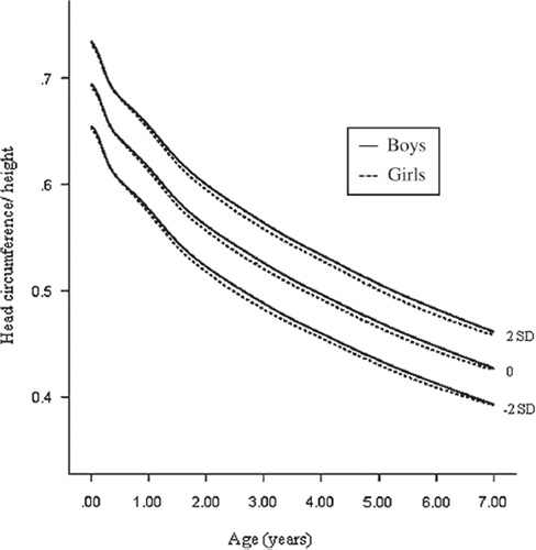
We also calculated HC-to-height ratio for age (). This ratio was highest in the early months, and it declined quite constantly during the whole age period.
Discussion
In this study we report a positive secular change in HC between the cohorts of Finnish children born 1953–1964 and 1986–2008. Our finding is consistent with previous studies in Sweden, the UK, and Japan (Citation10–15). In Sweden (Citation14), the updated mean HC reference values were 0.6–1.2 SDS above the previous reference values from birth to 48 months of age. Increase in HC most likely reflects a true secular change in HC, but some methodological factors may contribute. For instance, in Sweden it was speculated that some of the increment was due to the use of a tighter steel measurement instrument for HC (Citation14).
Through this study we demonstrated how outdated cut-off points may lead to misclassification of children. Theoretically, an uncorrected secular trend of +0.4 SDS in mean HC without a change in SD for HC would mean that only 0.8% of children remained below the lower −2 SDS limit and as many as 5.5% were above the upper +2 SDS limit, instead of the expected 2.3% at both ends. Our observations were consistent with such a rate of misclassification by the mean ±2 SD limits.
In addition to the secular increase in mean HC we also noted a reduction in SD of the HC in the 1986–2008 reference. This further increases the rate of misclassification of outdated limits used for screening of abnormalities in the growth of HC. This results in severe underdiagnosis of conditions with microcephaly, but on the other hand it compensates the secular increase in the mean head circumference. Hence, HC growth references must be periodically updated.
Our results also highlight the importance of using national HC references in Finland, because on average the HC in Finnish children are clearly larger than those published in the multiethnic WHO HC reference or in the US-based CDC 2000 HC reference (Citation19,Citation20). For details, please see Supplementary Figure 2.
The strength of this study was the large, population-based, and representative sample of HC measurements of children seen in recent years at child health clinics. Head and brain growth takes place mainly in the first 2–3 years (Citation21–24). Therefore we think that HC growth charts from 0 to 7 years are sufficient for screening purposes.
Head circumference-to-height ratio is informative in some growth disorders. For instance, in hypochondroplasia, the HC-to-height ratio depicts macrocephaly better than the HC alone (Citation25). Consistent with former findings, a slight difference between boys and girls was found in our data, with boys having a larger ratio. In Japan, secular trends in both height and HC growth have been reported, but the HC-to-height ratio has remained unchanged (Citation13). Such a comparison was not possible in our study, because data for HC-to-height ratios were not available from the 1953–1964 reference.
Conclusion
In HC growth a positive secular change over the past 40–50 years was found in Finnish children. Ignoring this change in HC will lead to significant misclassification and unnecessary referral of children to specialist care because of a false suspicion of macrocephaly. Furthermore, some children with true microcephaly escape our attention when outdated HC references are used. We provide updated HC reference data as a part of the updates of the Finnish growth references.
http://www.informahealthcare.com/ann/doi/10.3109/07853890.2011.558519
Download PDF (510.4 KB)Declaration of interest: The study was supported by EVO funds. The authors declare no other conflicts of interest.
References
- Kantero RL, Tiisala R.Studies on growth of Finnish children from birth to 10 years. V. Growth of head circumference from birth to 10 years. A mixed longitudinal study. Acta Paediatr Scand Suppl. 1971;220:27–32.
- Hallman N, Backstrom-Jarvinen L, Kantero RL, Tiisala R. Child growth and development. Presentation of the material in the longitudinal ‘model child’ study. Ann Paediatr Fenn. 1966;12:8–12.
- Bartholomeusz HH, Courchesne E, Karns CM. Relationship between head circumference and brain volume in healthy normal toddlers, children, and adults. Neuropediatrics. 2002;33:239–41.
- Lemons JA, Schreiner RL, Gresham EL. Relationship of brain weight to head circumference in early infancy. Hum Biol. 1981;53:351–4.
- Lindley AA, Benson JE, Grimes C, Cole TM 3rd, Herman AA. The relationship in neonates between clinically measured head circumference and brain volume estimated from head CT-scans. Early Hum Dev. 1999;56:17–29.
- Dolk H. The predictive value of microcephaly during the first year of life for mental retardation at seven years. Dev Med Child Neurol. 1991;33:974–83.
- Nelson KB, Deutschberger J. Head size at one year as a predictor of four-year IQ. Dev Med Child Neurol. 1970; 12:487–95.
- Abuelo D. Microcephaly syndromes. Semin Pediatr Neurol. 2007;14:118–27.
- Zahl SM, Wester K. Routine measurement of head circumference as a tool for detecting intracranial expansion in infants: what is the gain? A nationwide survey. Pediatrics. 2008;121:e416–20.
- Ishikawa T, Furuyama M, Ishikawa M, Ogawa J, Wada Y. Growth in head circumference from birth to fifteen years of age in Japan. Acta Paediatr Scand. 1987;76:824–28.
- Anzo M, Takahashi T, Sato S, Matsuo N. The cross-sectional head circumference growth curves for Japanese from birth to 18 years of age: the 1990 and 1992–1994 national survey data. Ann Hum Biol. 2002;29:373–88.
- Ounsted M, Moar VA, Scott A. Head circumference charts updated. Arch Dis Child. 1985;60:936–9.
- Tsuzaki S, Matsuo N, Saito M, Osano M. The head circumference growth curve for Japanese children between 0–4 years of age: comparison with Caucasian children and correlation with stature. Ann Hum Biol. 1990;17:297–303.
- Wikland KA, Luo ZC, Niklasson A, Karlberg J. Swedish population-based longitudinal reference values from birth to 18 years of age for height, weight and head circumference. Acta Paediatr. 2002;91:739–54.
- Wright CM, Booth IW, Buckler JM, Cameron N, Cole TJ, Healy MJ, . Growth reference charts for use in the United Kingdom. Arch Dis Child. 2002;86:11–14.
- Saari A, Sankilampi U, Hannila ML, Kiviniemi V, Kesseli K, Dunkel L. New Finnish growth references for children and adolescents aged 0 to 20 years: Length/height-for-age, weight-for-length/height, and body mass index-for-age. Ann Med. 2010 Sep 21 (Epub ahead of print).
- WHO Multicentre Growth Reference Study Group. WHO Child Growth Standards based on length/height, weight and age. Acta Paediatr Suppl. 2006;450:76–85.
- Stasinopoulos DM, Rigby RA. Generalized additive models for location scale and shape (GAMLSS) in R. J Stat Softw. 2007;23:1–46.
- WHO Multicentre Growth Reference Study Group. WHO child growth standards: head circumference-for-age, arm circumference-for-age, triceps skinfold-for-age and subscapular skinfold-for-age—methods and development. World Health Organisation, Geneva 2007.
- Kuczmarski RJ, Ogden CL, Guo SS, Grummer-Strawn LM, Flegal KM, Mei Z, . 2000 CDC Growth Charts for the United States: methods and development. Vital Health Stat 11. 2002;(246):1–190.
- Courchesne E, Chisum HJ, Townsend J, Cowles A, Covington J, Egaas B, . Normal brain development and aging: quantitative analysis at in vivo MR imaging in healthy volunteers. Radiology. 2000;216:672–82.
- Sgouros S, Goldin JH, Hockley AD, Wake MJ, Natarajan K. Intracranial volume change in childhood. J Neurosurg. 1999;91:610–16.
- Dekaban AS. Changes in brain weights during the span of human life: relation of brain weights to body heights and body weights. Ann Neurol. 1978;4:345–56.
- Nellhaus G. Head circumference from birth to eighteen years. Practical composite international and interracial graphs. Pediatrics. 1968;41:106–14.
- Saunders CL, Lejarraga H, del Pino M. Assessment of head size adjusted for height: an anthropometric tool for clinical use based on Argentinian data. Ann Hum Biol. 2006;33:415–23.

