Abstract
Background. Global ischemia (GI) electrocardiogram (ECG), wide-spread ST depression with inverted T waves maximally in leads V4–5, and lead aVR ST elevation (STE), is a marker of an adverse outcome in patients with non-ST elevation acute coronary syndromes (ACS), perhaps because this pattern is indicative of left main stenosis. The prognostic value of this ECG pattern has not been established.
Aims. The distribution of ECG changes and the prognostic value of the GI ECG were studied.
Methods. ECGs of consecutive patients admitted with suspected ACS (n = 1,188) were classified into seven ECG categories: STE, Q waves without STE, left bundle branch block, left ventricular hypertrophy, GI ECG, other ST depression and/or T wave inversion, and other findings.
Results. The GI ECG pattern predicted a high rate (48%) of composite end-points (mortality, re-infarction, unstable angina, resuscitation, or stroke) at 10-month follow-up compared to the other ECG categories (36%) (HR 1.78; CI 95% 1.31–2.41; P < 0.001). In multivariate analysis, the GI ECG pattern was associated with a higher rate of composite end-points (HR 1.40; CI 95% 1.02–1.91; P = 0.035). The multivariate analysis furthermore identified age, creatinine level, and diabetes as independent predictors of prognosis.
Conclusions. The GI ECG pattern predicted an unfavorable outcome, when compared to other ECG patterns in patients with ACS.
| Abbreviations | ||
| ACS | = | acute coronary syndrome |
| CI | = | confidence interval |
| ECG | = | electrocardiogram |
| HR | = | hazard ratio |
| LBBB | = | left bundle branch block |
| LM | = | left main |
| LVH | = | left ventricular hypertrophy |
| RBBB | = | right bundle branch block |
| TACOS | = | Tampere Acute COronary Syndrome |
Key messages
The global ischemia ECG pattern with wide-spread ST depression, maximally in leads V4–5 with inverted T waves and ST elevation in lead aVR, predicts poor prognosis compared to other ECG patterns in patients with acute coronary syndrome.
Further studies are needed to confirm whether coronary angiography should be considered in urgent cases with ECG signs of global ischemia.
Introduction
The electrocardiogram (ECG) is the most accessible and widely used diagnostic tool for patients with symptoms suggestive of acute myocardial ischemia. Although the presence of acute ischemic changes on the admission ECG has been associated with a higher risk of cardiac events, the prognostic value of the various ECG presentations of acute myocardial ischemia remains elusive (Citation1–5).
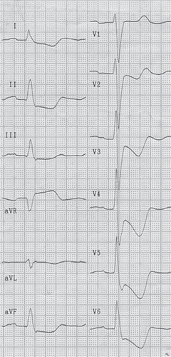
As left main (LM) coronary artery disease is associated with high mortality, early diagnosis is important (Citation6). Accordingly, ST depression and lead aVR ST elevation have been established as ECG markers of an adverse outcome in non-ST elevation acute coronary syndrome (ACS) (Citation7–10). ST depression with inverted T waves in the precordial leads in patients without tachycardia was associated with LM disease in rather small studies (Citation11–13). In one of these studies, one-quarter of the patients showing this ECG pattern proved to have severe three-vessel disease, while three-quarters had LM or LM-equivalent disease on coronary angiography (Citation13). The ECG pattern with wide-spread ST depression and inverted T waves maximally in leads V4–5 has been ascribed to circumferential subendocardial ischemia (Citation14). Lead aVR ST elevation is a typical finding in these patients (). The prognostic value of this ECG pattern, the ‘global ischemia ECG’ pattern, consisting of wide-spread ST depression and inverted T waves maximally in leads V4–5 and lead aVR ST elevation, in comparison to other ECG manifestations of ACS, has not been studied.
Figure 1. ECG (50 mm/s) shows the global ischemia ECG pattern: ST depression and inverted T waves maximally in leads V4–5 and ST elevation in lead aVR.
In the present study we investigated the distribution and prognostic impact of seven predefined ECG patterns in ECGs from patients admitted with ACS. The ECG patterns were: ST elevation, pathological Q waves, left bundle branch block (LBBB), left ventricular hypertrophy (LVH), global ischemia ECG, other ST depression and/or T wave inversion, and other findings, including normal ECG.
Material and methods
Patients
Patients presenting with presumptive diagnosis of ACS at admission to the emergency department were consecutively included in the study. The study was performed at Tampere University Hospital, and 1,188 patients were included between 1 January 2002 and 31 March 2003. A total of 343 presented with ST elevation myocardial infarction, 655 with non-ST elevation myocardial infarction, and 190 with unstable angina. The study end-point was a composite of mortality, re-infarction, unstable angina, resuscitation, or stroke in hospital and during 10-month follow-up. The detailed description of the protocol of the TACOS (Tampere Acute COronary Syndrome) study has been reported previously (Citation15).
The Ethics Committee at Tampere University Hospital approved the study protocol. The patients gave their written informed consent for participation.
ECG analysis
The incidence at hospital admission and the patient prognosis based on the ECG patterns were studied. The investigators analyzed the patient ECG recorded either pre-hospitally or in the emergency department showing maximal ischemic changes. If the ECG in the referral unit was normal, but a follow-up ECG in the emergency department showed ST deviation, the second one was used for analysis. No ECGs recorded during hospital stay—for example, in the coronary care unit or in the catheterization laboratory—were used. All the ECGs were analyzed by two investigators (KCN and MJE) blinded to the clinical data. The patients were classified into seven predefined ECG categories: 1) ST elevation (elevation of the ST segment ≥2 mm in two contiguous precordial leads or ≥1 mm in two contiguous limb leads); 2) pathological Q waves without ST elevation (defined as A: in leads V1–3 any Q wave ≥30 ms in duration; B: in leads I, II, aVL, aVF, V4–6 a Q wave ≥1 mm and ≥30 ms in duration in ≥2 adjacent leads; and C: in leads V1–2 R wave duration >40 ms and R/S ratio >1 in the absence of pre-excitation, right ventricular hypertrophy, or right bundle branch block (RBBB)); 3) typical LBBB; 4) LVH without ST elevation except in leads aVR and/or V1 (LVH was defined according to the Sokolow–Lyon criteria (Citation16) and/or the Cornell voltage-duration product (Citation17); 5) global ischemia ECG (ST depression ≥0.5 mm in ≥6 leads, maximally in leads V4–5 with inverted T waves and ST elevation ≥0.5 mm in lead aVR) (); 6) other ST depression and/or T wave inversion; and 7) other findings, including normal ECG.
The classification into the ECG categories was based solely on the actual ECG. No comparison to previous ECGs was done.
The ST segment, determined by drawing a line between subsequent PR segments, and measured 0.06 s after the J point, was considered elevated or depressed if it was 0.5 mm or more above or below the isoelectric line, respectively. The T wave was considered positive or negative if it was 1 mm or more above or below the isoelectric line, measured more than 120 ms after the J point with the aid of a hand-held magnifying lens.
Statistical analysis
Categorical variables were expressed as numbers of patients or percentages and continuous variables as medians followed by interquartile range. We used the chi-square test or Fisher's exact test for categorical variables and the Mann–Whitney test for numerical variables. A two-tailed P value of <0.05 was considered statistically significant. Confidence intervals (CI) were calculated at the 95% significance level. Composite end-point data between ECG categories were plotted as Kaplan–Meier curves. Comparison between the ECG groups was made using the log rank statistic. Hazard ratios (HR) were calculated by Cox regression analysis. Variables with P < 0.20 in the Cox univariate analysis were included in the multivariate Cox regression model. Age and gender adjustment was included. All calculations were performed with the SPSS 16.0 statistical package.
Results
Our study showed differences between groups of patients stratified according to ECG categories both in base-line characteristics and in-hospital findings ( and ). Among the seven categories, ST elevation proved to be the most frequent, followed by old Q waves without ST elevation (). Patients with global ischemia ECG, LBBB, and LVH were older than those from the four other categories, while patients with global ischemia ECG more often had hypertension, diabetes, prior angina, and severe anginal symptoms. They were also more often on aspirin, beta-blocker, nitrate, and diuretic medication. Systolic dysfunction based on echocardiographic ejection fraction measurement was more often seen in patients with LBBB and old Q waves. Patients with other ST depression and/or T wave inversion had the lowest troponin levels.
Figure 2. Distribution of ECG changes of all consecutive patients admitted with acute coronary syndrome. Rates are based on the TACOS study, n = 1,188.

Table I. Base-line characteristics according to electrocardiographic classification.
Table II. In-hospital findings, treatment, and outcome by electrocardiographic categories. Medication at discharge.
Coronary angiography during the hospital stay was performed in 560 patients (47%). The patients with global ischemia ECG had more severe disease on coronary angiography compared to the other ECG categories (). All the patients with global ischemia ECG, in whom angiography was performed, showed significant coronary artery disease, and this ECG pattern was associated with angiographic three-vessel disease in 71%. LM disease either isolated or in association with one-, two-, or three-vessel disease was present in 25% of the patients. The corresponding numbers for other ST depression and/or T wave inversion was only 22% for three-vessel disease and 3% for LM disease. Revascularization during hospital stay was more frequent in patients with global ischemia ECG than in patients from the other ECG categories.
In univariate analysis, in-hospital mortality rate was highest among patients with LBBB and global ischemia ECG (18% and 14%, respectively; P = 0.004). The incidence of in-hospital composite end-points was lowest in patients with LVH, ST elevation, and ST depression and/or T wave inversion (12%, 16%, and 14%, respectively; P = 0.009).
The global ischemia ECG pattern predicted a high rate of composite end-points (48%) at 10-month follow-up compared to all the other ECG categories (36%) (HR 1.78; CI 1.31–2.41; P < 0.001) (). In multivariate analysis, global ischemia ECG pattern, age, creatinine level at presentation, and diabetes were identified as independent predictors for poor prognosis at 10-month follow-up ().
Figure 3. The rate of composite end-points at 10-month follow-up according to the ECG at admission presented by the Kaplan–Meier curve (dashed line: global ischemia ECG, solid line: all other ECG categories).
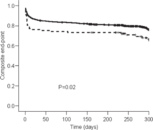
Table III. Variables retained in the final multivariate Cox proportional hazards model examining the rate of composite end-points at 10-month follow-up.
Discussion
This study adds new interesting data about prognostic and therapeutic differences between distinct ECG findings on hospital admission in patients with ACS. Our study shows that the global ischemia ECG pattern, consisting of concomitant ST depression with inverted T waves maximally in leads V4–5 and ST elevation in lead aVR (), is associated with worse prognosis than other ECG patterns.
Our study population represents consecutive patients of all the three categories of ACS, ST elevation and non-ST elevation acute myocardial infarction, and unstable angina. Patients in randomized clinical trials tend to be younger than those in unselected patient cohorts. In a pooled analysis of large randomized trials of ACS therapies, only 18% of the 34,266 patients enrolled were ≥75 years old (Citation18). Of the more than 11,000 patients included in the multinational prospective Global Registry of Acute Coronary Events (GRACE) study, more than 30% of the patients were ≥75 years old (Citation19). In the present study the median age for the patients in the seven ECG categories varied between 68 and 77 years. Hence, this represents a study population typically encountered in everyday clinical work. The GRACE registry study and our study showed similar proportions of patients with ST elevation in the ECG at presentation. Interestingly, we found that more than one-third of the patients with ST depression presented with the global ischemia ECG pattern. Normal ECG was found in 13% of the patients included in this study. This probably reflects that patients with unstable angina without elevated biomarkers of myocardial injury (n = 190) were included. Previous studies have reported incidences of up to 18.5% with normal ECG in patients with acute myocardial infarction (Citation3).
According to previous studies in non-ST elevation ACS, all the three global ischemia ECG components, ST depression, T wave inversion, and lead aVR ST elevation, were associated with higher mortality. Despite that the global ECG pattern has not previously been investigated, the different components of the pattern have all been studied separately and support our findings. ST depression and T wave inversion in leads V4–6 have been found to predict 1-year mortality independently (Citation1). Another study found that the presence, magnitude, and extent of ST depression were associated with increased mortality in patients with non-ST elevation myocardial infarction (Citation5). Interestingly, ST depression in two or more lateral (I, aVL, V5, or V6) leads proved to be the only ECG variable that predicted death after adjusting for base-line predictors. Patients with lateral ST depression had higher rates of death and severe heart failure than did the remaining patients, even though they had similar enzyme levels. In contrast, ST depression not involving the lateral leads did not predict poor outcome. The authors did not include T waves in their analyses. One could speculate that the poor outcome in patients with lateral ST depression was associated with the global ischemia ECG pattern described in our study.
ST elevation in lead aVR in the setting of ACS has been established as a marker of severe coronary artery disease and worse outcome. In a small study, Yamaji et al. compared the ECG findings of patients with acute LM obstruction with the findings of patients with acute left anterior descending or right coronary artery obstruction (Citation20). Lead aVR ST elevation >0.5 mm was markedly more frequent in the patients with LM obstruction than in the two other groups, and the patients who died during follow-up had higher levels of ST elevation in lead aVR than the survivors. The authors did not focus on possible ST depression. The HERO-2 investigators recently reported aVR ST elevation ≥1 mm to be associated with higher 30-day mortality in both anterior and inferior acute ST elevation myocardial infarction (Citation21). After adjusting for summed ST elevation and ST depression in other leads, associations with higher mortality were found with aVR ST elevation of ≥1.5 mm for anterior and of ≥1 mm for inferior ST elevation myocardial infarction. Notably, when adjustment was made for clinical factors, the association between aVR ST elevation and 30-day mortality lost its significance. This underlines the importance of recognizing the complete ECG pattern in myocardial ischemia and not only changes in one lead when assessing patient risk. In a recent systematic review article, the absence of aVR ST elevation appeared to exclude LM stenosis as the underlying cause in non-ST elevation myocardial infarction (Citation22).
Sclarovsky and associates introduced the concept of T wave inversion in combination with lateral ST depression as a risk marker in ACS without ST elevation (Citation11). They studied 32 consecutive patients who had horizontal or downward-sloping ST depression with peaked (n = 21) or inverted (n = 11) T waves. In the group with inverted T waves, the in-hospital mortality was 27%, whereas none of the patients with positive T waves died in the hospital. In addition, seven out of ten patients with inverted T waves had significant LM disease on angiography, while two out of ten patients had three-vessel disease.
We have earlier reported that patients (n = 25) with transient ST depression and an inverted T wave maximally in leads V4–5 during anginal pain had higher in-hospital mortality (24%) than patients (n = 25) with ST depression and a positive T wave (0%) (Citation13). In that study, all the patients with ST depression and inverted T waves also had ST elevation of at least 0.5 mm in lead aVR.
The exact electro/pathophysiologic mechanisms of the global ischemia ECG are not known. When myocardial ischemia is confined primarily to the subendocardium, the overall ST vector typically faces the inner ventricular layer and the ventricular cavity, such that the surface ECG leads show ST depression (Citation23). This subendocardial ischemic pattern represents the typical ECG finding during exercise tests, as energy demands are highest and blood supply most precarious in the inner layers of the myocardium (Citation24). In these cases, extensive ischemia impairs relaxation of the left ventricle, resulting in increase of the left ventricular end-diastolic pressure (Citation25). Inducing global left ventricular ischemia in dogs by hydraulic constriction of the LM resulted in a significant decrease in the endocardial-to-epicardial flow ratio and a significant increase of left ventricular end-diastolic pressure (Citation26). Also, inducing elevation of the left ventricular pressure by pacing in patients with significant coronary artery disease was associated with ST depression in the ECG (Citation27). Hence, we speculate that the global ischemia ECG represents the electrical effects of severe subendocardial ischemia, which generates an ST vector that points away from the apical/lateral leads V4–5 and towards lead aVR. In the present study, of the patients with global ischemia ECG, in which angiography was performed, almost three-quarters had angiographic three-vessel disease, while one-quarter had LM disease either isolated or in association with one-, two-, or three-vessel disease.
The present and above-mentioned studies reveal that different manifestations of ST/T changes have significantly different prognostic implications. Still, in the modern era of high technology, the ECG has a central role in clinical decision-making in ACS. We think that much is to be gained by extending the ECG analysis beyond ST elevation and non-ST elevation categories.
Limitations
There are quite a few study limitations to be reported in this study. The proportion of patients having coronary angiography was less than 50%. Accordingly, correlation between ECG and angiographic data cannot be reliably calculated. However, our primary aim was to study differences in outcome between the ECG categories, not to correlate with angiographic findings. Another possible limitation is the fact that the magnitude of ST elevation in lead aVR was not used in our statistical analyses. This could be included in future prospective trials.
Base-line data regarding Killip class and heart rate were not included in the statistical analyses. Neither were data on delay from symptom onset to ECG recording. Echocardiographic findings were not available for all patients.
Conclusion
We have identified a high-risk ECG pattern in patients categorized as non-ST elevation ACS. The ECG pattern with wide-spread ST depression, maximally in leads V4–5, with inverted T waves and ST elevation in lead aVR was present in 8% of ‘all-comers’ with ACS. This global ischemia ECG pattern predicted poor prognosis compared to other ECG patterns and was independently associated with an adverse outcome in multivariate analysis. From the therapeutic point of view, it is justified to conclude that future studies are needed to test whether urgent cases with ECG signs of severe coronary artery disease should have coronary angiography on an emergency basis. Besides the high rate of need for urgent revascularization there is a high probability for a composite of mortality, re-infarction, unstable angina, resuscitation, or stroke in hospital or at follow-up compared to other ECG patterns.
Acknowledgements
This study was supported financially by The Pirkanmaa Regional Fund of the Finnish Cultural Foundation, Tampere, Finland.
Declaration of interest: The authors report no conflicts of interest. The authors alone are responsible for the content and writing of the paper.
References
- Atar S, Fu Y, Wagner GS, Rosanio S, Barbagelata A, Birnbaum Y. Usefulness of ST depression with T-wave inversion in leads V(4) to V(6) for predicting one-year mortality in non-ST-elevation acute coronary syndrome (from the electrocardiographic analysis of the global use of strategies to open occluded coronary arteries IIB trial). Am J Cardiol. 2007;99:934–8.
- Cannon CP, McCabe CH, Stone PH, Rogers WJ, Schactman M, Thompson BW, . The electrocardiogram predicts one-year outcome of patients with unstable angina and non-Q wave myocardial infarction: Results of the TIMI III registry ECG ancillary study. Thrombolysis in myocardial ischemia. J Am Coll Cardiol. 1997;30:133–40.
- Miller WL, Sgura FA, Kopecky SL, Asirvathan SJ, Williams BA, Wright RS, . Characteristics of presenting electrocardiograms of acute myocardial infarction from a community-based population predict short- and long-term mortality. Am J Cardiol. 2001;87:1045–50.
- Savonitto S, Ardissino D, Granger CB, Morando G, Prando MD, Mafrici A, . Prognostic value of the admission electrocardiogram in acute coronary syndromes. JAMA. 1999;281:707–13.
- Barrabes JA, Figueras J, Moure C, Cortadellas J, Soler-Soler J. Prognostic significance of ST segment depression in lateral leads I, aVL, V5 and V6 on the admission electrocardiogram in patients with a first acute myocardial infarction without ST segment elevation. J Am Coll Cardiol. 2000;35:1813–9.
- Caracciolo EA, Davis KB, Sopko G, Kaiser GC, Corley SD, Schaff H, . Comparison of surgical and medical group survival in patients with left main equivalent coronary artery disease. Long-term CASS experience. Circulation. 1995;91: 2335–44.
- Kaul P, Fu Y, Chang WC, Harrington RA, Wagner GS, Goodman SG, . Prognostic value of ST segment depression in acute coronary syndromes: insights from PARAGON-A applied to GUSTO-IIb. PARAGON-A and GUSTO IIb Investigators. Platelet IIb/IIIa Antagonism for the Reduction of Acute Global Organization Network. J Am Coll Cardiol. 2001;38:64–71.
- Holmvang L, Clemmensen P, Lindahl B, Lagerqvist B, Venge P, Wagner G, . Quantitative analysis of the admission electrocardiogram identifies patients with unstable coronary artery disease who benefit the most from early invasive treatment. J Am Coll Cardiol. 2003;41:905–15.
- Savonitto S, Cohen MG, Politi A, Hudson MP, Kong DF, Huang Y, . Extent of ST-segment depression and cardiac events in non-ST-segment elevation acute coronary syndromes. Eur Heart J. 2005;26:2106–13.
- Barrabés JA, Figueras J, Moure C, Cortadellas J, Soler-Soler J. Prognostic value of lead aVR in patients with a first non-ST-segment elevation acute myocardial infarction. Circulation. 2003;108:814–9.
- Sclarovsky S, Rechavia E, Strasberg B, Sagie A, Bassevich R, Kusniec J, . Unstable angina: ST segment depression with positive versus negative T wave deflections—clinical course, ECG evolution, and angiographic correlation. Am Heart J. 1988;116:933–41.
- Nikus KC, Eskola MJ. The ECG in a mechanical obstruction of the ostium of the left main coronary artery. Int J Cardiol. 2002;86:327–9.
- Nikus KC, Eskola MJ, Virtanen VK, Vikman S, Niemelä KO, Huhtala H, . ST-depression with negative T waves in leads V4-V5—a marker of severe coronary artery disease in non-ST elevation acute coronary syndrome: A prospective study of angina at rest, with troponin, clinical, electrocardiographic, and angiographic correlation. Ann Noninvasive Electrocardiol. 2004;9:207–14.
- Sclarovsky S. Angina at rest and acute myocardial ischaemia. Sclarovsky S. Electrocardiography of acute myocardial ischaemic syndromes. 1st. London, UK: Martin Dunitz Ltd; 1999. 1–29.
- Nikus KC, Eskola MJ, Virtanen VK, Harju J, Huhtala H, Mikkelsson J, . Mortality of patients with acute coronary syndromes still remains high: A follow-up study of 1188 consecutive patients admitted to a university hospital. Ann Med. 2007;39:63–71.
- Sokolow M, Lyon TP. The ventricular complex in left ventricular hypertrophy as obtained by unipolar precordial and limb leads. Am Heart J. 1949;37:161–86.
- Norman JE Jr, Levy D. Improved electrocardiographic detection of echocardiographic left ventricular hypertrophy: results of a correlated data base approach. J Am Coll Cardiol. 1995;26: 1022–9. Erratum in: J Am Coll Cardiol. 1996;27:516.
- Alexander KP, Newby LK, Cannon CP, Armstrong PW, Gibler WB, Rich MW, . Acute coronary care in the elderly, part I: Non-ST-segment-elevation acute coronary syndromes: a scientific statement for healthcare professionals from the American Heart Association Council on Clinical Cardiology: in collaboration with the Society of Geriatric Cardiology. Circulation. 2007;115:2549–69.
- Steg PG, Goldberg RJ, Gore JM, Fox KA, Eagle KA, Flather MD, . Baseline characteristics, management practices, and in-hospital outcomes of patients hospitalized with acute coronary syndromes in the Global Registry of Acute Coronary Events (GRACE). Am J Cardiol. 2002; 90:358–63.
- Yamaji H, Iwasaki K, Kusachi S, Murakami T, Hirami R, Hamamoto H, . Prediction of acute left main coronary artery obstruction by 12-lead electrocardiography. ST segment elevation in lead aVR with less ST segment elevation in lead V(1). J Am Coll Cardiol. 2001;38:1348–54.
- Wong CK, Gao W, Stewart RA, Benatar J, French JK, Aylward PE, . aVR ST elevation: an important but neglected sign in ST elevation acute myocardial infarction. Eur Heart J. 2010;15:1845–53.
- Kühl JT, Berg RM. Utility of lead aVR for identifying the culprit lesion in acute myocardial infarction. Ann Noninvasive Electrocardiol. 2009;14:219–25.
- Cook RW, Edwards JE, Pruitt RD. Electrocardiographic changes in acute subendocardial infarction. I. Large subendocardial and large nontransmural infarcts. Circulation. 1958;18:603–12.
- Nikus K, Pahlm O, Wagner G, Birnbaum Y, Cinca J, Clemmensen P, . Electrocardiographic classification of acute coronary syndromes: A review by a committee of the international society for holter and non-invasive electrocardiology. J Electrocardiol. 2010;43:91–103.
- Grossman W. Evaluation of systolic and diastolic function of the ventricles and myocardium. Baim DS, Grossman W. Grossman's cardiac catheterization, angiography and intervention. 6th. Philadelphia, Pa: Lippincott Williams and Wilkins; 2001. 367–90.
- Visner MS, Arentzen CE, Parrish DG, Larson EV, O'Connor MJ, Crumbley AJ 3rd, . Effects of global ischemia on the diastolic properties of the left ventricle in the conscious dog. Circulation. 1985;71:610–9.
- Aroesty JM, McKay RG, Heller GV, Royal HD, Als AV, Grossman W. Simultaneous assessment of left ventricular systolic and diastolic dysfunction during pacing-induced ischemia. Circulation. 1985;71:889–900.