Abstract
Objective. Patients with hypertensive left-ventricular hypertrophy (LVH) have lower coronary flow reserve (CFR). Whether carvedilol can improve CFR of patients with hypertensive LVH is unknown. We aimed to investigate the effects of carvedilol on CFR in patients with hypertensive LVH. Methods. Sixty-three patients were randomly divided into two groups for treatment with carvedilol or metoprolol. The peak diastolic coronary flow velocity in the left anterior descending coronary artery at rest and at maximal vasodilation with dipyridamole infusion was recorded by transesophageal echocardiography (TEE), then CFR was calculated at baseline and at the end of 6 months of therapy. Left-ventricular mass index (LVMI) was calculated by 2-D echocardiography. Endothelium-dependent and -independent reactivity of the brachial artery was measured. Levels of plasma endothelin-1 (ET1), nitric oxide (NO) and other metabolites were monitored and analyzed before and after 6-month therapy. Results. Both blood pressure and heart rate decreased significantly in the two treatment groups after therapy (p<0.05). With carvedilol treatment, LVMI was lower (p<0.05), endothelium function of the brachial artery was higher (p<0.05), and peak diastolic coronary flow velocity at rest and at maximal vasodilation after dipyridamole infusion was significantly higher (p<0.05) than with metoprolol treatment, which led to a significantly higher CFR (p<0.05). Changes in CFR and LVMI with carvedilol treatment were inversely correlated (R2=0.474, p=0.036). With carvedilol treatment, plasma level of ET-1 was lower, but that of NO was significantly higher than with metoprolol treatment (both p<0.05). Conclusions. The CFR of patients with hypertensive LVH but not coronary artery disease could increase with 6-month carvedilol therapy.
Introduction
Most patients with hypertensive left-ventricular hypertrophy (LVH) show symptoms of angina, which is difficult to distinguish from atherosclerotic heart disease. The myocardium in hypertensive LVH may have undergone ischemia.
Many studies have reported lower coronary flow reserve (CFR) in patients with hypertensive LVH than in patients without hypertensive LVH (Citation1–3).
Carvedilol, a third-generation beta-blocker, is a multiple-acting compound with non-selective beta-adrenoceptor and selective alpha-1-adrenoceptor blocking activity in the calcium channel. Carvedilol has many other properties distinct from second-generation beta-adrenoreceptor blockers, such as anti-oxidation (Citation4,Citation5), anti-proliferation of smooth muscle cells (Citation6,Citation7), alleviation of endothelium dysfunction (Citation8) and radical free-oxygen scavenging and iron chelating (Citation9). In addition, carvedilol has more pronounced antioxi-dative effects and inhibitory effects than the second-generation beta-adrenoreceptor blockers on the sympathetic nervous system. It has related blood viscosity in patients after acute myocardial infarction (Citation10,Citation11). The recently published large-scale carvedilol or metoprolol European trial (COMET) demonstrated that carvedilol reduces total mortality to a greater extent than does metoprolol (Citation12). Furthermore, carvedilol could improve the CFR of patients with coronary artery disease (Citation13). However, whether carvedilol can improve CFR of patients with hypertensive LVH is unknown.
We aimed to investigate by transesophageal echocardiography (TEE) whether carvedilol could reverse CFR in patients with hypertensive LVH.
Materials and methods
Study population
We studied 63 patients with hypertensive LVH (35 men; mean age 61.2± 12.6 years) who were admitted to the Department of Cardiovascular Medicine, Shandong University Qilu Hospital, China.
Inclusion and exclusion criteria
The patients satisfied the following inclusion criteria: (i) systolic blood pressure (BP) 140-160 mmHg or diastolic BP 90-100 mmHg; (ii) hypertensive LVH (Devereux criteria: left-ventricular mass index ≥ 134 g/m2 for men and ≥ 110 g/m2 for women) (Citation14); (iii) subjective or objective signs of ischemia during exercise stress (19 patients with symptoms of angina and 44 with descended ST segment); and (iv) no evidence of coronary artery disease after coronary angiography. Patients with secondary hypertension, diabetes mellitus, or esophageal, liver or kidney disease were excluded. Patients with a heart rate (HR) <50 beats/min, significant brady-arrhythmia, atrio-ventricular block, presence of stenotic valvular heart disease, hypertrophic obstructive cardiomyopathy, and active myocarditis were also excluded.
We randomly assigned the 63 patients into two groups for treatment with carvedilol or metoprolol. After a 2-week drug washout, patients received oral doses twice daily of 10 mg carvedilol tartrate (Astra Zeneca, China) or 50 mg metoprolol (Qilu Pharmaceutical Co, Jinan ShanDong P.R China). Patients with side-effects were dropped from the study. At the end of 6-month therapy, 28 patients remained in the carvedilol group and 29 in the metoprolol group. The research was a single-centre, randomized, open, endpoint-blinded, parallel-group study. All recruited patients received endpoint-blinded treatment.
Each subject gave their written informed consent to be in the study, which was approved by the Ethics Committee of Shandong University Qilu Hospital.
Patients visited physicians every 2 weeks for BP and HR monitoring during the study. Before and after treatment with carvedilol or metoprolol, patients underwent TEE, assessment of endothelium function of the brachial artery and transthoracic echocardiography. Blood samples were drawn for monitoring levels of endothelin-1 (ET-1), nitric oxide (NO), fasting glucose (Gs), total cholesterol (TC), total triglyc-erides (TGs), low-density lipoprotein cholesterol (LDL-C) and high-density lipoprotein cholesterol (HDL-C). In addition, the plasma concentration of ET-1 was determined by use of a radioimmunoas-say kit (Dongya Immuno-technology Institute, Beijing, China). Plasma level of NO was tested by the enzymic method (Jingmei Biotech Co. Ltd. Shenzhen, China).
Echocardiography
Echocardiography involved use of a SONOS-5500 (Philips Medical Systems. The Netherlands) equipped for transthoracic examination (2-4 MHz), TEE (5-7 MHz) and peripheral vessel examination (7-12 MHz). Imaging depth was adjusted for each patient to obtain the echocardiographic image that included the whole left ventricle. Harmonic imaging was used with optimal adjustment of gain to ensure the best-quality images and adequate delineation of endo-cardial borders. Images were stored on magneto-optical disks for off-line analysis.
2-D Echocardiography
2-D echocardiography was used to measure the thickness of the interventricular septum (IVS), posterior left ventricular wall (LVPW) and left ventricular end-diastolic diameter (LVEDD) as described (Citation14). LVMI was calculated as follows (Citation15):
LVM= 1.04[(IVS + LVEDD + LVPW)3-(LVEDD)3-13.6 g;
LVMI=LVM/body surface area (BSA);
BSA (m2) =0.061 × height (cm) + 0.0128 × weight (kg)-0.1529.
Transesophageal echocardiography
Each patient underwent TEE after 6-h fasting, with the throat anesthetized by use of 2% tetracaine. The coronary artery was visualized by use of a transducer just above the aortic leaflets. The ultrasound probe was placed at the aortic root to show the cut section of the aortic valve after turning the probe wafer to 30°. At the same time, the probe was turned slowly to show the Y-shaped bifurcation of the left coronary artery, the circumflex branch and the left anterior descending branch. The blood velocity of the initial part of left anterior descending coronary artery was measured by pulsed-wave Doppler at rest and at hyperemia after dipyridamole injection (0.56 mg/kg intravenously for 5 min). Peak diastolic velocity (PDV), and mean diastolic velocity were evaluated by tracing the contour of the Doppler pattern in five to seven consecutive cardiac cycles. CFR was calculated as follows: CFR=PDVh/PDVa, where PDVh is mean hyperemic diastole coronary blood flow velocity after dipyridamole injection and PDVa is mean diastole coronary flow velocity of baseline. BP, HR and ECG (electrocardiogram) were monitored continuously. Each patient was carefully monitored during the pharmacological agent infusion to detect any adverse events.
Endothelial function of brachial artery
Endothelium-dependent and -independent reactivity of the brachial artery was assessed as described (Citation16). Arterial diameter was measured at rest and after reactive hyperemia (with increased flow causing endothelium-dependent vasodilation), again at rest, and finally after sublingual administration of glyceryl trinitrate, an endothelium-independent vasodilator. Brachial artery diameter was recorded 1 min after rapid deflation of a cuff that was held inflated around the patient's forearm, with BP>250 mmHg for 4.5 min. After 15 min, the second baseline measurement was obtained. Nitroglycerin, 0.5 mg, was administered sublingually, and after an additional 4.5 min, final brachial artery diameter was measured. Ultrasound images were recorded on videotape. Then flow-mediated arterial dilations and nitroglycerin-induced dilations were calculated as percentage change from the pre-ischemic and nitroglycerin baseline values, respectively.
Statistical analysis
Data are expressed as mean±SE. Statistical analysis involved use of SPSS 13.0 (SPSS Inc., Chicago, IL, USA). Comparison of variables involved use of analysis of variance (ANOVA). Comparisons between the two treatment groups at the end of 6 months were analyzed by two-way repeat-measures ANOVA, as appropriate. A p<0.05 was considered statistically significant. The correlation between changes in CRF and LVMI before and after treatment in the two groups was analyzed by Pearson correlation coefficient.
Results
The characteristics of patients are in . The two treatment groups did not differ in age, sex, etiology of hypertrophic ventricle or BP.
Table I. Baseline characteristics of the study population treated with carvedilol and metoprolol (n=57).
All procedures were well tolerated, apart from the common side-effects found with beta-receptor block-ers. In the carvedilol group, data for four patients were dropped from analysis because of headache (Citation1), vertigo (Citation1) and poor compliance (Citation2). In the meto-prolol group, data for two patients were dropped from analysis because of vertigo (Citation1) and poor compliance (Citation1). Data for the remaining 57 patients (28 in carvedilol group and 29 in metoprolol group) were analyzed.
Effect of carvedilol and metoprolol on BP, HR and metabolic parameters
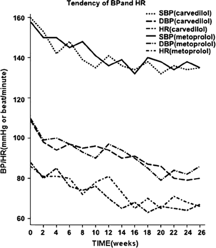
HR and BP decreased significantly after 6-month therapy in both metoprolol and carvedilol groups as compared with at baseline (p<0.05), with no significant difference between the two groups (). Plasma levels of TC, HDL-C, LDL-C, TGs or FGs did not change before or after carvedilol or metoprolol treatment (p=NS). However, in the carvedilol group, the plasma level of ET-1 significantly decreased, from 79.11 ± 11.02 to 50.04± 10.09 pg/ml (p=0.05). NO level significantly increased after carvedilol therapy, from 62.69± 10.42 to 85.78± 11.23 µmol/l (p<0.05). Conversely, in the metoprolol group, the plasma levels of ET-1 and NO did not significantly change from baseline after 6-month treatment ().
Table II. Effect of 6-month carvedilol and metoprolol treatment on metabolic variables in patients with hypertensive LVH (n=57).
Figure 1. Changes in systolic and diastolic blood pressure and heart rate during treatment with carvedilol and metoprolol for 26 weeks. Comparison of systolic blood pressure (SBP) and diastolic blood pressure (DBP) and heart rate (HR) between baseline and 6-month treatment with cavedilol and metoprolol monitored every 2 weeks.
Effect of carvedilol and metoprolol on CFR
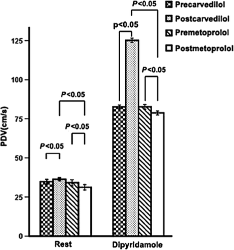
In the carvedilol group, the PDV of the anterior descending coronary artery at rest was significantly higher post-treatment than at baseline (p<0.05), and the peak hyperemic diastolic blood flow velocity after dipyridamole treatment was higher (p<0.05) ( and ). However, in the metoprolol group, both PDV at rest and after dipyridamole treatment decreased significantly after therapy (p<0.05).
Figure 2. Changes in coronary artery flow velocity in patients with hypertensive left-ventricular hypertrophy during treatment with carvedilol for 26 weeks. At baseline, the peak diastolic and systolic blood flow velocity of the anterior descending coronary artery was 55 and 25 cm/s, respectively (A), and increased to 125 and 60 cm/s, respectively, during maximal hyperemia with intravenous dipyridamole treatment (B). After carvedilol treatment, at baseline the peak diastolic and systolic blood flow velocity was 52 and 20 cm/s, respectively (C), and increased to 160 and 60 cm/s with dipyridamole injection (D). After carvedilol treatment, the ratio of the systolic peak hyperemic flow velocity to baseline flow velocity increased from 2.27 to 2.67 and the ratio of the diastolic peak hyperemic flow velocity to baseline flow velocity increased from 2.4 to 3.0.
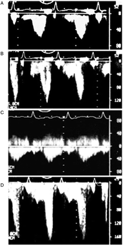
Figure 3. Changes in peak diastolic blood flow velocity (PDV) of anterior descending coronary artery after carvedilol and metoprolol treatment in hypertensive patients with left-ventricular hypertrophy (LVH). PDV at rest and hyperemia increased significantly after carvedilol treatment.
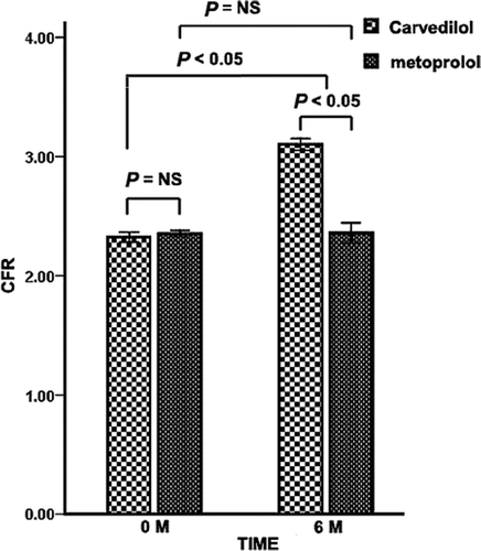
CFR was significantly higher after carvedilol therapy than at baseline (2.31±0.31 vs 3.16±0.67, p=0.039); in the metoprolol group, CFR showed no significant change from baseline (). The two treatment groups significantly differed in CFR at 6 months (3.16±0.67 vs 2.46±0.58, p<0.05).
Figure 4. Changes in coronary flow reserve (CFR) before and after 6-month carvedilol and metoprolol therapy in patients with hypertensive left-ventricular hypertrophy (LVH). CFR increased significantly after carvedilol treatment. NS, not significant; M, month.
Effect of carvedilol and metoprolol on LVMI
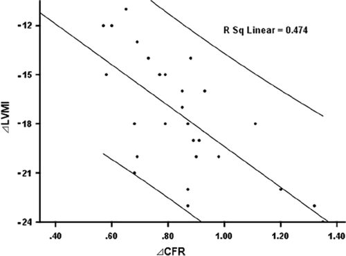
LVMI was attenuated significantly after therapy as compared with at baseline only in the carvedilol group (144.41±43.21 g/m2 vs 128.7±21.14 g/m2, p=0.043), with significant differences between the groups after therapy (p<0.05) (). In the carvedilol group, changes in CFR and LVMI were inversely correlated (R2 = 0.474, p=0.036).
Figure 5. Correlation between change in LV mass index (LVMI) and coronary flow reserve (ΔCFR) after carvedilol treatment in patients with hypertensive LVH. ΔCFR, change in CFR during carvedilol treatment; ΔLVMI, variance in LV mass index during carvedilol treatment. ΔLVMI and ΔCFR have negative correlation after carvedilol treatment.
Effect of carvedilol and metoprolol on brachial arterial dilation
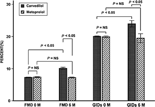
For both treatment groups, brachial artery diameter did not significantly change after therapy In the carvedilol group, flow-mediated arterial dilation increased significantly, from 7.21±2.90% to 9.81±2.79% (p<0.05), as did glyceryl trinitrate-induced dilation, from 20.50±7.21% to 23.87±6.78% (p<0.05) (). The metoprolol group showed no significant changes in endothelium-dependent and -independent arterial dilation (p=NS). Changes in brachial arterial dilation in the two groups were significant (p<0.05).
Figure 6. Change in flow-mediated artery dilation (FMD) and glyceryl trinitrate-induced dilation (GID) in brachial artery after 6-month treatment with carvedilol and metoprolol in patients with hypertensive LVH. NS, not significant. M, month.
Discussion
In this study, we used TEE to determine whether carvedilol could improve CFR in patients with hypertensive LVH. The flow velocity of the left anterior descending coronary artery after dipyridamole injection was increased only by 2.31-fold at baseline in the patients, which is much lower than normal. Therefore, the CFR of patients with hypertensive LVH was lower than normal, as was reported previously (Citation17). Both carvedilol and metoprolol significantly reduced the BP and HR of patients with hypertensive LVH. The effects seem to be attributed to the beta-receptor blockade effect of carvedilol and metoprolol, which may be explained by negative inotropy and negative conduction of beta-receptor blockade. Interestingly, despite similar lowered BP and HR by the two beta-blockers, carvedilol additionally increased CFR, attenuated LVMI, improved endothelium function and regulated ET-1 and NO levels.
CFR is defined as the ratio of hyperemic to basal average peak coronary flow velocity (Citation17). In the carvedilol group, the mean PDV of maximal hyper-emia after administration of dipyridamole increased more significantly than that of the baseline state after treatment, which led to a significantly higher CFR. However, in the metoprolol group, both PDV at maximal hyperemia after administration of dipyri-damole and that of the baseline state decreased. So CFR in the metoprolol group did not significantly change with treatment. The different effects of carve-dilol and metoprolol on CFR must be attributed to the different properties of the two beta-adrenoreceptor blockers.
Carvedilol, a third-generation beta-adrenoreceptor blocker, is a multiple-acting compound with nonse-lective beta-adrenoceptor and selective alpha-1-adrenoceptor blocking activity, calcium channel blocking; it has many other properties distinct from other second-generation beta-adrenoreceptor block-ers such as metoprolol. The distinctive properties are anti-oxidation, anti-proliferation of smooth muscle cells, alleviating endothelium dysfunction, radical free-oxygen scavenging and iron chelating.
In the absence of epicardial artery stenosis, the CFR of the distal anterior descending artery is a reliable marker of microvascular function (Citation18). Many reported pathological changes (Citation19) of hypertension with LVH could result in disturbances of myocardial microcirculation, which lead to lower CFR (Citation20–22). First, impaired endothelium-dependent vascular dilation (Citation23) may induce microcirculation coronary dysfunction, thus leading to myocardial ischemia. Our study demonstrated that the plasma levels of ET-1 and NO of patients with hypertensive LVH were out of balance. That of ET-1, the most important vascular constricting factor, was increased and that of NO, the most important vascular dilating factor, was decreased. This observation may be related to damaged endothelium-dependent vascular dilation of patients with hypertensive LVH. Carvedilol could have regulated the levels of ET-1 and NO. Furthermore, it improved endothelium-dependent vasodilatation. All of these results suggest that carvedilol might play a protective role in the endothelium system of patients with hypertensive LVH by regulating the endothelium-derived factors ET-1 and NO. At the same time, reactive oxygen species (ROS), including the superoxide anion, could react with NO and reduce NO bioavailability (Citation24). A decrease in the relative bioavailability of NO impairs endothelium-dependent vasorelaxation of the artery. The anti-oxidation of carvedilol may have had a close relation to improved endothelium-dependent vasodilatation in the study. However, ROS levels were not determined. The potential mechanisms need further study.
Second, coronary artery remodeling and CFR are closely related. The wall of minimal coronary artery, especially the tunica media, becomes thicker gradually in patients with hypertensive LVH (Citation25,Citation26). Smooth-muscle-cell hypertrophy and hyperplasia in the arterioles resulting from prolonged hypertension reduce the caliber of the lumen, with a larger ratio of wall to lumen, thus decreasing CFR. The remodeling decreases coronary arteriolar compliance and plays an important role in affecting the CFR of patients with hypertensive LVH (Citation27–29). So glyceryl trinitrate (GTN)-mediated dilation of the brachial artery of patients with hypertensive LVH is lower than normal. GTN, a donor of NO, acts directly on vascular muscle and not on endothelium (Citation30). We showed that GTN-mediated dilation of the brachial artery was greater in the carvedilol group than in the metoprolol group. Carvedilol might reduce the remodeling of the coronary artery. The reduction may be attributed to anti-proliferation of smooth muscle cells by carvedilol, but arterial intimal-media thickness (IMT) was not detected. A final analytical conclusion is still unclear.
Third, extravascular factors also harm the CRF of patients with hypertensive LVH by perhaps by decreasing expansion of coronary arterioles (Citation31). Myocardial cells in patients with hypertensive LVH show hypertrophy, hyperplasty and disordered myo-neme (Citation32,Citation33), so extra-myocardium collagen levels and matrix also increase (Citation34). All of the extravascular factors result in higher stiffness of the myocardium. In our study, carvedilol significantly attenuated LVMI significantly. Carvedilol improved the CFR of patients with hypertensive LVH, possibly through attenuating LVMI, although the negative correlation between changes in CRF and in LWMI was weak. In fact, little is known about the role of carvedilol in reversing the cardiachypertrophy. Whether the agent can reduce myonemes needs further study.
In summary, this study disclosed that carvedilol could elevate the CFR of patients with hypertensive LVH as compared with metoprolol. The different effect on the CFR of patients with hypertensive LVH was related to the special functions of carvedilol.
Limitations of the study
We did not investigate ROS level and the IMT of arteries, which may have a strong relation to CFR. Further mechanisms of carvedilol affecting CFR need further investigation.
Second, we monitored the endothelial function of the brachial artery, not the coronary artery in this trial. In fact, the two arteries differ in structure, arrangement, development and origin. So the response of the two arteries to altered NO and ET-1 levels must differ. However, examining the endothelial function of the coronary artery is more difficult than examining that of the brachial artery, so we monitored the endothelial function of the brachial artery instead of coronary artery, which might have increased systematic error.
Third, the research was an open trial. Such a trial is easy and feasible to carry out, but it could have incurred unavoidable bias.
Acknowledgements
The study group recognizes the time and dedicated effort of all patients and the supporting staff at the Clinical Research Unit. The authors gratefully acknowledge the technical assistance of Dr Hai-Yan Qu and technician Rong Wang.This work was supported by The National Basic Research Program of China (973 Program, 2006CB503803), the HI-TECH Technique and Development Program of China (863 Program, 2007AA02Z448), and grants from the National Natural Science Foundation of China (No. 30728025).
Declaration of interest: The authors report no conflicts of interest. The authors alone are responsible for the content and writing of the paper.
References
- Nemes A, Neu K, Forster T, Kovacs Z, Csanady M. Coronary flow velocity reserve is diminished in hypertensive left ventricular hypertrophy. Kardiol Pol. 2005;62:1–5.
- Caliskan M, Erdogan D, Gullu H, Ciftci O, Yildirim I, Baycan S, . Association between serum gamma-glutamyltransferase levels and coronary microvascular function in hypertensive patients. J Hypertens. 2007;25:2497–2503.
- Chen D, Lin J, Chen J. In patients with hypertensive left ventricular hypertrophy and coronary heart disease, coronary flow reserve is similarly impaired. Chin Med Sci J. 1995; 10:151–157.
- Kim W, Jeong MH, Cha KS, Hyun DW, Hur SH, Kim KB, . Effect of anti-oxidant (carvedilol and probucol) loaded stents in a porcine coronary restenosis model. Circ J. 2005; 69:101–106.
- Cargnoni A, Ceconi C, Bernocchi P, Boraso A, Parrinello G, Curello S, . Reduction of oxidative stress by carvedilol: Role in maintenance of ischaemic myocardium viability. Cardiovasc Res. 2000;47:556–566.
- Nakajima T, Ma J, Iida H, Iwasawa K, Jo T, Omata M, . Inhibitory effects of carvedilol on calcium channels in vascular smooth muscle cells. Jpn Heart J. 2003;44:963–978.
- Fujio H, Nakamura K, Matsubara H, Kusano KF, Miyaji K, Nagase S, . Carvedilol inhibits proliferation of cultured pulmonary artery smooth muscle cells of patients with idiopathic pulmonary arterial hypertension. J Cardiovasc Pharmacol. 2006;47:250–255.
- Vyssoulis GP, Marinakis AG, Aznaouridis KA, Karpanou EA, Arapogianni AN, Cokkinos DV, . The impact of third-generation beta-blocker antihypertensive treatment on endothelial function and the prothrombotic state: Effects of smoking. Am J Hypertens. 2004;17:582–589.
- Oettl K, Greilberger J, Zangger K, Haslinger E, Reibnegger G, Jürgens G. Radical-scavenging and iron-chelating properties of carvedilol, an antihypertensive drug with antioxidative activity. Biochem Pharmacol. 2001;62:241–248.
- Jonsson G, Abdelnoor M, Seljeflot I, Arnesen H, Hostmark AT, Kjeldsen SE, . The antioxidative effects of long-term treatment are more pronounced for carvedilol than for atenolol in post-myocardial infarction patients. J Cardiovasc Pharmacol. 2007;49:27–32.
- Jonsson G, Fossum E, Kjeldsen SE, Høieggen A, Os I, Eide I, . Lower plasma noradrenaline and blood viscosity on carvedilol vs atenolol in men with recent myocardial infarction. Blood Press. 2002;11:377–384.
- Koepfli P, Wyss CA, Namdar M, Klainguti M, von Schulthess GK, Lüscher TF, . Beta adrenergic blockade and myo-cardial perfusion in coronary artery disease: Differential effects in stenotic versus remote myocardial segments. J Nucl Med. 2004;45:1626–1631.
- Kveiborg B, Major-Petersen A, Christiansen B, Torp-Pedersen C. Carvedilol in the treatment of chronic heart failure: Lessons from the Carvedilol Or Metoprolol European Trial. Vasc Health Risk Manag. 2007;3:31–37. Review.
- Bank AJ, Kelly AS, Thelen AM, Kaiser DR, Gonzalez-Campoy JM. Effects of carvedilol versus metoprolol on endothelial function and oxidative stress in patients with type 2 diabetes mellitus. Am J Hypertens. 2007;20:777–783.
- Eichhorn EJ, Bristow MR. The Carvedilol Prospective Randomized Cumulative Survival (COPERNICUS) trial. Curr control trials Cardiovasc Med. 2001;2:20–23.
- Walther G, Nottin S, Karpoff L, Pérez-Martin A, Dauzat M, Obert P. Flow-mediated dilation and exercise-induced hyper-aemia in highly trained athletes: Comparison of the upper and lower limb vasculature. Acta Physiol (Oxf). 2008;193:139–150.
- Caliskan M, Erdogan D, Gullu H, Ciftci O, Yildirim I, Baycan S, . Association between serum gamma-glutamyltransferase levels and coronary microvascular function in hypertensive patients. J Hypertens. 2007;25:2497–2503.
- Galderisi M, Cicala S, D’Errico A, de Divitiis O, de Simone G. Nebivolol improves coronary flow reserve in hypertensive patients without coronary heart disease. J Hypertens. 2004;22:2201–2208.
- Rozenberg VD, Nepomnyashchikh LM. Pathomorphology of variant relationships between left ventricle and interventri-cular septum in hypertensive heart. Bull Exp Biol Med. 2007;144:103–107.
- Palmieri V, Storto G, Arezzi E, Pellegrino T, Mancini M, Di Minno G, . Relations of left ventricular mass and systolic function to endothelial function and coronary flow reserve in healthy, new discovered hypertensive subjects. J Hum Hypertens. 2005;19:941–950.
- Galderisi M, Cicala S, De Simone L, Caso P, Petrocelli A, Pietropaolo L, . Impact of myocardial diastolic dysfunction on coronary flow reserve in hypertensive patients with left ventricular hypertrophy. Ital Heart J. 2001;2:677–684.
- Plantinga Y, Ghiadoni L, Magagna A, Giannarelli C, Penno G, Pucci L, . Peripheral wave reflection and endothelial function in untreated essential hypertensive patients with and without the metabolic syndrome. J Hypertens. 2008;26:1216–1222.
- Higashi Y, Noma K, Yoshizumi M, Kihara Y. Endothelial function and oxidative stress in cardiovascular diseases. Circ J. 2009;73:411–418.
- Harrison D, Griendling KK, Landmesser U, Hornig B, Drexler H. Role of oxidative stress in atherosclerosis. Am J Cardiol. 2003;91:7A–11A.
- Mörtsell D, Malmqvist K, Held C, Kahan T. Irbesartan reduces common carotid artery intima-media thickness in hypertensive patients when compared with atenolol: The Swedish Irbesartan Left Ventricular Hypertrophy Investigation versus Atenolol (SILVHIA) study. J Intern Med. 2007;261:472–479.
- Tedesco MA, Natale F, Mocerino R, Tassinario G, Zhang YJ, Xiang MX, . Renal resistive index and cardiovascular organ damage in a large population of hypertensive patients. J Hum Hypertens. 2007;21:291–296.
- de Gregorio C, Micari A, Grimaldi P, Bragadeesh T, Arrigo F, Coglitore S. Behavior of both epicardial and intramural coronary artery flow velocities in various models of myocardial hypertrophy: Role for left ventricular outflow tract obstruction. Am Heart J. 2005;149:1091–1098.
- Takiuchi S, Rakugi H, Fujii H, Kamide K, Horio T, Nakatani S, . Carotid intima-media thickness is correlated with impairment of coronary flow reserve in hypertensive patients without coronary artery disease. Hypertens Res. 2003;26:945–951.
- Galderisi M, de Simone G, D’Errico A, Sidiropulos M, Viceconti R, Chinali M, . Independent association of coronary flow reserve with left ventricular relaxation and filling pressure in arterial hypertension. Am J Hypertens. 2008;21:1040–1046.
- Khalil A, Sareen R, Mallika V, Chowdhury V. Non-invasive evaluation of endothelial function, arterial mechanics and nitric oxide levels in children of hypertensive parents. Indian Heart J. 2008;60:34–38.
- Shirwany A, Weber KT. Extracellular matrix remodeling in hypertensive heart disease. J Am Coll Cardiol. 2006;48:97–98.
- Flamant M, Placier S, Dubroca C, Esposito B, Lopes I, Chatziantoniou C, . Role of matrix metalloproteinases in early hypertensive vascular remodeling. Hypertension. 2007;50:212–218.
- Grimm D, Huber M, Jabusch HC, Shakibaei M, Fredersdorf S, Paul M, . Extracellular matrix proteins in cardiac fibrob-lasts derived from rat hearts with chronic pressure overload: Effects of beta-receptor blockade. J Mol Cell Cardiol. 2001;33:487–501.
- Zhang YJ, Xiang MX, San J, Cheng G, Wang SS. Effect of matrine and carvedilol on collagen and MMPs activity of hypertrophy myocardium induced by pressure overload. J Zhejiang Univ Sci B. 2006;7:245–250.