Abstract
Dyxin is a novel LIM domain protein acting as a transcriptional cofactor with GATA transcription factors. Here, we characterized dyxin as a p38 mitogen-activated protein kinase (MAPK) regulated gene, since combined upstream MAPK kinase 3b and wild-type p38α MAPK gene transfer increased left ventricular dyxin mRNA and protein levels in vivo. We also studied cardiac dyxin expression in experimental models of pressure overload and myocardial infarction (MI) in vivo. Angiotensin II infusion increased left ventricular dyxin mRNA levels (9.4-fold, p<0.001) rapidly at 6 h followed by induction of protein levels. Furthermore, simultaneous administration of p38 MAPK inhibitor SB203580 abolished angiotensin II-induced activation of dyxin gene expression. During the post-infarction remodeling process, increased dyxin mRNA levels (7.7-fold, p<0.01) were noted at day 1 followed by the increase in proteins levels at 2 weeks after MI (1.5-fold, p<0.05). Moreover, direct wall stretch by using isolated rat heart preparation as well as direct mechanical stretch of cardiomyocytes in vitro activated dyxin gene expression within 1 h. Our results indicate that dyxin expression is rapidly upregulated in response to mechanical load, this increase being at least partly mediated by p38 MAPK. These results suggest that dyxin may play an important role in regulating hypertrophic process.
Introduction
A limited number of transcription factors are proposed to coordinate cardiac development and the differentiation of myocytes including the GATA family of transcription factors. Interestingly, many of these cardiac transcription factors are re-employed in the adult heart in hypertrophied myocardium, where they are thought to have fundamental roles in regulating a number of cardiac genes, hypertrophic process and cardiac remodeling. Several transcription factors are in turn activated through phosphorylation and dephosphorylation events mediated by intracellular signal transducers such as mitogen-activated protein kinase (MAPK) (Citation1,Citation2). Accordingly, two of the terminal branches of the MAPK cascades, p38 MAPK and extracellular signal regulated kinase (ERK) pathways, have important roles in GATA-4 activation through post-translational modification (Citation1). Our recent p38 MAPK microarray study, performed by adenovirus-mediated gene transfer of wild-type (WT) p38α and constitutively active upstream MAPK kinase 3b (MKK3bE), identified several novel p38 MAPK target genes, including dyxin, also known as LIM and cysteine-rich domain 1 (LMCD1) (Citation3).
Dyxin is a LIM domain protein acting as a transcriptional cofactor (Citation4), highly expressed in skeletal muscle and also in the heart. The protein contains a cysteine-rich domain in the amino-terminal region and two LIM domains in the carboxyterminal region, which offer possibilities for protein interactions (Citation5). Dyxin interacts with GATA1/4/6 proteins, which regulate specific aspects of hematopoietic and cardiovascular development. Dyxin acts as a coactivator of GATA1/4-mediated gene transcription, but its interaction with GATA-6 involves repression of GATA-6 DNA binding, which is a novel mechanism to restrict GATA-6 function in cardiac development (Citation4).
GATA-4 plays an essential role in the transcriptional regulation of gene expression in response to pathophysiological stress and various hypertrophic stimuli including pressure overload and mechanical stretch (Citation1,Citation2,Citation6). Because previous studies have revealed interaction between dyxin and GATA-4 (Citation4), dyxin might act as a transcriptional cofactor during hypertrophic growth (Citation1,Citation2). Consequently, we characterized here the mechanisms regulating cardiac dyxin expression. First, we established the role of p38 MAPK as upstream regulator of cardiac dyxin expression. Next, dyxin expression in response to angiotensin II (Ang II)-induced hypertension and acute pressure overload produced by arginine8-vasopressin (AVP) as well as during cardiac remodeling process after experimental myocardial infarction (MI) in rats was studied. Finally, we measured dyxin gene expression during the mechanical stretch-induced hypertrophy by using neonatal rat ventricular myocytes to determine whether stretch directly activates dyxin gene expression.
Material and methods
Recombinant adenovirus vector of dyxin
The coding region of rat dyxin (accession number NM_001008562) was cloned into the HindIII-EcoRV site of pShuttle-CMV vector (Qbiogene Inc., Illkirch, Cedex-France). An adenoviral vector was prepared according to the manufacturer's instructions with some modifications. Briefly, the shuttle vector was linearized with PmeI and transformed into BJ5183-Ad-1 cells by electroporation. The adenoviral vector including dyxin gene was then linearized with PacI and transfected into QBI-293A cells (Qbiogene Inc.) using Lipofectamine 2000 (Invitrogen). Adenoviruses carrying the dyxin gene were amplified using freeze–thaw cycles and infecting fresh QBI-293A cells with the extracted virus. High concentrated viruses were purified using 15%:30%:40% iodixanol (OPTIPREP™, Axis-Shield PoC AS, Oslo, Norway) density gradient centrifugation (100 000g, +4°C overnight). The purified viruses were diluted in glycerol buffer (15 mM Tris pH 8.0, 150 mM NaCl, 0.15% BSA and 50% glycerol) and in 90% glycerol (1:1:1, virus gradient:buffer:glycerol) and stored at −70°C. The multiplicity of the infection for dyxin adenovirus virus was titrated using the AdEasy™ Viral Titer Kit according to the manufacturer's instructions.
Expression and purification of dyxin for antibody production
For dyxin protein production, the cDNA coding for rat dyxin was subcloned into the NdeI-BamHI site of pET-15b expression vector (Novagen) containing a (His)6-tag at the aminoterminus. The construct was transformed into the E. coli BL21(DE3) cells and grown in LB-medium in the presence of ampicillin (100 μg/ml) at +37°C. Protein expression was induced by 1 mM isopropyl b-D-thiogalactoside (IPTG) for 3 h, after which the cells were harvested, washed and stored at −20°C. The cells were suspended in lysis buffer (50 mM Tris–HCl, 500 mM NaCl, 8 M urea, pH 8.0), and shaken and sonicated 6 × 10 s (200–300 V). The soluble protein fraction was obtained by centrifuging at 10 000g for 20 min at +4°C.
Recombinant protein containing a His-tag at the aminoterminus was purified on a Ni–NTA–agarose (Qiagen) according to procedures recommended by the manufacturer. Briefly, the agarose was stirred with the soluble fraction of the E. coli supernatant for 2 h. The matrix was washed in the buffer (50 mM Tris–HCl, 500 mM NaCl, 8 M urea, pH 6.3). Elution was carried out in the buffers containing 50 mM Tris–HCl, 500 mM NaCl, 8 M urea, 200 mM imidazole, pH 5.9 and pH 4.5.
Eluted proteins were concentrated with Centricon® Centrifugal Filter device (Millipore) and separated with preparative SDS–PAGE. The gel was stained by 0.25 mM KCl (Citation7) and dyxin was cut from the gel and eluted in 2 ml buffer containing 20 mM Tris–HCl pH 8.0, 0.01% SDS, 1 mM CaCl2 at +37°C overnight, after which the buffer was changed into phosphate-buffered saline (PBS) by PD-10 (GE Healthcare). The antibody against dyxin was prepared in rabbits by Davids Biotechnology (Regensburg, Germany). Dyxin protein was injected three different times for immunization and rabbit serum antibody was purified by precipitation and affinity purification. The specificity of the dyxin antibody was tested with the pure dyxin protein used as antigen, and also in cultured neonatal rat cardiac myocytes using dyxin protein as a positive control.
Animals
Male 2–3-month-old Sprague–Dawley rats weighting 250–320 g from the colony of the Center of Experimental Animals at the University of Oulu, Finland, were used. All rats were kept in individual plastic cages with free access to tap water and normal rat chow. A 12-h light and 12-h dark environmental light cycle was maintained. The experimental design was approved by the Animal Care and Use Committee of the University of Oulu.
Recombinant adenovirus vectors and adenoviral gene transfer in vivo
Adenovirus containing the coding regions of the constitutively active MKK3b (RAdMKK3bE) and WTp38α (RAdp38α) and recombinant replication-deficient adenovirus RAdlacZ were transferred into the left ventricle as a myocardial injection. Eight-week-old rats were anesthetized and single injections of 2–8 × 10⁸ pfu of adenovirus in a 100 μl volume were made into the left ventricular free wall by the method described previously (Citation3).
Pressure overload in conscious rats
Rats were anesthetized and instrumented for vehicle and drug infusions as previously described (Citation8,Citation9). The experiments were started in conscious animals by measurement of mean arterial pressure and heart rate for 30 min before baseline hemodynamic measurements were made. Then AVP (Peninsula Laboratories) at a dose of 0.05 μg/kg per minute or vehicle (0.9% NaCl) was infused i.v. at 37.5 μl/min. In separate series of experiments Ang II (33 μg/kg/h), losartan (400 μg/kg/h) or their combination and vehicle was infused through subcutaneously implanted osmotic minipumps (Alzet) in male rats as described previously (Citation10). p38 MAPK kinase inhibitor SB203580 (1 mg/kg/day) was administered i.p. with or without Ang II. During Ang II infusion, mean arterial pressure increases within 3 h and remains significantly elevated throughout the 2-week period (Citation11).
Tissues were prepared for mRNA measurement at the end of infusions as previously described (Citation9). All cardiac tissue samples were blotted dry, weighed, immersed in liquid nitrogen, and stored at −70°C until assayed.
Myocardial infarction
Myocardial infarction was produced by ligation of the left anterior descending coronary artery (LAD) (Citation12). The rats were anesthetized, a left thoracotomy and pericardial incision were performed, and the LAD was ligated as previously described (Citation13). The sham-operated rats underwent the same surgical procedure without the ligation of LAD.
Mechanical stretch ex vivo and in vitro
For mechanical stretch ex vivo, rats were decapitated and the hearts were quickly removed and arranged to retrograde perfusion by the Langendorff method (Citation14,Citation15). The beating hearts were subjected to an acute elevation of wall stretch by increasing the size of a fluid-filled balloon in the left ventricle and perfused under these conditions (left ventricular systolic pressure between 130 and 150 mmHg) for 1 or 2 h, as described previously (Citation16,Citation17). At the end of the experiments, the left ventricles were immersed in liquid nitrogen and stored in −70°C until assayed.
For mechanical stretch in vitro, neonatal rat ventricular myocytes were prepared from 2–4-day-old rats as previously described (Citation18). Briefly, after digestion of ventricular tissue with collagenase (2 mg/ml), cell suspension was pre-plated for 30–45 min in Dulbecco's modification of Eagle's medium/Ham's F12 medium (DMEM/F12) containing 10% fetal bovine serum (FBS). The non-attached myocyte-enriched cell fraction was plated at the density of 2 × 10⁵/cm2 on flexible bottomed collagen I-coated six-well elastomere plates (Bioflex, Flexcell International Corporation, Hillborough, NC, USA), and cultured overnight with DMEM/F12 10% FBS, and thereafter in complete serum-free medium (CSFM). The cells were exposed for 1–48 h to cyclic mechanical stretch (Citation18). Experiments were carried out in two separate sets each having their own controls: (i) 1, 4 and 12 h, and (ii) 24 and 48 h of stretch. The vacuum varied in 2-s cycles at a level sufficient to promote cyclic 10–25% elongation of the cardiomyocytes at the point of maximal distension of the culture surface. Mechanical stretch has been shown to stimulate B-type natriuretic peptide (BNP) transcription (Citation19). After experiments, the cells were washed twice with PBS and quickly frozen at −70°C.
Isolation and analysis of RNA
Total RNA from left ventricular tissues was isolated by the guanidine thiocyanate-CsCl method (Citation9,Citation20), and total RNA from cultured myocytes was isolated with TRIzol Reagent according to the manufacturer's protocol (Invitrogen) by using Phase Lock Gel system (Eppendorf). For Northern blot analyses, 20 μg of total RNA from left ventricular tissue were separated by agarose–formaldehyde gel electrophoresis and transferred to nylon membranes (Osmonics Inc.). PCR-amplified Northern probes corresponding to bases 257–766 of rat dyxin (accession number NM_001008562), 76–509 of rat BNP (accession number M25297) and rat 18S, were random primerlabeled with [α32P]dCTP and the membranes were hybridized and washed at +60°C as described previously (Citation20). The membranes were exposed to Phosphor screens (GE Healthcare Life Sciences). The results were visualized and quantified with Molecular Imager FX Pro Plus equipment (Bio-Rad Laboratories) using Quantity One software (Bio-Rad Laboratories). The signals of dyxin mRNA were normalized to 18S in each sample to correct potential differences in loading and/or transfer.
Quantitative RT–PCR analysis
The cDNA was synthesized from RNA (First-Strand cDNA Synthesis Kit, GE Healthcare Life Sciences) derived from rat tissues after myocardial infarction and from cardiomyocytes after mechanical stretch using reverse transcriptase. The quantitative PCR reactions were performed with an ABI 7700 Sequence Detection System (Applied Biosystems) using Taqman chemistry as described previously (Citation3). The sequences of the forward (F), reverse (R) and for Fluorogenic probes (P) for RNA detection were as follows: Dyxin (F) 5′-CCTCGAGTGCAAAAGATGTCC-3′, (R) 5′-CGCAGACAAGCCACTCCTCT-3′, (P) 5′-Fam-TGGGCCAGCAGCAGTCCGC-Tamra-3′ and 18S (F) 5′-TGGTTGCAAAGCTGAAACTTAAAG-3′, (R) 5′-AGTCAAATTAAGCCGCAGGC-3′, (P) 5′-Fam-CCTGGTGGTGCCCTTCCGTCA-Tamra-3′. The results were normalized to 18S RNA quantified from the same samples.
Protein extraction
The LV tissue samples were broken in liquid nitrogen and the fractions containing total protein, cytoplasmic proteins or nuclear proteins were extracted as previously described (Citation3,Citation13,Citation15). The left ventricular tissue samples were broken in liquid nitrogen and homogenized in lysis buffer consisting of 20 mmol/l Tris–HCl (pH 7.5), 10 mM NaCl, 0.1 mM EDTA, 0.1 mM EGTA, 1 mM β-glycerophosphate, 1 mM Na3VO4, 2 mM benzamidine, 1 mM phenylmethylsulfoxide (PMSF), 50 mM NaF, 1 mM dithiothreitol (DTT) and 10 μg/ml each of leupeptin, pepstatin and aprotinin. The tissue homogenates were centrifuged at 2000 rpm in +4°C for 1 min.
To separate the total protein fraction, 5 × nuclear extraction buffer (NEB) (100 mM Tris–HCl [pH 7.5], 750 mM NaCl, 5 mM EDTA, 5 mM EGTA, 5% Triton X100, 12 mM sodium pyrophosphate, 5 mM β-glycerophosphate, 5 mM Na3VO4) was added to the tissue homogenate following by centrifugation at 12 500 rpm for 20 min. The supernatant was frozen in liquid nitrogen and stored in −70°C until assayed. The cytoplasmic proteins of the homogenate from the first centrifugation were separated by centrifugation at 12 500 rpm for 20 min The supernatant was frozen in liquid nitrogen and stored in −70°C until assayed.
For the membrane protein extracts, pellet from the cytoplasmic protein extraction was resuspended in 1 × NEB, sonicated and soluble fraction was separated by centrifugation at 12 500 rpm for 20 min. The supernatant was frozen in liquid nitrogen and stored in −70°C until assayed. To extract the nuclear protein fraction, the supernatant from the first centrifugation was incubated on ice for 15 min NP-40 was added, and the nuclei were collected by centrifugation at 12 500 rpm for 30 s. The pellet was resuspended in a solution containing 20 mM HEPES (pH 7.9), 0.4 M NaCl, 1 mM EDTA, 1 mM EGTA, 1 mM Na3VO4, 2 mM benzamidine, 1 mM PMSF, 50 mM NaF, 1 mM DTT, 3 μg/ml 1-chloro-3-tosylamido-7- phenyl-2-butanone (TPCK), 3 μg/ml l-1-tosylamido-2-phenylethyl chloromethyl ketone (TLCK), and 10 μg/ml each of leupeptin, pepstatin and aprotinin. The samples were incubated at +4°C for 30 min, centrifuged at 12 500 rpm for 5 min and the resulting supernatants frozen in liquid nitrogen and stored in −70°C until assayed. All protein concentrations were determined by Bio-Rad protein assay.
Western blotting
Western blots were performed using anti-dyxin polyclonal antibody (Davids Biotechnology, Regensburg, Germany), anti-Akt, antiphospho-p38 and anti-p38 obtained (Cell Signaling Technology, Beverly, MA). For Western blot, 30 μg of protein was loaded onto an SDS–PAGE gel and transferred to nitrocellulose filters. The membranes were blocked in 5% non-fat milk for 1 h and incubated with anti-dyxin polyclonal antibody (1:2500 in 0.5% milk-TBS-Tween) overnight. After incubation, horseradish peroxidase-linked goat anti-rabbit (dilution 1:2000) was used as a secondary antibody. All protein levels were detected by ECL Plus (GE Healthcare Life Sciences).
Histological analysis
For immunohistochemical analysis, the hearts were fixed in 10% buffered formalin solution. Transversal sections of the left ventricle were embedded in paraffin, and 5-μm-thick sections were cut. Sections were deparaffinized in xylene and dehydrated in graded ethanol, and to identify cellular localization of dyxin, incubated with specific anti-dyxin polyclonal antibody prepared in rabbits at the dilution of 1:1000. The primary antibody was detected by a peroxidase conjugated Dako REAL™ EnVision™ Detection System, Peroxidase/DAB+ kit (Dako Denmark) according to manufacturer's instructions, and the samples were counterstained with hematoxylin.
Statistics
The results are expressed as mean±SEM. For the comparison between two groups, Student's t-test was used. For multiple comparisons, data were analyzed with one-way analysis of variance (ANOVA) followed by least significant difference (LSD) post-hoc test. A value of p<0.05 was considered statistically significant.
Results
Effect of adenovirus-mediated gene transfer of p38 MAPK on dyxin expression
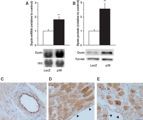
Comparable to the results obtained by the DNA microarray analysis of combined MKK3bE and WTp38α gene transfer (Citation3), the p38 MAPK overexpression increased significantly both dyxin mRNA and protein levels in the left ventricle, as measured by Northern blot and Western blot analyses, respectively (). To identify the cell types expressing dyxin in the adult rat heart, immunohistochemistry with a specific dyxin antibody was performed. In the LacZ-treated control hearts, dyxin was mainly expressed in the endothelial cells, whereas a fairly weak signal was detected in the cytoplasm of cardiac myocytes and vascular smooth muscle cells (). However, enhanced cytoplasmic immunostaining was observed in myocytes in the inflammatory area surrounding the adenovirus injection site in both LacZ control hearts () and MKK3bE plus WTp38α-treated hearts () without clear differences between the two groups. Interestingly, in MKK3bE plus WTp38α-treated hearts dyxin was localized also to the nuclei of the cardiac myocytes (). We have previously studied cell types affected by p38 MAPK overexpression in the rat left ventricle in vivo and p38 MAPK was found to affect inflammatory cells, endothelial cells and cardiomyocytes (Citation3).
Figure 1. Adenovirus-mediated overexpression of p38 MAPK. Dyxin mRNA levels in rat left ventricles (A). Results (mean±SEM, n=9–10) are expressed as a ratio of dyxin mRNA to 18S mRNA as determined by Northern blot analysis. White columns, LacZ; black columns, p38 MAPK. Western blot analysis showing dyxin protein levels in rat left ventricles (B). Results are mean±SEM, n=8–10. White columns, LacZ; black columns, p38 MAPK. Immunohistochemistry with a specific dyxin antibody (400× magnification) revealed strong staining in the cytoplasm of endothelial cells in LacZ-treated control hearts (C), whereas only weak signal was observed in the cytoplasm of the vascular smooth muscle cells and cardiac myocytes except the inflammatory area surrounding the adenovirus injection site (D). MKK3bE + WTp38α-treated hearts show dyxin staining to be localized also to the nucleus of the myocytes (E). The endothelium in D and E is indicated with arrowhead **p<0.01 vs LacZ (Student's t-test).
Effect of pressure overload on dyxin expression in the left ventricle
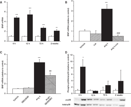
To characterize the effect of pressure overload on cardiac dyxin expression, a model of Ang II-induced hypertension in conscious rats was used (Citation11). Left ventricular dyxin mRNA levels were highest at 6 h and remained elevated up to 12 h during Ang II infusion, administered via osmotic minipumps (). The increase in mRNA levels during Ang II infusion was followed by induction of dyxin protein levels (). Western blot analysis revealed increased cytoplasmic and membrane dyxin protein levels, whereas no change in nuclear dyxin protein levels in response to Ang II infusion was noted (). Simultaneous infusion of Ang II type 1 receptor (AT1-receptor) antagonist losartan completely abolished the Ang II-induced activation of left ventricular dyxin gene expression at 6 h (). To study further the mechanism regulating dyxin gene expression, p38 MAPK inhibitor SB203580 was administrated simultaneously with Ang II. Administration of SB203580 reduced Ang II-induced elevation of left ventricular dyxin gene expression by 50% (p<0.05) at 6 h (). To characterize the model at the level of gene expression, we measured BNP mRNA levels in the left ventricle, since upregulation of BNP gene represents a typical genetic reprogramming in the heart subjected to acute hemodynamic overload (Citation9,Citation20). Ang II caused induction of BNP gene at 6 h of Ang II infusion, and increase in BNP mRNA levels in the left ventricle remained upregulated up to 2 weeks (). AT1-receptor blockade by losartan completely abolished Ang II-induced changes in the cardiac BNP gene expression at 6 h, and when given alone, losartan did not modify BNP mRNA levels (). SB203580 only partially (42%, p<0.01) reduced Ang II-induced changes in left ventricular BNP gene expression at 6 h, suggesting slight inhibition of Ang II-induced hemodynamic load (). We also measured p38 MAPK phosphorylation in response to Ang II treatment, and activation of p38 MAPK phosphorylation correlated with the early induction of dyxin mRNA levels (). The rapid induction of cardiac dyxin gene expression in response to acute pressure overload was confirmed by using AVP infusion in conscious normotensive rats (Citation8,Citation9). Increase in left ventricular dyxin mRNA levels was seen already after 1 h of AVP infusion followed by significant elevation of protein levels at 4 h (data not shown).
Figure 2. The effect of Ang II administration (33 μg/kg/h) on left ventricular dyxin mRNA levels in rats (A). Results are mean±SEM (n=6–8). ***p<0.00l, **p<0.0l vs vehicle (Student's t-test). White columns, vehicle; black columns, Ang II. Dyxin protein levels in the rat left ventricles (B). Results are mean±SEM (n=7–8). ***p<0.00l vs vehicle (Student's t-test). White columns, vehicle; black columns, Ang II. The effect of Ang II administration on dyxin nuclear, cytoplasmic and membrane protein levels at 3 days of Ang II infusion (C). Representative Western blots are shown. The effect of Ang II and AT1-receptor blockade by losartan (Los) on left ventricular dyxin mRNA levels at 6 h (D). Results are mean±SEM (n=6–7). ***p<0.00l vs vehicle. †††p<0.001 vs Ang II (ANOVA). The effect of Ang II and p38 MAPK inhibitor SB203580 on left ventricular dyxin mRNA levels at 6 h (E). Results are mean±SEM (n=4–7). ***p<0.00l vs vehicle. †p<0.05 vs Ang II (ANOVA). All mRNA results are expressed as a ratio of dyxin mRNA to 18S mRNA as determined by Northern blot analysis.
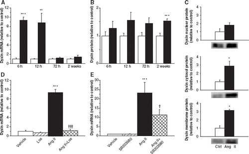
Figure 3. The effect of Ang II administration (33 μg/kg/h) on left ventricular BNP mRNA levels in rats (A). ***p<0.00l, **p<0.0l vs vehicle (Student's t-test). White columns, vehicle; black columns, Ang II. The effect of Ang II and AT1-receptor blockade by losartan (Los) on left ventricular BNP mRNA levels at 6 h (B). Results are mean±SEM (n=6–7). ***p<0.00l vs vehicle, †††p<0.001 vs Angll (ANOVA). The effect of Ang II and p38 MAPK inhibitor SB203580 on left ventricular BNP mRNA levels (C) at 6 h. Results are mean±SEM (n=4–7). ***p<0.00l vs vehicle. ††p<0.0l vs Ang II (ANOVA). All mRNA results are expressed as a ratio of BNP mRNA to 18S mRNA as determined by Northern blot analysis. The effect of Ang II administration (33 μg/kg/h) on the activation of p38 MAPK in rats (D). Western blot analysis showing total-p38 and phospho-p38 protein levels in the rat left ventricles. Results are mean±SEM (n=3–4). **p<0.0l vs vehicle (Student's t-test). White columns, vehicle; black columns, phospho-p38/total-p38.
Cardiac remodeling after myocardial infarction and dyxin expression
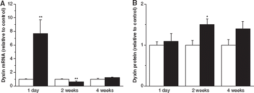
To study the effect of post-infarction cardiac remodeling on cardiac dyxin gene expression, ligation of left ascending coronary artery was performed in rats, producing left ventricular dilation, thinning of the anterior wall and hypertrophy of the posterior wall (Citation13). Dyxin mRNA levels were markedly upregulated in the left ventricle at day 1 following MI when compared to sham-operated animals, whereas a significant decrease in dyxin gene expression was seen at 2 weeks, returning to baseline levels at 4 weeks after MI (). Dyxin protein levels increased at 2 weeks following MI () indicating that also post-infarction cardiac remodeling process regulates dyxin expression.
Figure 4. The effect of myocardial infarction on left ventricular dyxin mRNA levels (A). Results (mean±SEM, n=5–8) are expressed as a ratio of dyxin mRNA to 18S mRNA as studied by real-time quantitative RT-PCR. The effect of myocardial infarction on left ventricular dyxin protein levels (B) as studied by Western blot. Results are mean±SEM (n=7). White columns, sham-operated; black columns, myocardial infarction. **p<0.0l, *p<0.05 vs sham-operated animals (Student's t-test).
Effect of mechanical stretch on dyxin expression
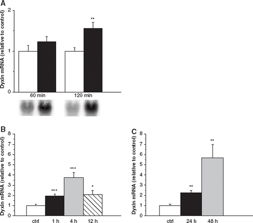
Because cardiac overload in vivo activated left ventricular dyxin expression, we evaluated the effect of direct left ventricular wall stress on dyxin gene expression by using an ex vivo stretch model in isolated perfused rat hearts (Citation14,Citation15). The increase in left ventricular wall stress ex vivo for 1 h did not significantly increase dyxin gene expression, but in response to 2 h of left ventricular wall stress, a significant increase in dyxin gene expression was noted (). Because immunohistochemistry showed that both myocytes and endothelial cells were stained with dyxin, we next examined whether mechanical stretch regulates dyxin explicitly in cardiac myocytes. The dyxin gene expression was measured at 1, 4, 12, 24 and 48 h after start of cyclic mechanical stretch in neonatal ventricular myocytes in vitro. Cyclic mechanical stretch elevated dyxin mRNA levels already after 1 h, peaking at 4 and 48 h ().
Figure 5. The effect of Stretch on left ventricular dyxin mRNA levels using ex vivo Langendorff method (A). Results (mean±SEM, n=6) are expressed as a ratio of dyxin mRNA to 18S mRNA as determined by Northern blot analysis. **p<0.0l vs control (Student's t-test). Dyxin mRNA levels after mechanical Stretch in vitro in neonatal rat ventricular myocytes. Experiments were carried out in two separate sets each having their own controls: (i) 1, 4 and 12 h, and (ii) 24 and 48 h of Stretch (B). Results are mean±SEM (n=8). Dyxin mRNA levels after mechanical Stretch in vitro in neonatal rat ventricular myocytes as studied by real-time quantitative RT-PCR (C). Results are mean±SEM (n=6) and expressed as a ratio of dyxin mRNA to 18S mRNA. White columns, control; black columns, Stretch. ***p<0.001, **p<0.01, *p<0.05 vs control (Student's t-test).
Discussion
The LIM protein family is considerably heterogeneous, linked to a variety of fundamental biological processes including cytoskeletal organization, cell lineage specification and organ development (Citation21). Previously, only few LIM proteins, such as human heart LIM protein, have been implicated in pathophysiological processes in the heart (Citation22). Dyxin is a LIM domain protein acting as a transcriptional cofactor (Citation4), whose biological role and downstream mediators have not been elucidated. In the present study, we characterized the mechanisms regulating dyxin expression in the heart.
This study demonstrates that cardiac dyxin gene expression is rapidly upregulated in response to pressure overload, similarly to well-known markers of early hypertrophic genetic response, e.g. components of the activator protein-1 (AP-1) transcription factor complex (c-fos, Jun-B, Fra-1) and early growth response (Egr-1) (Citation23,Citation24). Ang II-induced upregulation of dyxin gene expression in left ventricle was abolished by AT1-receptor antagonist losartan, which most likely can be explained by the inhibition of Ang II-induced increase in mean arterial pressure. The rapid elevation of left ventricular dyxin mRNA levels was followed by increased dyxin protein levels. This different time-dependent upregulation of mRNA and protein levels may related to the involvement of post-transcriptional regulation of dyxin gene expression and/or enhanced stability of dyxin mRNA. Moreover, many transcription factors, including GATA-4, are regulated through post-translational modifications such as phosphorylation (Citation1,Citation2). Our results also indicate that dyxin gene expression is rapidly upregulated in response to MI in vivo as well as to direct myocyte stretch in vitro indicating that mechanical load is a critical regulator of cardiac dyxin gene expression. Moreover, the observed biphasic activation of dyxin gene expression suggests that dyxin may have a role both in the early and later phases of the hypertrophic response in cardiomyocytes. Consequently, this biphasic activation of dyxin gene expression may indicate that besides being critical regulator of the early genetic response to hemodynamic overload dyxin also may contribute to subsequent extracellular matrix remodeling in cardiac hypertrophy.
Nothing is known about the mechanisms and signaling pathways regulating cardiac dyxin expression. Our results are first to show that p38 MAPK regulates cardiac dyxin expression. The role of p38 MAPK as regulator of dyxin expression in the adult heart was established here by using two experimental approaches. Adenovirus-mediated overexpression of p38 MAPK resulted in increased dyxin mRNA and protein levels. During Ang II infusion, activation of p38 MAPK phosphorylation correlated with the early induction of dyxin mRNA levels. Furthermore, pharmacological inhibition of p38 MAPK reduced in part Ang II-induced activation of dyxin gene expression. Previously several transcription factors have been shown to be activated by p38 MAPK in the adult heart in vivo, including GATA-4, AP-1, serum response factor (SRF) and nuclear factor κB (NF-κB) (Citation3). The transcriptional activity of GATA-4 and many other transcription factors is regulated via phosphorylation by p38 MAPK pathway (Citation25). However, whether p38 MAPK directly activates dyxin remains to be studied. Collectively, our results indicate that dyxin mRNA levels are upregulated rapidly in response to pressure overload, and this increase is, at least in part, mediated by p38 MAPK. However, it is likely that also other kinases such as glycogen synthase kinase 3β may regulate dyxin during hypertrophic process. Furthermore, we cannot exclude that distinct cell-specific effects are caused by myocyte stretch and adenoviral overexpression of p38 MAPK.
The biological role and transcriptional cofactors of dyxin are poorly understood. As transcriptional regulator, dyxin has been shown to interact with GATA1/4/6 proteins (Citation4). Based on our immunohistochemical stainings, dyxin is expressed in endothelial cells in the normal adult rat heart, which suggests possible interaction with GATA-6, being expressed in endothelial cells of normal tissues (Citation26). Our finding that dyxin is expressed in endothelium and cardiomyocytes of adult rat heart in similar manner to GATA-6 and GATA-4, respectively, is in agreement with previous observation that dyxin interacts with these members of GATA-family (Citation4). Because dyxin expression was increased in several models of cardiac overload, it is intriguing to speculate that the activity of GATA-factors may be regulated partly via interaction with dyxin during these processes. Because GATA-4 is the member of GATA-factors that acts as crucial regulator of gene expression in cardiac myocytes, we suggest that dyxin may interact with GATA-4 in loaded heart.
A recent report showed that interaction between GATA-6 and dyxin inhibited GATA-6 DNA binding, resulting in repression of GATA-6 activation (Citation4). On the other hand, in several reported GATA–LIM interactions, LIM proteins such as cysteine-rich proteins (CRP-1 and -2), act as coactivators of GATA-mediated gene transcription (Citation27). We have previously shown under identical experimental conditions to our present study that p38 MAPK overexpression (Citation3), and direct mechanical stretch increase GATA-4 binding (Citation18). Accordingly, it is likely that dyxin may act as a coactivator rather than repressor of GATA-4 mediated gene expression in cardiac myocytes. Nevertheless, since GATA-4 interacts with both coactivators and corepressors, the exact role of dyxin as a transcriptional co-regulator of GATA-4 in response to hypertrophic stimuli remains to be studied. Moreover, our results show that Ang II-induced upregulation of dyxin resulted in increased cytoplasmic and membrane but not nuclear levels of dyxin suggesting that dyxin is not translocated to the nucleus or its nuclear export is induced, and thus unable to activate or repress GATA-4 binding and function. However, although it has been shown that dyxin acts as a repressor of GATA function in heart (Citation4), it remains to be established whether this interaction has functional and structural consequences in mechanical load-induced cardiac hypertrophic response or on risk of cardiovascular events.
In conclusion, our present study provides new information of dyxin being a novel p38 MAPK regulated target associated with mechanical load induced hypertrophic response. Although the precise functional role of dyxin in cardiac hypertrophy is yet unclear, we hypothesize that dyxin may play a critical role in regulating hypertrophic gene expression by interacting with GATA-4. A major impact of this study was to indicate for the first time that dyxin is upregulated in response to pressure load in the heart and this upregulation is at least partly mediated by p38 MAPK.
Acknowledgments
We thank Marja Arbelius, Pirjo Korpi, Kaisa Penttilä, Sirpa Rutanen, Erja Tomperi, Mirja Vahera and Kati Viitala for expert technical assistance.
Funding
This study was supported by the Academy of Finland, the Finnish Foundation of Cardiovascular Research, the Sigrid Jusélius Foundation and the Research and Science Foundation of Farmos.
Declaration of interest: The authors have no conflicts of interest that are directly relevant to the content of this study.
References
- Akazawa H, Komuro I. Roles of cardiac transcription factors in cardiac hypertrophy. Circ Res. 2003;92:1079–1088.
- Oka T, Xu J, Molkentin JD. Re-employment of developmental transcription factors in adult heart disease. Semin Cell Dev Biol. 2007;18:117–131.
- Tenhunen O, Rysä J, Ilves M, Ruskoaho H, Leskinen H. Identification of cell cycle regulatory and inflammatory genes as predominant targets of p38 MAPK in the heart. Circ Res. 2006;99:485–493.
- Rath N, Wang Z, Lu MM, Morrisey EE. LMCD1/Dyxin is a novel transcriptional cofactor that restricts GATA6 function by inhibiting DNA binding. Mol Cell Biol. 2005;25:8864–8873.
- Bespalova IN, Burmeister M. Identification of a novel LIM domain gene, LMCD1, and chromosomal localization in human and mouse. Genomics. 2000;63:69–74.
- Pikkarainen S, Tokola H, Kerkelä R, Ruskoaho H. GATA transcription factors in the developing and adult heart. Cardiovasc Res. 2004;63:196–207.
- Prussak CE, Almazan MT, Tseng BY. Peptide production from proteins separated by sodium dodecylsulfate polyacrylamide gel electrophoresis. Anal Biochem. 1989;178:233–238.
- Romppanen H, Puhakka J, Földes G, Szokodi II, Vuolteenaho O, Tokola H, Toth M, Ruskoaho H. Endothelin-1-independent and angiotensin II-independent induction of adrenomedullin gene expression. Hypertension. 2001;37:84–90.
- Magga J, Marttila M, Mäntymaa P, Vuolteenaho O, Ruskoaho H. Brain natriuretic peptide in plasma, atria, and ventricles of vasopressin- and phenylephrine-infused conscious rats. Endocrinology. 1994;134:2505–2515.
- Földes G, Suo M, Szokodi I, Lako-Futo Z, de Chatel R, Vuolteenaho O, . Factors derived from adrenals are required for activation of cardiac gene expression in angiotensin II-induced hypertension. Endocrinology. 2001;142:4256–4263.
- Suo M, Hautala N, Földes G, Szokodi I, Toth M, Leskinen H, . Posttranscriptional control of BNP gene expression in angiotensin II-induced hypertension. Hypertension. 2002;39:803–808.
- Pfeffer MA, Pfeffer JM, Fishbein MC, Fletcher PJ, Spadaro J, Kloner RA, . Myocardial infarct size and ventricular function in rats. Circ Res. 1979;44:503–512.
- Tenhunen O, Soini Y, Ilves M, Rysä J, Tuukkanen J, Serpi R, . p38 Kinase rescues failing myocardium after myocardial infarction: Evidence for angiogenic and anti-apoptotic mechanisms. FASEB J. 2006;20:1907–1909.
- Szokodi I, Kinnunen P, Tavi P, Weckström M, Toth M, Ruskoaho H. Evidence for cAMP-independent mechanisms mediating the effects of adrenomedullin, a new inotropic peptide. Circulation. 1998;97:1062–1070.
- Tenhunen O, Sarman B, Kerkelä R, Szokodi I, Papp L, Toth M, . Mitogen-activated protein kinases p38 and ERK 1/2 mediate the wall stress-induced activation of GATA-4 binding in adult heart. J Biol Chem. 2004;279:24852–24860.
- Thienelt CD, Weinberg EO, Bartunek J, Lorell BH. Loadinduced growth responses in isolated adult rat hearts. Role of the AT1 receptor. Circulation. 1997;95:2677–2683.
- Hautala N, Tenhunen O, Szokodi I, Ruskoaho H. Direct left ventricular wall stretch activates GATA4 binding in perfused rat heart: Involvement of autocrine/paracrine pathways. Pflugers Arch. 2002;443:362–369.
- Pikkarainen S, Tokola H, Majalahti-Palviainen T, Kerkelä R, Hautala N, Bhalla SS, . GATA-4 is a nuclear mediator of mechanical stretch-activated hypertrophic program. J Biol Chem. 2003;278:23807–23816.
- Liang F, Wu J, Garami M, Gardner DG. Mechanical strain increases expression of the brain natriuretic peptide gene in rat cardiac myocytes. J Biol Chem. 1997;272:28050–28056.
- Magga J, Vuolteenaho O, Tokola H, Marttila M, Ruskoaho H. Involvement of transcriptional and posttranscriptional mecha-nisms in cardiac overload-induced increase of B-type natriuretic peptide gene expression. Circ Res. 1997;81:694–702.
- Bach I. The LIM domain: Regulation by association. Mech Dev. 2000;91:5–17.
- Zheng B, Wen JK, Han M, Zhou AR. hhLIM protein is involved in cardiac hypertrophy. Biochim Biophys Acta. 2004;1690:1–10.
- Karin M, Liu Z, Zandi E. AP-1 function and regulation. Curr Opin Cell Biol. 1997;9:240–246.
- Shaulian E, Karin M. AP-1 as a regulator of cell life and death. Nat Cell Biol. 2002;4:E131–136.
- Kyriakis JM, Avruch J. Mammalian mitogen-activated protein kinase signal transduction pathways activated by stress and inflammation. Physiol Rev. 2001;81:807–869.
- Kamnasaran D, Guha A. Expression of GATA6 in the human and mouse central nervous system. Brain Res Dev Brain Res. 2005;160:90–95.
- Chang DF, Belaguli NS, Iyer D, Roberts WB, Wu SP, Dong XR, . Cysteine-rich LIM-only proteins CRP1 and CRP2 are potent smooth muscle differentiation cofactors. Dev Cell. 2003;4:107–118.