Abstract
Background. The aim is to evaluate whether cardiovascular (CV) risk stratification in newly diagnosed hypertensive patients according to the European Society of Hypertension (ESH) guidelines, can predict the evolution of target organ damage (TOD) using routine examinations in clinical practice during 1 year. Methods. Prospective study of recently diagnosed untreated hypertensives. At the moment of inclusion and 1 year later, urinary albumin excretion rate (UAER), blood analysis, electrocardiogram, retinography, self-monitored blood pressure (BP) and ambulatory BP measurement were performed. TOD was defined following the ESH guidelines and evaluated as having favorable or unfavorable evolution. Results. Four hundred and seventy-nine hypertensive patients were included (58.8 years; 43.4% women). The baseline prevalence of TOD was: high UAER (2.4%), left ventricular hypertrophy (LVH) (20.7%), advanced lesion of the fundus oculi (FO) (10.2%). After 1 year, no differences were found between the final systolic and diastolic BP neither in the high/very high nor in the low/moderate CV risk groups. Patients with low/moderate CV risk had less unfavorable TOD evolution, LVH (9.2% vs 41.7%; p <0.001), FO advanced damage (0.99% vs 14.3%; p <0.001), high UAER (0.3% vs 5.1%; p <0.005) and amount of TOD (9.2% vs 44.0%; 0<0.001) than those with high/very high CV risk. The odds ratios of favorable TOD evolution adjusted for BP change and antihypertensive drug treatment were (low/moderate vs high/very high CV risk); 5.14 (95% confidence interval, CI, 3.99–6.64) for LVH; 12.42 (6.67–23.14) FO advanced damage; 10.71 (3.67–31.22) high UAER and 13.99 (10.18–19.22) for amount of TOD. Conclusions. It is possible to detect variations in TOD in hypertensive patients with a 1-year follow-up using the examinations available in routine clinic practice. The risk determined by the ESH guidelines predicts the evolution of TOD at 1 year.
Introduction
Target organ damage (TOD) in hypertensive patients is useful for stratifying cardiovascular (CV) risk (Citation1). Its presence allows CV risk to be individualized, in the sense that it represents a phenotypic expression of vulnerability to the effects of hypertension (HT) and other risk factors (Citation2). Population risk tables do not include TOD, and their predictive capacity for individual risk is, therefore, around 50%, which means that approximately half of the individuals presenting CV events present a low or moderate calculated risk (Citation3). Moreover, TOD is not static, and undergoes modifications with time, with greater amount of TOD increasing the CV risk for each individual (Citation4). Thus, in hypertensive patients, the appearance of new TOD and the persistence or progression of previously existing damage are associated with a greater CV risk (Citation5–7), and indicate that the treatment of HT and other risk factors is insufficient. Conversely, the disappearance or regression of previously existing TOD lead to lower CV risk, and are indicators of good control of HT and other risk factors (Citation7,Citation8). Therefore, the detection of TOD is not only important to stratify CV risk among hypertensive patients at diagnosis, but also to evaluate its evolution, by monitoring during follow-up, as an expression of the degree to which HT and CV risk are being controlled.
The effectiveness of the different examination methods used to detect the presence of TOD varies, as well as their availability (Citation1). Evidence shows that the determination of microalbuminuria (MA), estimated kidney function (glomerular filtration rate; GFR) and electrocardiogram (ECG), together with a blood analysis, allow CV risk stratification exhibiting similar results to those obtained through echocardiogram and carotid echography (Citation9). In advanced lesions (hemorrhages, soft exudates, hard exudates), the fundus oculi (FO) examination by retinography allows additional stratification of 10% of the hypertensive patients (Citation10). These examinations are available in most countries, and are generally routine procedures in the evaluation of HT.
The primary aim of this study is to evaluate whether CV risk stratification in newly diagnosed hypertensive patients, according to the European Society of Hypertension (ESH) guidelines, can predict the favorable or unfavorable evolution of TOD at 1-year follow-up, as well as to identify those cases in which the control of HT is more important. The secondary aim is to assess whether routine examinations conducted in clinical practice can detect changes in the evolution of TOD after 1 year of follow-up.
Patients and method
Study population
The VAMPAHICA study (acronym in Spanish of ‘assessment of the self monitoring of blood pressure (BP) in the diagnosis of isolated clinical hypertension’) has been previously described (Citation11). Briefly, this was a multi-center study, involving 14 primary care centers in the Girona Healthcare Region (Catalonia), with a total of 140 collaborators. The studied population included all the patients visiting the healthcare professionals participating in the study. Participants were untreated hypertensive patients, recently diagnosed, and with at least 1 year of follow-up.
Study design
This was a prospective cohort study. Patients who met the following criteria were included: (i) aged 15–75 years; (ii) clinically diagnosed HT, defined as the average of two BP readings, separated by 2 min, measured on 3 different days with results of ≥140 and/or ≥90 mmHg; (iii) recently diagnosed hypertensive patients never treated for HT; (iv) patients providing correct self-BP monitoring (SBPM) and ambulatory BP monitoring (ABPM). Exclusion criteria were the following: obvious inability to perform SBPM; diabetes mellitus; secondary HT; previous CV disease; renal insufficiency (serum creatinine >2 mg/dl); liver insufficiency; alcoholism or severe psychiatric disease; endocrine or severe hematological disease; other severe diseases or limitations, which, in the physician's opinion, were a reason for exclusion. Diabetic patients were excluded to avoid confusing diabetic lesions with hypertensive lesions in the examination of the FO.
The study was approved by the Clinical Research Ethics Committee of the Healthcare Institute, Girona, and all the participants signed an informed consent document.
Determination of BP and control of HT
Determination of clinic BP (CBP). Clinical HT was diagnosed based on measurements taken by the nurses. After sitting down for 5 min, two measurements were performed at an interval of 2 min. This was performed on 3 different days. If the difference between the readings on the same day was greater than 5 mmHg, a third measurement was performed to obtain the mean. The CBP value was the mean of all the measurements. All measurements were performed using Omron 705 CP and Omron 705 IT monitors, under the standard conditions that are recommended by international organizations (Citation1), with an armband adapted to the circumference of each patient's arm.
SBPM procedure. This was performed on all the patients included in the study. Each participant was trained by an expert nurse regarding the steps required to obtain adequate readings, confirming twice that the process was carried out correctly in her presence. All the measurements were performed using Omron 705 CP and Omron 705 IT monitors. It was carried out with an armband adapted to the circumference of each patient's arm. Patients were given a written guide containing the instructions. During 3 working days, two readings were performed in the morning before breakfast, and two more at night before dinner, in both cases at an interval of 2 min, and after sitting down for 5 min. Patients wrote down the readings in a form they were given for this purpose. In order to check the reliability of the data, they also provided the readings obtained through the monitor. The first day's readings were discarded in average calculations.
ABPM procedure. Twenty-four-hour ABPM was conducted 2–3 weeks after the SBPM. The researcher who carried out the latter was not aware of the results of the former readings. The following validated automatic oscillometric monitors were used: SpaceLabs 90207 and Spacelabs 90217 (Redmond, WA). All the monitors were programmed to carry out readings every 20 min during the day period (08:01–23:00 h) and every 30 min during the sleep period (23:01–08:00 h), on a weekday, or according to normal activity if the patient did not work. The sleep period was defined as being between 23:00 and 08:00 h.
SBPM was performed once a year, as well as ABPM. Measurement of CBP was performed each time the patient came to the doctor's or nurse's office in relation to HT.
Treatment
Patients were treated at the discretion of the family doctor, following the guidelines of the health region (based on ESH Guidelines).
Data collection, variables and follow-up
Data collection. A case report form specially designed for this study was used. The variables included in this study were: gender, age, body mass index (BMI), tobacco consumption, systolic and diastolic CBP, systolic and diastolic SBPM, systolic and diastolic ABPM, total cholesterol, high-density lipoprotein (HDL)-cholesterol, low-density lipoprotein (LDL)-cholesterol, blood glucose, left ventricular hypertrophy (LVH), urinary albumin excretion rate (UAER), GFR according to the MDRD (Modification of Diet in Renal Disease) formula, advanced lesions on FO and antihypertensive treatment at the follow-up visit. All data were entered into a computer database.
Initial study and follow-up. At baseline and every year, all the participants included in the study underwent a physical examination, blood analysis, standard 12-lead ECG and retinography. The retinography was carried out using a retinograph equipped with a non-mydriatic digital camera (Canon CR6-45NM, Camera EOS D30) and was evaluated by an experienced physician unaware of the patient's details. For the detection of UAER, a morning urine sample was analyzed. If positive, the presence of leukocytes, red blood cells or nitrites was ruled out using a reactive strip. Once the cause of the anomaly found on the test strip (if any) had been examined and treated, an early morning urine test to determine the albumin/creatinine ratio was repeated 15 days later. Two out of three consecutive test results were required to be positive in order to make the diagnosis. Smokers were defined as patients who had consumed tobacco in the last 6 months.
Any of the following alterations were considered TOD: serum creatinine >1.2 mg/dl or 1.3 mg/dl in women and men, respectively; LVH by electrocardiographic criteria (Cornell criteria, modified by Dalfó et al. (Citation12) or Sokolow–Lyon criteria); presence of high UAER defined using the ESH values for normal (Citation1); alteration to kidney function, expressed as estimated GFR <60 ml/min using the MDRD formula (Citation13). The presence of advanced lesions of the FO, such as soft exudates or hard exudates and hemorrhages, were also included.
Stratification of CV risk. The CV risk of each patient was calculated according to the ESH Guidelines except for LVH. In this case, we used the Cornell voltage criteria amended by Dalfó et al. (Citation12) because of higher yields in our population. We calculated the number of TOD in each patient and used it as variable in the analysis. After 1 year of follow-up, all cases of TOD were classified as: persistent if they still met the established criteria; new if they did not meet criteria at the baseline visit but were positive after 1 year; and in regression if they were present at the baseline visit and did not meet criteria after 1 year. It was considered favorable when a TOD disappeared or remained negative, and when the number of total TODs decreased or remained zero. Unfavorable evolution was considered when new TOD was detected, persisted and when the total number of TODs did not changed or increased. In the analysis, two groups of CV risk were considered: low or moderate, and high or very high.
Statistical analysis
After carrying out a descriptive analysis of the hypertensive cohort, the differences in the means and/or percentages for the variables of interest were tested. The difference in percentages was contrasted using non-parametric contrast (by means of a chi-square in a contingency table) and the means were contrasted by Student's t-test for independent samples (contrasting the homogeneity of the variance by means of Levene's F-test).
A multivariate analysis was carried out. Specifically, for each individual, both the baseline and 1-year measurements were considered. Response variables were dichotomous denoting, for each TOD, appearance or persistence and disappearance or never. Models were adjusted for cholesterol, LDL-cholesterol, HDL-cholesterol, triglycerides, UAER, GFR, BMI, treatment with antihypertensive drugs, CBP at baseline and after 1 year. Random effects were entered in the models in order to check unobserved individual heterogeneity and possible dependence arising from considering two dimensions per individual; p <0.05 was considered statistically significant and all tests were two-tailed. The contrasts of means and ratios were performed using the statistical software SPSS v.15. The causal model estimates were performed using the free access software R v. 2.11.0.
Results
Baseline and final data of the cohort
shows the baseline characteristics and the characteristics after a 1-year follow-up. Of the 479 hypertensive patients included in the study, 46.7% were women and the average age was 58.8 years (SD =10.57). Follow-up periods were conducted after 1.18 years (SD 0.99), and the number of consultations by nurse or physician were 2.33 (SD =1.06). The only treatment recommended to 44.2% of the patients was lifestyle modifications, whereas the rest (55.8%) were taking antyhypertensive treatment at the follow-up visit. Recommended drugs were: 11.4% diuretics, 7.7% beta-blockers, 31.0% angiotensin-converting enzyme inhibitor, 0.6% calcium antagonist, 3.1% angiotensin receptors blockers and others in 2.0%.
Table I. Baseline and final data after 1-year follow-up in hypertensive patients.
Risk stratification, baseline and final follow-up associated variables
Baseline CBP, total cholesterol and LDL-cholesterol significantly contributed to CV risk stratification (). After 1-year follow-up, no differences were detected in fasting glucose, total cholesterol, HDL-cholesterol and LDL-cholesterol neither among hypertensive patients with baseline low/ moderate risk, nor in those with high/very high risk. Hypertensive patients with high/very high CV risk reached a final systolic and diastolic CBP, systolic and diastolic SBPM or systolic and diastolic ABPM (day) similarly to patients with low/moderate CV risk ().
Table II. Baseline variables according to cardiovascular risk ESH guidelines.
Table III. Baseline and final variables of the cohort according to cardiovascular risk ESH guidelines stratification.
Risk stratification and TOD evolution. Patients at baseline included in the low or moderate risk groups, experienced a significantly favorable evolution of the TOD in comparison with those included in the high or very high CV risks ( and , and and ). After adjusting for variation in systolic and diastolic CBP and treatment with antihypertensive drugs, patients with low/moderate CV risk presented higher odds ratio (OR) of presenting a favorable evolution of TOD (). Adjusted results showed that patients with LVH or advanced FO lesions had more chances to reduce CV risk ().
Table IV. Absence, regression, persistence or appearance of TOD after 1 year, according to cardiovascular risk at baseline.
Table V. Target organ damage evolution after 1 year, according to CV risk at baseline of hypertensive patients.
Figure 1. Unfavorable evolution of target organ damage (TOD) after 1 year according to baseline cardiovascular (CV) risk. ESH, European Society of Hypertension LVH: left ventricular hypertrophy; FO: fundus oculi; UAER: urinary albumin excretion rate; GFR: glomerular filtration rate. *p <0.001 **p <0.005.
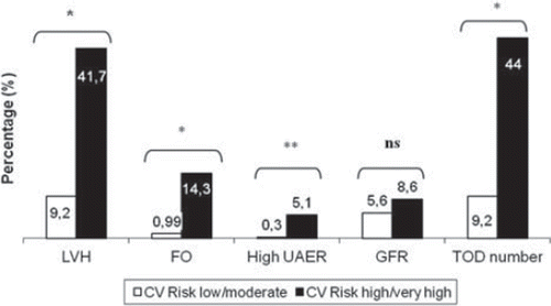
Figure 2. Absence, regression, persistence or appearance of target organ damage (TOD) after 1 year according to cardiovascular (CV) risk at baseline (%). *p <0.001.
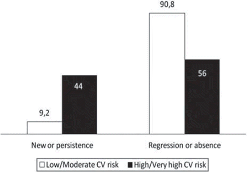
Table VI. Odds ratio of TOD's favorable evolution, according to baseline cardiovascular risk (low/moderate risk vs high/very high risk) of hypertensive patients.
Table VII. Multivariate analysis of cardiovascular risk change group of hypertensive patients.
Discussion
The primary objective was to evaluate whether CV risk stratification in newly diagnosed hypertensive patients, according to the ESH guidelines, can predict the favorable or unfavorable evolution of TOD at 1-year follow-up with the common examinations conducted in clinic practice. Our results show that, despite having similar BP reduction and similar antihypertensive drug treatment (specially with angiotensin-converting enzyme inhibitors), patients whose baseline CV risk is low or moderate experience more favorable evolution of TOD than those whose baseline risk is high or very high. The estimated baseline CV risk according to the ESH tables (including advanced lesions of the FO), is associated with significant differences in the evolution of TOD changes at follow-up. Patients with high or very high CV risk are more likely to have a lower percentage of favorable outcomes, and are more susceptible to experience an unfavorable evolution, thereby requiring careful control of BP and other risk factors.
Changes in the different types of TOD are observed in newly diagnosed hypertensive patients after 1 year. Results show that there are differences according to baseline CV risk of each patient, with the exception of renal function. The definition of favorable or unfavorable evolution includes the continued absence or presence of TOD as well as the appearance or disappearance of the TOD. This definition is better suited to one of the goals of the antihypertensive treatment, which is not to simply provoke the disappearance of TOD, but also to avoid its appearance or persistence.
In our study, after 1 year, only changes in LVH and FO advanced lesions were associated with favorable changes in CV risk. It is likely that with a longer follow-up period this study would show that the evolution of other TODs is also associated with favorable CV risk re-stratification.
In our study, not all the patients received antihypertensive drug treatment. Patients with high or very high CV risk received more drug treatment than those with low/moderate CV risk. However, it was not significant when compared with lower CV risk patients. In fact, although systolic and diastolic final BP, as well as the percentage of patients with controlled BP, were similar between groups at risk, a higher percentage of hypertensive patients with high/very high risk had a BP ≥160/100 mmHg. So, it is possible that if more patients with high/very high risk had been treated with drugs until reaching a normal BP, the TOD's evolution would have been more favorable. In this sense, other authors previously reported that BP is more difficult to control in patients with TOD (Citation14), and that an early intensive treatment reduces the risk of CV events (Citation15). Our study shows that for a similar reduction in BP, and for a similar drug treatment, patients with high or very high CV risk are more prone to experience an unfavorable evolution than those with low or moderate CV risk. In our study, patients in the cohort have been treated following their family physician criteria, which led to a low percentage of pharmacological treatments and poor BP control, especially in patients with high or very high CV risk. ESH Guidelines recommend starting drug treatment after 3 months if BP is not controlled or before 3 months if TOD is present. Several hypertensive patients have not followed these guidelines. This last point allowed us to show that managing hypertensive patients without taking into account the CV risk is associated with poorer outcomes in those whose CV risk is high or very high.
Identification and follow-up of TOD in hypertensive subjects by means of routine examinations in clinical practice, easily accessible and available, allows their evolution to be monitored, providing information on whether the antihypertensive therapy (Citation16,Citation17), the approach to other risk factors (Citation18) and the patient's compliance (Citation19) are adequate. It also allows the identification of a situation of “isolated ambulatory resistance” or “inverse white coat effect” in hypertensive patients who apparently have a well-controlled BP at the medical visit (Citation20).
Retinography enables high-quality images of the FO to be obtained, facilitating the identification of exudates and hemorrhages, and stratification of risk (Citation10). Although examination of the FO is not currently considered routine (Citation1), the growing availability of retinography in healthcare, and the prevalence of advanced lesions (exudates or hemorrhages) in newly diagnosed hypertensive patients, with a prevalence of around 10% (Citation21), support its use.
Nevertheless, we would like to discuss some limitations of our study. First, the evolution of LVH was made using a dichotomous criteria (presence or absence) and quantitative criteria such as voltage or Cornell product. If the Cornell product and its changes had been calculated, the effectiveness of the ECG would have been higher (Citation22). Second, of all the TOD included only LVH and high UAER showed a regression associated with better CV prognosis (Citation5–7). Even though it is possible that normalization of GFR and the disappearance of advanced lesions of the FO represented a desirable endpoint in hypertensive subjects, to date, it has not been demonstrated that a favorable evolution in these factors is associated with a better CV prognosis. Finally, although the number of TOD is associated with a greater CV risk (Citation4), probably because it translates into a more extensive effect on different types of vessels, its overall validity as an indicator has not yet been demonstrated.
Our results show that by using the examinations available in routine clinical practice, it is possible to detect variations in TOD in hypertensive patients with a 1-year follow-up. These data support the recommendation of systematic detection of TOD in the initial study of hypertensive patients, and regular follow-up. The risk determined by the ESH Guidelines predicts favorable or unfavorable evolution of TOD, so it is helpful when it comes to treating these patients. As stated by the ESH Guidelines, patients with high or very high CV risk should be treated faster and more intensely than those with low CV risk, especially when taking into account that a lack of BP control for 1 year is associated with a poor outcome of TOD in the first group.
Acknowledgements
This study was part financed by grant PI03/346 of the Instituto Carlos III Health Research Fund. Ministry of Science and Innovation. Government of Spain.
List of researchers in the VAMPAHICA study: EAP Anglès: Antonio Rodríguez Poncelas (HC), Anna Tura Suñer, Manuel Roman Pomares, Gemma Caparrós Boixés, Elena Cardús Gòmez, Maria Sanmartin, Carme Comalada Daniel, Cati Ferriol Busquets, Eugènia Díaz Giraldos, Núria Alsina, Gabriel Coll de Tuero. EAP Can Gibert del Pla: Joaquim Franquesa Salvador (HC) Pilar Franco Comet. M00AA Angels Sieira Ribot, Pilar Font i Roura, Jacqueline Llaveria Fernández, Margarida Puigvert Vilalta, Carmen Peruga Pascua, Aida Fortuny i Borsot, Dolors Boix Pujol. EAP Cassà de la Selva: Marta Beltrán Vilella (HC), Glòria Ribas Miquel, Neus Ferré Morell, Josep Ma Gifré Hipòlit, Anna Serra Joaniquet, Sònia Rubau Camps, Elena Navarro Pou, Marta Raset Pimas, Jordi Vilanó Vives, Ruth Arnau Torres, Mercè Ribot Igualada, Cèlia Esteban Romero, Carolina Roig Buscató, Jacobo Martínez Rodríguez, Susana Vargas Vila, Susana Trèmols Iglesias, Marian Fernández Yañez, Elena Amorós Guillem, Raquel Jiménez Quiñónez. EAP Celrà. Ma Jesús Gelado Ferrero (HC), Artemi Rosell Ferrer, Julio Gil Rubio, Pere Peya Fusellas, Irene Peré Solavilla, Marta Quirch Nuñez. EAP Hostalrich-Breda: Antonio Ubieto Lope (HC), Anna Escura Reixach, Montse Pomes Casas, Sílvia Sánchez Fraile, Tamara García Ulloa, Sandra Ortiz Alonso. EAP La Bisbal: Helena Badia Capdevila (HC), Dolors Gelabert Ribas, Mercè Agustí Sánchez. EAP La Jonquera: Jordi Isart Rafecas (HC),Lorenzo de la Peña López, Jaume Domenech Doménech, Mercè Fores Viñeta, Xavier Lecumberri Acedo, Conchita Valls Doménech, Dolors Perez Rodríguez, Pilar Pujol Adrados,Angels Lopez Sabater,Anna Costa Porxas. EAP Llançà: Manolo de la Cruz López, Conxita Rojo Ratera, Isabel Fernandez Martín,Carme Montenegro Famada, Margarita Rodriguez Gisado, Montserrat Mallol Castello. EAP Montilivi: Narcís Salleras Marcó(HC), Júlia Massana Masgrau, Pedro Ferrer Jiménez, Laia Sánchez Solanilla, Núria Gispert-Sauch Puigdevall, Lota Font Bertrana, Anna Ma Pérez Gutierrez, Dolors Perpinà Bosch, Anna García Chumillas, Eva Vega García, Núria Pugiver Viu, Anna Rebarter Rius, Dolors Melció Soler. EAP Palafrugell: Emili Mas Parareda (HC), Esther Vilert Garrofa,Clara Carrasco Rauret, Montse Verdaguer Clavera, Rosa Pascual, Pilar Rovira Camino, Margarita Mauri Junqué, Josep Bargalló Roigé. EAP Peralada: Lluís Martinez Via (HC), Judit Noguera Suquet, Ferran Vaquero Belmonte, José Vallejo Gracia, Ramon Tarrés Gimferrer, Tei Marsillach Daunis, Jero Dorado Diaz, Joan Pagès Pérez, Pere Sors Cuffí.EAP Salt: Victòria Sala Fita (HC), Miquel Quesada Sabaté, Artur Marquès Vidal, Fernando Montesinos Vicente, Helena Comas Soler, Carmen Jimenez Ruiz, Silvia Cairó Corominas. EAP Sarrià de Ter: Ramon Creus Bosch (HC), Jordi Taberner Mundet, Mercè Algans Coll, Emili Marco Segarra, Carme Rigau Lleal,Mireia LLoveras Garriga, Emilia Rustullet Felip, Dolors Antequera Lopez.EAP Sils: Josep Ma. Garrido Martin (HC), Mercè Lluch Burget, Montse Torra Pla, Marta Cortés López, Pilar Solà Bohigas. [HC, Head of center; EAP, Equipo de Atención Primária (Primary Health Care Service in that location)].
Declaration of interest: The authors report no conflicts of interest. The authors alone are responsible for the content and writing of the paper.
References
- Mancia G, de Backer G, Dominiczak A, Cifkova R, Fagard R, Germano G, . 2007 Guidelines for the Management of Arterial Hypertension. The Task Force for the Management of Arterial Hypertension of the European Society of Hypertension (ESH) and of the European Society of Cardiology (ESC). J Hypertens. 2007;25:1105–1187.
- Cuspidi C, Valerio C, Sala C, Esposito A, Masaidi M, Negri F, . Prevalence and correlates of multiple organ damage in a never treated hypertensive population: Role of ambulatory blood pressure. Blood Press Monit. 2008;13:7–13.
- Magnus P, Beaglehole R. The real contribution of the major risk factors to the coronary epidemics: Time to end the “only-50%”. Arch Intern Med. 2001;161:2657–2660.
- Olsen MH, Wachtell K, Bella JN, Palmieri V, Gerdts E, Smith G, . Heart aortic valve sclerosis and albuminuria predict cardiovascular events independently in hypertension. A Losartan Intervention for Endpoint-reduction in Hypertension (LIFE) substudy. Am J Hypertens. 2005;18:1430–1436.
- Levy D, Salomon M, D’Agostino RB, Belanger AJ, Kannel WB. Prognostic implications of baseline electrocardiographic features and their serial changes in subjects with left ventricular hypertrophy. Circulation. 1994;90:1786–1793.
- Muiesan ML, Salvetti M, Paini A, Monteduro C, Galbassini G, Bonzi B, . Inappropriate left ventricular mass changes during treatment adversely affects cardiovascular prognosis in hypertensive patients. Hypertension. 2007;49:1077–1083.
- Ibsen H, Olsen MH, Wachtell K, Borch-Johnsen K, Lindholm LH, Mogensen CE, . Reduction in albuminuria translates to reduction in cardiovascular events in hypertensive patients: Losartan intervention for endpoint reduction in hypertension study. Hypertension. 2005;45:198–202.
- Ruilope LM, Schmieder RE. Left ventricular hypertrophy and clinical outcomes in hypertensive patients. Am J Hypertens. 2008;21:500–508.
- Leoncini G, Ratto E, Viazzi F, Conti N, Falqui V, Parodi A, . Global risk stratification in primary hypertension: The role of the kidney. J Hypertens. 2008;26:427–432.
- Foguet Q, Rodríguez A, Saez M, Ubieto A, Beltrán M, Barceló MA, . Usefulness of optic fundus examination with retinography in initial evaluation of hypertensive patients. Am J Hypertens. 2008;21:400–405.
- Coll de Tuero G, Boreu QF, Rodríguez-Poncelas A, Creus R, Sanmartín M, Salleras N, ; VAMPAHICA Study Group. Assessment of self-monitoring of blood pressure in the diagnosis of isolated clinic hypertension. Blood Press. 2006;15:227–236.
- Dalfó A, Lopez-Contreras J, Gil M. Electrocardiographic diagnosis of left ventricular hypertrophy (LVH). Proposal of modification of Cornell criteria. Am J Hypertens. 1997;10:206A.
- Levey AS, Bosch JP, Lewis JB, Greene T, Rogers N, Roth D. A more accurate method to estimate glomerular filtration rate from serum creatinine: A new prediction equation. Modification of Diet in Renal Disease study group. Ann Intern Med. 1999;130:461–470.
- Brown MJ, Castaigne A, de Leeuw PW, Mancia G, Palmer CR, Rosenthal T, . Influence of diabetes and type of hypertension on response to antihypertensive treatment. Hypertension. 2000;35:1038–1042.
- Julius S, Kjeldsen SE, Weber M, Brunner HR, Ekman S, Hansson L, . Outcomes in hypertensive patients at high cardiovascular risk trated with regimens based on valsartan or amlodipine: The VALUE randomised trial. Lancet. 2004;363:2022–2031.
- Peer N, Steyn K, Dennison CR, Levitt NS, Nyo MT, Nel JH, . Determinants of target organ damage in black hypertensive patients attending primary health care services in Cape Town: The Hi-Hi Study. Am J Hypertens. 2008;21:896–902.
- Martina B, Nordmann A, Dieterle T, Sigle JP, Bengel G, Kiefer G, . Impact of baseline echocardiography on treatment outcome in primary care patients with newly detected arterial hypertension: A randomized trial. Am J Hypertens. 2006;19:1150–1155.
- Schillaci G, Pasqualini L, Vaudo G, Lupattelli G, Pirro M, Gemelli F, . Effect of body weight changes on 24-hour blood pressure and left ventricular mass in hypertension: A 4-year follow-up. Am J Hypertens. 2003;16:634–639.
- Vaidya A, Bentley-Lewis R, Jeunemaitre X, Adler GK, Williams JS. Dietary sodium alters the prevalence of electrocardiogram determined left ventricular hypertrophy in hypertension. Am J Hypertens. 2009;22:669–673.
- Cuspidi C, Parati G. Masked hypertension: An independent predictor of organ damage. J Hypertens. 2007;25:275–279.
- Van den Born BJ, Hulsman CAA, Hoekstra JBL, Schlingemann RO, Van Montfrans GA. Value of routine funduscopy in patients with hypertension: Systematic review. Br Med J. 2005;331:73–78.
- Okin PM, Devereux RB, Jern S, Kjeldsen SE, Julius S, Nieminen MS, . Losartan Intervention for Endpoint reduction in hypertension Study Investigations. Regression of electrocardiographic left ventricular hypertrophy by losartan versus atenolol: The Losartan Intervention for Endpoint reduction in Hypertension (LIFE) Study. Circulation. 2003;108:684–690.