Abstract
Aim. The aim of the present study was to evaluate the association of central blood pressure (BP) with organ damage and risk of future hypertension in a cohort of young to middle-aged patients. Methods. We studied 305 subjects screened for stage 1 hypertension to determine which subjects developed hypertension needing therapy according to current guidelines. Central BP was obtained from radial artery tonometry. Organ damage was the presence of left ventricular hypertrophy and/or microalbuminuria. Results. In a multiple logistic regression including ambulatory 24-h BP, central mean BP was associated with presence of end-organ damage (p = 0.003). In the subjects divided according to whether their central mean BP was above or below the median, subjects with high central mean BP presented an earlier impairment of arterial distensibility and developed sustained hypertension more frequently compared with those with low central mean BP (p < 0.001). In logistic analyses, central mean BP was an independent predictor of future hypertension (p < 0.001) and remained associated with outcome when 24-h BP was included in the same model (p = 0.006). Conclusions. In young to middle-aged subjects in the early stage of hypertension, central mean BP is a useful adjunct to brachial BPs to better define the individual risk profile.
Key Words::
Introduction
The predictive capacity of blood pressure (BP) measurement with standard sphygmomanometry for cardiovascular (CV) morbidity and mortality is well established. However, BP determined at a given site of the arterial tree may not be representative of the BP values at other sites (Citation1); brachial systolic BP and pulse pressure (PP) are greater than their corresponding aortic values, due to the phenomenon known as pressure amplification (Citation1). The variability of the brachial–central systolic BP difference can be very wide among patients with similar brachial BP, ranging from 2 to 33 mmHg (Citation2). It was also demonstrated that the same antihypertensive drugs have differential effects on brachial and central BP (Citation3,Citation4). In the last few decades, several authors examined the role of central BP values as predictors of target organ damage (TOD) and it was shown that central values were stronger determinants of vascular hypertrophy (Citation5,Citation6), left ventricular hypertrophy (LVH) (Citation6,Citation7) and glomerular filtration rate (Citation6) than brachial BP values. Central BP has also been found to be a better predictor of CV events (Citation6,Citation8,Citation9) and kidney disease (Citation10), than brachial measurements (Citation8,Citation9). However, in a recent meta-analysis of five studies, central PP was associated with a marginally higher relative risk of clinical events than brachial PP (Citation11). In all the above studies, central BP was compared with brachial BP measured in the office and only few studies compared central BP with ambulatory 24-h BP. Ambulatory BP is a better predictor of CV disease than clinic BP and showed a good correlation with central hemodynamics (Citation12). However, the predictive value of 24-h BP vs central BP has been tested longitudinally in only one study of older adults and no such comparison has been performed in young individuals.
The aim of the present study was to investigate the role of central BP as predictor of future hypertension in a cohort of young to middle-aged subjects from the HARVEST study who underwent ambulatory BP monitoring and central BP assessment. Another purpose was to evaluate the association of the two pressures with the presence of TOD.
Methods
The study participants took part in the HARVEST (Hypertension and Ambulatory Recording VEnetia STudy), a long-term prospective observational study, initiated in April 1990. Patients’ recruitment was obtained with the collaboration of the local general practitioners who were instructed during local meetings. Young to middle-aged (18–45 years old) subjects, screened for stage I hypertension, who had never been treated for hypertension, were enrolled. Those with diabetes, renal impairment, cardiac diseases or secondary form of hypertension, were excluded. Secondary forms of hypertension were excluded on the basis of a complete history and physical examination and by routine diagnostic procedures. These included serum potassium, urinalysis, plasma renin activity, plasma and urinary aldosterone, and urinary catecholamines. To exclude the presence of renovascular disease further, all patients underwent a Doppler examination of the renal arteries or renal scintigraphy. The baseline data included medical and family history, a questionnaire of current use of coffee, alcoholic beverages, smoking status and physical activity habits (Citation13–15). All subjects underwent physical examination, anthropometry and routine blood chemistry. Brachial office BP at entry was the mean of six measurements obtained with a mercury sphygmomanometer, during two visits, performed 2 weeks apart. At the enrolment, patients also underwent 24-h BP monitoring, using the A&D TM2420 model 7 (A&D, Tokyo, Japan) or ICR Spacelabs 90207 monitor (Spacelabs, Redmond, WA) devices. Both of these devices were previously validated (Citation16,Citation17) and were shown to provide comparable results (Citation18). Baseline TOD was evaluated with echocardiography and 24-h urine collection according to the previously published procedures (Citation13–15,Citation19). Left ventricular mass (LVM) was calculated according to the Devereux formula (Citation20) and was normalized to body surface area. LVH was defined as a LVM index ≥ 125 g/m2 for men and ≥ 110 g/m2 for women (Citation21). Microalbuminuria (MA) was detected from 24-h urine collections and was defined as an albumin excretion rate (AER) ≥ 30 mg/24-h (Citation22). TOD was defined as the presence of LVH and/or MA.
Follow-up visits were scheduled at 1, 2, 3 and 6 months and thereafter at 6-month intervals. During the initial period of observation, subjects were given general information about non-pharmacological measures by the HARVEST investigators, following the suggestions of current guidelines on the management of hypertensive patients. To ensure homogeneous counseling by doctors participating in the study, training in current international guidelines was provided to them throughout the study duration. If after at least 6 months of implementation of non-pharmacological measures, the participant's BP was above the “operational threshold level”, the patient was rescheduled for a visit within 2–4 weeks and the average BP was calculated. If BP was still above the limit, the patient reached the “end-point” and was given antihypertensive drug treatment; otherwise he or she was checked at monthly intervals. The BP “operational threshold level” was established on the basis of the criteria adopted by international guidelines at the time of patients’ evaluation. Thus, we followed the recommendations of the 1999 WHO/ISH guidelines (Citation23), and the 2003 and 2007 ESC/ESH guidelines (Citation21,Citation24). The median follow-up, for the present study, was 9 years. Other details on follow-up procedures in the HARVEST were reported elsewhere (Citation13–15).
Central blood pressure assessment
Four HARVEST centers agreed to participate in the arterial elasticity study. Arterial elasticity assessment was performed in 354 subjects, on average 6.8 years after enrolment (interquartile range: 2.8–9.9 years). Forty-nine subjects with unsatisfactory (n = 10) or incomplete assessment (n = 39) were excluded leaving 305 subjects for analysis. Central BP was assessed from brachial pressure waveform, recorded at the radial artery with applanation tonometry. We used the DAT System (SN1002-960604- 12, Specaway, Sydney, Australia), connected to the Millar tonometry (SPC-301; Millar Instruments, Huston, Texas, USA). After acquiring 30 sequential waveforms at the radial artery, a software system, which incorporated a validated transfer function, was used to generate an average peripheral and corresponding ascending aortic pressure-waveform (Citation25). From the radial pulse wave analysis, using a generalized transfer function, derived from the invasive measurements, we obtained central BP values (Citation25–28). The device also returned the radial augmentation index (AIx) as the ratio of the difference between the pressure at the second systolic shoulder and diastolic BP to that between diastolic BP and pressure at the first inflection point: AIx = 100 × (P2−DBP)/(P1−DBP) (Citation29). Pulse wave velocity (PWV) was determined by simultaneous applanation tonometry and electrocardiography recording and calculated as the ratio of the distance between the two recording sites (radial and carotid artery) and the time of travel of pulse wave over this distance (Citation26). Aortic (central) systolic and diastolic BPs were respectively the maximum and the minimum pressure of the aortic waveform, aortic mean BP was calculated according to the following formula:
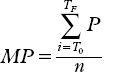
where MP is mean pressure (mmHg); ∑, sum; TF, end of the waveform (ms); T0, start of the waveform (ms); Pi, pressure points; and n = numbers of pressure points.
Vascular elasticity was measured by arterial pulse waveform analysis using an HDI/Pulse Wave CR2000 (Hypertension Diagnostics, Inc, Eagan, NY) (Citation30). This technique involves 30-s recording of radial artery waveforms by applanation tonometry. The tonometry unit contained an array of pressure transducers capable of measuring the relative intra-arterial pulse-amplitude with high accuracy. The tonometer was centered over the radial artery obtaining the optimal waveform by pneumatic stabilization. Brachial BP was measured oscillometrically in the opposite arm. A beat-marking algorithm determined the beginning of the systole, peak systole, onset of diastole and end-diastole for each beat during the measurement period. To obtain arterial compliance, a model was used that divides the total systemic arterial compliance into large artery (or capacitative, C1) and small artery (or oscillatory, C2) compliance (Citation30). All data were collected from untreated patients.
Statistical methods
PP was calculated as the difference between systolic BP and diastolic BP, brachial office mean BP and 24-h mean BP were calculated as diastolic BP + 1/3 PP. Data are presented as mean ± standard deviation (SD) unless specified. Differences between means were assessed by two-tailed Student's t-test for unpaired observations and by one-way analysis of variance (ANOVA), adjusting for age and sex. To avoid the occurrence of spuriously significant results, the Bonferroni adjustment was used. Logistic regression models for a binary outcome, unadjusted and adjusted for age, sex, body mass index (BMI), lifestyle factors, parental hypertension, time to reach the end-point and brachial heart rate (HR), were used to define the relationship between baseline TOD or development of hypertension and the different pressures, and the related odds ratios (OR) were provided. As central mean BP was the best predictor of future hypertension (p < 0.001) compared with systolic BP (p = 0.01) or diastolic BP (p = 0.04), central mean BP was used in all comparative analyses with office BP and 24-h BP.
Patients were defined as having high or low central BP according to whether their central mean BP was above or below, respectively, the median in the group (103.0 mmHg). In addition, subjects were divided into mean BP tertiles of brachial office BP, 24-h BP and central pressures. A two-tailed probability value < 0.05 was considered significant. All analyses were performed using Systat versions 10 and 11 (SPAA Inc., Evanston, IL, USA).
Ethical considerations
The study was approved by the HARVEST Ethics Committee and by the Ethics Committee of the University of Padova. A written informed consent was given by the participants.
Results
The study participants were more frequently males (73.8%). Their clinic brachial office BP at entry in the HARVEST study was 144.3 ± 11.1/91.7 ± 6.3 mmHg and at the time of arterial distensibility assessment was 137.2 ± 12.2/85.7 ± 7.5 mmHg. During the 9 years of follow-up, 156 patients (51%) developed hypertension requiring antihypertensive treatment (end-point subjects), while the rest of the group remained untreated. The baseline characteristics of the whole cohort, and of the end-point subjects and the subjects who remained untreated are reported in . End-point subjects were older and less active compared with those who remained untreated. No between-group statistically significant difference was found for gender, BMI, prevalence of smokers, alcohol and coffee drinkers, and parental hypertension. With regard to metabolic data, end-point patients had higher triglycerides than those who remained untreated. Also serum glucose, total cholesterol and AER were higher among the end-point patients but the differences did not attain the level of statistical significance after the Bonferroni correction. LVM index did not differ significantly between the two groups. End-point subjects presented higher baseline brachial office diastolic and mean BP, all 24-h and central BP values, in comparison with subjects who remained untreated (). No significant difference was observed between the two groups for office systolic BP, office and 24-h HR and all PP values. Correlations between brachial office, 24-h and central systolic, diastolic and mean BP values are reported in . For mean BP, all coefficients were < 0.3. Central BPs were unrelated to 24-h BPs. In multivariable logistic regression analyses, brachial office mean BP and 24-h mean BP were not associated with baseline TOD (p = n.s.), whereas central mean BP presented a significant association (p = 0.009). This association remained statistically significant also when office and 24-h mean BPs were forced into the logistic model (OR = 1.06, 95% CI 1.01–1.12), p = 0.003). None of the PP values was associated with baseline TOD.
Table I. Baseline characteristics in the whole group and in the participants divided according to whether they developed hypertension needing treatment (end-point) or remained untreated.
Table II. Baseline blood pressure values in the whole group and in the participants divided according to whether they developed hypertension needing treatment (end-point) or remained untreated.
Table III. Correlation between brachial office, average 24-h and central systolic, diastolic and mean BP values.
Arterial distensibility assessment
The 305 subjects were divided into two subgroups according to whether their central mean BP was below or above the median in the group (103.0 mmHg). Subjects with high central mean BP presented lower C1 and C2 compared with those with low central mean BP. C1 and C2 were 16.1 ± 5.3 ml/mmHgx10 and 7.8 ± 3.0 ml/mmHgx100, respectively, in subjects with low central mean BP, and were 14.3 ± 4.6 ml/mmHgx10 and 6.3 ± 2.5 ml/mmHgx100, respectively, in subjects with high central mean BP (p-value adjusted for age and sex = 0.025 for C1 and p = 0.001 for C2). Subjects with high central mean BP also presented a higher PWV and AIx. PWV was 9.7 ± 3.8 m/s and AIx was 23.5 ± 0.23% in subjects with high central mean BP; PWV was 8.1 ± 1.6 m/s and AIx was 7.2 ± 0.25% in subjects with low central mean BP (p-value adjusted for age and sex < 0.001 for both). In addition, subjects with high central mean BP reached the endpoint more frequently than those with low central mean BP (62.6% vs 36.6%, p < 0.001 adjusted for age and sex).
Follow-up
At the last follow-up visit, brachial office BP was 147.8 ± 12.0/97.1 ± 8.1 mmHg in the subjects who met the criteria for starting antihypertensive drug treatment (end-point) and was 132.3 ± 8.9/85.0 ± 6.3 mmHg in the patients who did not need treatment (all adjusted p < 0.001). The corresponding values for 24-h BP were 134.6 ± 9.6/86.3 ± 7.2 mmHg and 128.9 ± 9.2/79.4 ± 5.8 mmHg, respectively (all adjusted p < 0.001). In univariate analyses, development of hypertension requiring drug treatment was predicted by brachial office, 24-h and central mean BPs (all p < 0.001), whereas brachial office and 24-h PP were not predictive of outcome. The predictive value of central PP was of borderline statistical significance (p = 0.07). When the three mean BPs were included simultaneously in the same unadjusted model, brachial BP was not accepted by the model, whereas 24-h mean BP (p < 0.001) and central mean BP (p = 0.001) remained significant predictors of outcome. Brachial office, 24-h and central mean BPs were significant predictors of future hypertension also when included in a multivariable model separately (, models 1, 2 and 3). When the three pressures were included together in the same multivariable model, again only central mean BP and 24-h mean BP remained associated with outcome (, model 5). In the multivariable regressions, brachial PP and 24-h PP were not associated with outcome. Central PP maintained a borderline relationship in the multivariable model when central mean BP was incorporated (, model 4).
Table IV. Odds ratios and 95% confidence interval for development of hypertension needing antihypertensive treatment from multivariable adjusted logistic regressions.
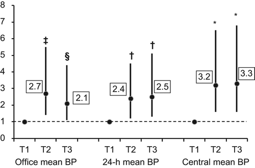
In the subjects divided into tertiles of mean BP, the top tertile of brachial office, 24-h and central mean BP had a 2.1, 2.5 and 3.3 increased adjusted risk, respectively, of developing hypertension needing antihypertensive therapy compared with the bottom tertile ().
Figure 1. Odds ratios of developing hypertension needing treatment in 305 stage 1 hypertensive subjects, divided into tertiles of brachial office, 24-h and central mean blood pressure. Office mean blood pressure: T1, 1st tertile (85.22–106.83 mmHg); T2, 2nd tertile (106.84–111.78 mmHg); T3, 3rd tertile (111.89–125.22 mmHg). Twenty-four-hour mean blood pressure: T1, 1st tertile (73.19–93.86 mmHg); T2, 2nd tertile (93.93–99.26 mmHg); T3, 3rd tertile (99.27–119.57 mmHg). Central mean blood pressure: T1, 1st tertile (77.00–99.20 mmHg); T2, 2nd tertile (99.30–105.56 mmHg); T3, 3rd tertile (105.57–133.00 mmHg). ‡p = 0.03 vs 1st tertile, §p = 0.004 vs 1st tertile †p < 0.01vs 1st tertile, *p < 0.001 vs 1st tertile.
Discussion
The main finding of our study was that mean central BP, measured non-invasively with applanation tonometry, was a useful adjunct to brachial BP measurements to better define the risk profile in young subjects screened for stage I hypertension. We observed that central BP remained a significant predictor of future hypertension on top of ambulatory BP. Central mean BP was the only BP measurement associated with the presence of baseline LVH or MA. In addition, patients with elevated central mean BP presented an early impairment of arterial distensibility parameters, as documented by the higher values of PWV and AIx and the lower values of C1 and C2 compared with patients with low central mean BP. At variance, central PP was not a significant predictor of outcome in this young population.
These data confirm our previous results obtained in 67 young subjects with isolated systolic hypertension (Citation31) and indicate that central BP is useful for identifying hypertensive patients at increased risk who may need an earlier antihypertensive treatment.
The clinical role of central BP has been highlighted in several clinical studies that showed that central BP had a stronger association with CV morbidity than brachial BP (Citation5–7). Recently, Vlachopoulos and colleagues analyzed the results of 11 longitudinal studies and concluded that central systolic BP, PP and AIx predicted CV events and total mortality (Citation11). However, the relationship between central BP and CV risk was not confirmed by Dart and colleagues (Citation32) who observed no predictive value of central systolic BP, PP and AIx for CV diseases in elderly female hypertensives. In that study, brachial BP was a significant predictor of outcome. These results can be explained by the changing relationship between central BP and brachial BP with aging. Age-related aortic stiffening reduces the difference between central and brachial systolic BP, which may account for the relatively better predictivity of brachial compared with central BP in elderly individuals. In contrast, in our young to middle-aged patients, central BP was much lower than brachial office BP (mean brachial office BP 109.2 mmHg, mean central BP 103.0 mmHg).
Another finding that may be related to the young age of our population is that in the present study central mean BP and not central PP was predictive of outcome, at variance with previous data obtained in older subjects (Citation11,Citation33,Citation34). Vlachopoulos et al. (Citation11) examined six relevant studies and observed that for a 10-mmHg increase in central PP there was a 3.7% risk increase of CV events. In another recent paper, Huang et al. (Citation34) compared central and 24-h BP measurements and found that only central PP was significantly predictive of all-cause mortality. However, it should be noted that in elderly individuals PP reflects increased arterial stiffness, whereas in young subjects it is mainly the result of heightened cardiac output.
Ambulatory BP monitoring is currently used in clinical practice for a better assessment of a hypertensive individual because it adds prognostic information to that provided by brachial office BP. Whether central BP may provide additional information on risk related to BP when used in conjunction with ambulatory BP is not well known. In adult to elderly uncomplicated hypertensives, Schultz and colleagues (Citation35) observed that peripheral arterial pressure either measured in the clinic or with ambulatory recording was poorly representative of central BP because of wide variation in PP amplification. Among patients from different categories of BP control, central BP measurement produced a reclassification of the risk related to BP. Only one study prospectively compared the predictive value of 24-h BP with that of central BP. In a population of subjects with a mean age of 52 ± 13 years (range 30–79 years), Huang and colleagues (Citation34) observed that office central BP was more valuable than office peripheral BP in the prediction of all-cause and CV mortalities. Ambulatory peripheral BP was superior to central BP in the prediction of CV mortality, but central PP better predicted all-cause mortality than 24-h systolic BP or PP. To the best of our knowledge, no such comparison has been performed in young populations. The present study indicates that assessment of central BP with applanation tonometry allows the identification of young hypertensive subjects with increased CV risk who may need earlier antihypertensive treatment.
Study limitations
In this analysis, we arbitrarily considered the subjects whose central mean BP was above the median in the group as having high central BP. Little information is available about the upper normal limit of central BP in young to middle-aged individuals. The difference between central BP and brachial BP declines progressively with aging and it is not known whether different normality limits should be identified in younger compared with older individuals. In addition, we used mean BP to compare the different BP measurements. We selected this parameter because central mean BP was the best predictor of future hypertension needing treatment, compared with central systolic or diastolic BP. Moreover, using mean BP we decreased the risk of collinearity between the three pressures. Another possible limitation is that central aortic BP was not measured directly but was estimated from non-invasive pressure waveforms with applanation tonometry. A potential pitfall of this technique is that the values for central pressures depend on the validity and applicability of the generalized transfer function used to generate the central aortic waveforms. In addition, the calibration of central aortic pressures depends on the accuracy of the brachial pressure measurements. However, the transfer function used by the DAT system to derive central BP has been validated in several studies, which showed a good correspondence between calculated and directly recorded central aortic BP (Citation28,Citation36). Another limitation is the higher prevalence of male subjects in our population, due to the effect of the natural selection. Because of the small number of female hypertensives, we must be cautious about conclusions of our longitudinal analysis in this gender.
Conclusion
The present results suggest that in young to middle-aged subjects, in the initial stage of hypertension, assessment of central BP is a useful adjunct to brachial BP measurement even if ambulatory BP monitoring is performed. Central BP may be useful for identifying subjects with a higher risk profile in whom early anti-hypertensive treatment should be started. Future studies in larger samples are needed to identify an objective operational threshold level between normal and abnormal central BP.
Declaration of interest: The authors report no conflicts of interest. The authors alone are responsible for the content and writing of the paper.
The study was funded by the University of Padova, Padova, Italy and by the Associazione “18 maggio 1370”, San Daniele del Friuli, Italy.
References
- Nichols W, O’Rourke MF, editors. McDonalds’ blood flow in arteries: Theoretical, experimental and clinical principles. 5th ed. Oxford: Oxford University Press; 2005.
- Sharman JE, Stowasser M, Fassett RG, Marwick TH, Franklin SS. Central blood pressure measurement may improve risk stratification. J Hum Hypertens. 2008;22: 838–844.
- Morgan T, Lauri J, Bertram D, Anderson A. Effect of different antihypertensive drug classes on central aortic pressure. Am J Hypertens. 2004;17:118–123.
- Williams B,Lacy PS,Thom SM,Cruickshank K,Stanton A,Collier D, .; CAFE Investigators; Anglo-Scandinavian Cardiac Outcomes Trial Investigators; CAFE Steering Committee and Writing Committee.Differential impact of blood pressure-lowering drugs on central aortic pressure and clinical outcomes: Principal results of the Conduit Artery Function Evaluation (CAFE) study. Circulation. 2006;113: 1213–1225.
- Roman MJ, Devereux RB, Kizer JR, Lee ET, Galloway JM, Ali T, . Central pressure more strongly relates to vascular disease and outcome than does brachial pressure: The Strong Heart Study. Hypertension. 2007;50:197–203.
- Wang K-L, Cheng H-M, Chuang SY, Spurgeon HA, Ting CT, Lakatta EG, . Central or peripheral systolic or pulse pressure: Which one best relates to target organs and future mortality?J Hypertens. 2009;27:461–467.
- Roman MJ, Okin PM, Kizer JR, Lee ET, Howard BV, Devereux RB. Relations of central and brachial blood pressure to left ventricular hypertrophy and geometry: The Strong Heart Study. J Hypertens. 2010;28:384–388.
- Jankowski P, Kawecka-Jaszcz K, Czarnecka D, Brzozowska-Kiszka M, Styczkiewicz K, Loster M, .; <Aortic Blood Pressure and Survival Study Group.Pulsatile but not steady component of blood pressure predicts cardiovascular events in coronary patients. Hypertension. 2008;51:848–855.
- Pini R, Cavallini MC, Palmieri V, Marchionni N, Di Bari M, Devereaux RB, . Central but not brachial blood pressure predicts cardiovascular events in an unselected geriatric population. The ICARe Dicomano study. J Am Coll Cardiol. 2008;51:2432–2439.
- Cohen DL, Townsend RR. Central blood pressure and chronic kidney disease progression. J Nephrol. 2011; 24 February 2011; doi:10.4061/2011/407801.
- Vlachopoulos C, Aznaouridis K, O’Rourke M, Safar ME, Baou K, Stefanadis C. Prediction of cardiovascular events and all-cause mortality with central haemodynamics: A systematic review and meta-analysis. Eur Heart J. 2010;31: 1865–1871.
- White WB, Lund-Johansen P, Weiss S, Omvik P, Indurkhya N. The relationships between casual and ambulatory blood pressure measurements and central hemodynamics in essential human hypertension. J Hypertens. 1994;12:1075–1081.
- Palatini P, Mormino P, Mos L, Mazzer A, Dorigatti F, Zanata G, .; <HARVEST Study Group.Microalbuminuria, renal function and development of sustained hypertension: A longitudinal study in the early stage of hypertension. J Hypertens. 2005;23:175–182.
- Palatini P, Graniero G, Mormino P, Nicolosi L, Mos L, Visentin P, . Relation between physical training and ambulatory blood pressure stage I hypertensive subjects. Results of the HARVEST Trial. Hypertension and Ambulatory Recording Venetia Study. Circulation. 1994;90: 2870–2876.
- Sartori M, Semplicini A, Siffert W, Mormino P, Mazzer A, Pegoraro F, . G-protein beta3-subunit gene 82ST allele and hypertension: A longitudinal study in young grade I hypertensive. Hypertension. 2003;42:909–914.
- Palatini P, Penzo M, Canali C, Pessina AC. Validation of the accuracy of the A & D TM-2420 model 7 for ambulatory blood pressure monitoring and effect of microphone replacement on its performance. J Amb Monitor. 1991;4:281–288.
- O’Brien E, Mee F, Atkins N, O’Malley K. Accuracy of the SpaceLabs 90207 determined by the British Hypertension Society protocol. J Hypertens. 1991;9:573–575.
- Palatini P, Mormino P, Canali C, Santonastaso M, De Venuto G, Zanata G, . Factors affecting ambulatory blood pressure reproducibility. Results of the Harvest trial. Hypertension. 1994;23:211–216.
- Palatini P, Winnicki M, Santonastaso M, Mos L, Longo D, Zaetta V, . Prevalence and clinical significance of isolated ambulatory hypertension in young subjects screened for stage 1 hypertension. Hypertension. 2004;44:170–174.
- Devereux RB, Lutas EM, Casale PN, Kigfield P, Eisenberg RR, Hammond IW, . Standardization of M-mode echocardiographic left ventricular anatomic measurement. J Am Coll Cardiol. 1984;4:1222–1230.
- Mancia G, De Backer G, Dominiczak A, Cifkova R, Fagar R, Germano G, ; Management of Arterial Hypertension of the European Society of Hypertension; European Society of Cardiology.2007 Guidelines for the Management of Arterial Hypertension: The Task Force for the Management of Arterial Hypertension of the European Society of Hypertension (ESH) and of the European Society of Cardiology (ESC). J Hypertens. 2007;25:1105–1187.
- Pinto-Sietsma SJ, Janssen WMT, Hillege HL, Navis G, DE Zeeuw D, de Jong PE. Urinary albumin excretion is associated with renal functional abnormalities in a non diabetic population. J Am Soc Nephrol. 2000;11:1882–1888.
- 1999 World Health Organization–International Society of Hypertension Guidelines for the Management of Hypertension. Guidelines Sub-Committee. Blood Press Suppl. 1999;1:9–43.
- 2003 European Society of Hypertension–European Society of Cardiology guidelines for the management of arterial hypertension. European Society of Hypertension–European Society of Cardiology Guidelines Committee. J Hypertens. 2003;21:1011–1053.
- Adji A, Hirata K, O’Rourke MF. Clinical use of indices determined non-invasively from the radial and carotid pressure waveforms. Blood Press Monit. 2006;11:215–221.
- Wilkinson IB, Fuchs SA, Jansen IM, Spratt JC, Murray GD, Cockcroft JR, . Reproducibility of pulse wave velocity and augmentation index measured by pulse wave analysis. J Hypertens. 1998;16:2079–2084.
- Gallagher D, Adji A, O’Rourke MF. Validation of the transfer function technique for generating central from peripheral upper limb pressure waveform. Am J Hypertens. 2004;17: 1059–1067.
- Chen CH, Nevo E, Fetics B, Pak PH, Yin FC, Maughan WL, . Estimation of central aortic pressure waveform by mathematical transformation of radial tonometry pressure. Validation of generalized transfer function. Circulation.1997; 95:1827–1836.
- Cameron JD, McGrath BP, Dart AM. Use of radial artery applanation tonometry and a generalized transfer function to determine aortic pressure augmentation in subjects with treated hypertension. J Am Coll Cardiol. 1998;32:1214–1220.
- Cohn JN, Finkelstein S, McVeigh G, Morgan D, LeMay L, Robinson J, . Noninvasive pulse wave analysis for the early detection of vascular disease. Hypertension. 1995;26: 503–508.
- Saladini F, Santonastaso M, Mos L, Benetti E, Zanatta N, Maraglino G, ; HARVEST Study Group; on behalf of the HARVEST Study Group.Isolated systolic hypertension of young-to-middle-age individuals implies a relatively low risk of developing hypertension needing treatment when central blood pressure is low. J Hypertens. 2011;29:1311–1319.
- Dart AM, Gatzka CD, Kingwell BA, Willson K, Cameron JD, Liang YL, . Brachial blood pressure but not carotid arterial waveforms predict cardiovascular events in elderly female hypertensives. Hypertension. 2006;47:785–790.
- Safar ME, Blacher J, Pannier B, Guerin AP, Marchais SJ, Guyonvarc'h PM, London GM. Central pulse pressure and mortality in end-stage renal disease. Hypertension. 2002;39: 735–738.
- Huang CM, Wang K-L, Cheng H-M, Chuang S-Y, Sung S-H, Yu W-C, . Central versus ambulatory blood pressure in the prediction of all-cause and cardiovascular mortalities. J Hypertens. 2011;29:454–459.
- Schultz MG, Gilroy D, Wright L, Bishop WLJ, Abhayaratna WP, Stowasser M, . Out-of-office and central blood pressure for risk stratification: A cross- sectional study in patients treated for hypertension. Eur J Clin Invest. 2012;42:393–401.
- Adji A, Hirata K, Hoegler S, O’Rourke MF. Noninvasive pulse waveform analysis in clinical trials: Similarity of two methods for calculating aortic systolic pressure. Am J Hypertens2007;20:917–922.