Abstract
Obstructive sleep apnoea (OSA) is significantly associated with the risk of stroke, and this association is independent of other risk factors, including hypertension, atrial fibrillation and diabetes mellitus. Therefore, additional pathogenic mechanisms may exist, which contribute to the increased risk of stroke. OSA is characterized by prolonged sympathetic overactivity; however the role of the sympathetic nervous system in regulating cerebral circulation remains a matter of controversy. Converging data indicate that brain perfusion is significantly distorted in OSA, with reported decreases in cerebral blood flow as well as intermittent surges in blood pressure and cerebral blood flow velocity. Based on recent research, there is accumulating evidence that sympathetic nerve activity is an important element in brain protection against excessive increases in perfusion pressure during blood pressure surges and flow during rapid eye movement sleep. The aim of this article was to review: (i) the current physiological knowledge related to the role of the sympathetic system in the regulation of cerebral blood flow, (ii) how the influence of the sympathetic system on cerebral vessels is affected by apnoea (increased PaCO2) and (iii) the potential significance of the pathological sympathetic system/PaCO2 interplay in OSA. Sympathetic system seems to be at least partially involved in pathogenesis of distorted haemodynamics and stroke in OSA patients. However, there are still several open questions that need to be addressed before the effective therapeutic strategies can be implemented.
Introduction
Obstructive sleep apnoea (OSA) is significantly associated with the risk of stroke, and this association is independent of other risk factors, including hypertension, atrial fibrillation and diabetes mellitus. Furthermore, increased severity of OSA is associated with an increased risk of stroke. The incidence of stroke is two- to threefold higher in patients suffering from OSA than in the general population (Citation1–3). Therefore, additional pathogenic mechanisms (other than typical cardiovascular risk factors) may exist, contributing to the increased risk of stroke. Converging data indicate that brain perfusion is significantly distorted in OSA, with reported decreases in cerebral blood flow (CBF) (Citation4) as well as intermittent surges in blood pressure (BP) and cerebral blood flow velocity (CBFV) (Citation5,Citation6). Furthermore, OSA is characterized by prolonged sympathetic overactivity (Citation7,Citation8). The role of the sympathetic nervous system in regulating cerebral circulation remains a matter of controversy (Citation9–11). Nevertheless, there is accumulating evidence that sympathetic nerve activity (SNA) might be an important element in brain protection against excessive increases in perfusion pressure during BP increases (Citation12,Citation13) and flow during rapid eye movement (REM) sleep (Citation14,Citation15). Thus, the role of the sympathetic system in the cerebral circulation seems to be different from and independent of its peripheral mode of action. Moreover, recent studies employing techniques based on infrared light (IR) such as near infrared spectroscopy (NIRS) and near-infrared transillumination/backscattering sounding (NIR-T/BSS) have suggested an increase in cerebral venous blood volume following sympathetic stimulation (Citation16–18). The aim of this article was to review: (i) the current physiological knowledge related to the role of SNA in the regulation of CBF, (ii) how the influence of SNA on cerebral vessels is affected by apnoea (increased PaCO2) and (iii) the potential significance of the pathological SNA/PaCO2 interplay in OSA.
SNA in the regulation of CBF
The cerebral arteries, arterioles and, to lesser extent, veins are richly innervated with sympathetic nerve fibres (Citation19,Citation20). Cerebral sympathetic innervation originates in the superior cervical ganglion, with a minor contribution from the stellate ganglia and possibly an intrinsic central adrenergic pathway (Citation21). α and β adrenergic receptors have been found in human cerebral vessels, including the middle cerebral artery (MCA) (Citation22), and sympathomimetic agents (adrenaline, noradrenaline and phenylephrine) contract human pial artery segments (Citation23). The effect of noradrenaline (NE) infusion into the internal carotid artery (ICA) on the amplitude of pial artery pulsation in intact rabbits is shown in . Initial vasoconstriction, seen as a decrease in pial artery pulsation, which represents the direct response to NE, is followed by passive transmission, seen as an increase in pial artery pulsation, when the BP exceeds the upper autoregulatory limit in the rabbit. Blood flow in the ICA remains diminished. Registration was performed using NIR-T/BSS, which allows for non-invasive monitoring of pial artery pulsation (Citation24). SNA induces vasoconstriction in the rabbit basilar artery (Citation25) and MCA (Citation26), while electrical stimulation of the cervical sympathetic ganglion in humans decreases CBFV in the MCA (Citation27). The influence of SNA on the large-diameter cerebral vessels requires further attention before we proceed with reviewing the results of transcranial Doppler (TCD) studies. Assuming the MCA diameter remains constant, TCD measurement of flow velocity in the MCA has been frequently used to infer changes in CBF from changes in CBFV. However, this assumption is disputed by some authors (Citation17,Citation28–30). Phenylephrine (PE) and NE infusion causes a reduction in the NIRS-derived estimate of frontal lobe oxygenation (ScO2), suggesting sympathetically-mediated cerebral vasoconstriction (Citation17,Citation31–33). The PE infusion-mediated reduction in ScO2 is accompanied by increased CBFV in the MCA (Citation17,Citation32,Citation34). CBF, measured in the ICA by duplex ultrasound allowing the simultaneous assessment of diameter and CBFV, provides some explanation to these seemingly contradicting results. After PE infusion, CBF measured in the ICA remains unchanged while CBFV in the MCA increases, which clearly suggests MCA vasoconstriction (Citation17). Furthermore, a head-to-head comparison between TCD and MRI has demonstrated that MCA diameter changes significantly during hypoxia (Citation30). Finally, there are differences between ICA and vertebral artery flow velocity responses to exercise (Citation35). Thus, the TCD measurement of CBFV in the MCA alone is likely to be unreliable in evaluating CBF, in particular in the sympathetic system and in OSA research. Only a combined assessment of ICA and vertebral artery flow velocity and anatomy (vessel diameter) allows CBF to be estimated correctly.
Figure 1. Influence of an injection of adrenaline into the ICA on the recorded variables (ascending phase): (Citation1) time indicator, (Citation2) amplitude of pial artery pulsation (NIR-T/BSS), (Citation3) blood flow in the left ICA (mL/min), (Citation4) mean BP ( mmHg), (Citation5) respiration, (Citation6) BP in the centripetal portion of the right ICA (mmHg). Initial vasoconstriction, seen as a decrease in pial artery pulsation, which represents the direct response to NE, is followed by passive transmission, seen as an increase in pial artery pulsation, when the BP exceeds the upper autoregulatory limit in the rabbit. Blood flow in the ICA remains diminished. Adapted with permission from Reference 24.
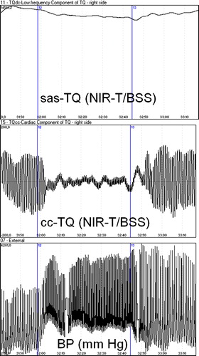
The handgrip test is often used to investigate the effects of sympathetic activation in humans. However, the obtained data are conflicting, with several authors reporting that CBFV remains unchanged during the handgrip test, while cerebrovascular resistance significantly increases (Citation18,Citation36,Citation37); others have claimed that CBFV increases (Citation38–40). These differences can potentially be explained by changes in PaCO2. Since PaCO2 is the most potent regulator of cerebral circulation, the precise control of PaCO2 is critical in the interpretation and understanding of the influence of SNA on CBF regulation. Ainslie et al. (Citation36) demonstrated that the controlled isocapnia handgrip test evokes an increase in cerebrovascular resistance, with unchanged CBFV. In contrast, during hypercapnia, cerebrovascular resistance does not change, while CBFV increases (Citation37). Nevertheless, studies by Rasmussen et al. (Citation39) and Seifert et al. (Citation40) showed controlled PaCO2, as well, yielding contradicting results. Furthermore, Seifert et al. (Citation40) demonstrated that the increase in CBFV is abolished with the muscarinic receptor blocker glycopyrrolate. The role of the parasympathetic system in CBF regulation in humans is beyond the scope of this review. The question remains open whether reported increases in CBFV are due to MCA vasoconstriction, or indeed reflect elevated CBF. shows a representative NIR-T/BSS beat-by-beat tracing during the handgrip test performed in our lab. It should be noted that the significant decrease in the amplitude of pial artery pulsation after starting the handgrip test clearly suggests pial artery vasoconstriction. Thus, the pial artery may efficiently counteract increased cerebral perfusion pressure, attenuating the elevation in CBF during an imposed hypertensive episode (Citation18).
Figure 2. Changes in the width of the subarachnoid space (upper tracing), pial artery pulsation (middle tracing) and mean arterial pressure (lower tracing) during the handgrip test. The start and end of the handgrip test are indicated by vertical lines. Taken with permission from Reference 18.
There is growing evidence that SNA plays an important protective role under conditions that threaten the integrity of cerebral blood vessels (Citation13). During acute, severe hypertension, increases in CBF are accompanied by a disruption of the blood–brain barrier (Citation41). Sympathetic stimulation attenuates the increase in CBF and permeability of the blood–brain barrier during acute BP elevation (Citation12). Large increases in BP occurring during REM sleep and REM sleep disrupted by apnoea are associated with increased SNA (Citation42,Citation43). In particular, increased SNA occurs during phasic REM sleep, when BP is unstable and cerebral microvessels may be vulnerable to large fluctuations in cerebral perfusion pressure (Citation15). After bilateral sympathectomy, CBF was significantly elevated with respect to the control group throughout the entire sleep cycle, with baseline CBF already exceeding the peak level observed in the control group (Citation14). Importantly, SNA does not seem to play any significant role with regard to CBF regulation during decreases in BP (Citation13,Citation44).
Sympathetic nerves also influence the ability of cerebral capacitance vessels to regulate intracranial pressure and cerebral blood volume. Cerebral blood volume increases have been shown during the handgrip test (Citation16,Citation18) and PE infusion (Citation17). On the other hand, Wilson et al. (Citation45) suggested that the cold pressor test may selectively decrease cerebral blood volume in grey matter. Furthermore, early animal studies showed that SNA stimulation leads to pial vein constriction (Citation46,Citation47). Further research is needed to elucidate the influence of sympathetic activation on cerebral venous blood volume, as the results reported to date by various teams remain inconsistent. Disturbances in venous blood volume/outflow maybe potentially harmful if maintained over long periods of time by inducing local ischaemia and/or oedema and leading to leukoaraiosis (Citation48) or promoting amyloid beta deposition (Citation49).
SNA influence on cerebral vessels is affected by PaCO2
Hypercapnia induces relatively uniform vasodilation in the entire arterial–arteriolar bed, with the arterioles playing a major role in the control of CBF (Citation50,Citation51). Cerebrovascular vasodilation caused by hypercapnia increases CBF (Citation52–54), decreases pial arteriolar bed resistance (Citation51,Citation55) and significantly increases the amplitude of pial artery pulsation (Citation56–58). The traditional view is that the ability to autoregulate CBF with changes in BP is significantly dependent on PaCO2. Several studies have shown that hypercapnia impairs dynamic cerebral autoregulation, as estimated by the cuff deflation method (Citation59) and transfer function analysis (Citation60–62). The most recent data, however, support the thesis that dynamic autoregulation is not affected by increased PaCO2 (Citation63). Nevertheless, hypercapnia impairs the pial artery response to elevation in CBF (Citation57).
The initial thinking that SNA may partially attenuate the CO2-induced increase in CBF (Citation64) was replaced by the opinion that, under physiological conditions, there is no evidence that basal levels of sympathetic vasoconstrictor outflow limit apnoea-induced cerebral vasodilatation (Citation65) or CO2 responsiveness (Citation44). However, the results of the abovementioned studies should be generalized into clinical sleep-disordered breathing like OSA with caution, since many sleep apnoeas are accompanied by more substantial hypoxemia and are of longer duration than in the Przybyłowski et al. study (Citation65). Furthermore, as discussed above, decreases in BP, as in the LeMarbre et al. study (Citation44), do not evoke substantial SNA directed toward the brain circulation. Interesting data comes from studies in elite breath-holding divers. Prolonged periods of hypoxia/ hypercapnia, lasting up to 10 min, are accompanied by extreme sympathetic activation, peripheral vasoconstriction, increased BP and CBF that offset reduced arterial oxygenation and maintain ScO2 (Citation66). Thus, the influence of even very high SNA on cerebral vessels can be overridden by increased PaCO2 and/or metabolic demand. This hypothesis is strengthened by the evidence that the PE-induced decrease in frontal lobe oxygenation can be offset by metabolic demand during moderately intense exercise (Citation32). The hypercapnia-induced decrease in MCA constriction during the handgrip test was discussed earlier (Citation36). To conclude, it seems that sympathetic activity does not significantly influence CO2-induced increases in CBF, but hypercapnia may override SNA-induced cerebral vasoconstriction. This is in line with the finding that SNA is withdrawn during hypercapnia in REM sleep, thus augmenting the CBF response (Citation67).
The potential significance of the pathological SNA/PaCO2 interplay in OSA
ScO2 during sleep is decreased in patients with moderate to severe OSA (Citation68,Citation69). Increased cerebral blood volume was reported in both of the abovementioned NIRS studies. The authors explained this result as an increase in CBF to compensate for decreased cerebral oxygenation. However, baseline CBFV is decreased in OSA patients, and differences in the response to hypercapnic challenge (5% CO2, 2 min) versus healthy volunteers are not statistically significant (Citation4). Other authors have reported impaired cerebrovascular reactivity in OSA (Citation70–72). Thus, it is unlikely that the increase in cerebral blood volume in OSA is solely due to an elevation in CBF. According to recent studies, sympathetic activation may increase cerebral venous blood volume (Citation17,Citation18). An increase in cerebral venous blood volume may be responsible or partially responsible for the elevated intracranial pressure in OSA reported during wakefulness and sleep (Citation73,Citation74). Increases in intracranial pressure are highest in NREM stage 2 and 3 sleep and in REM sleep (Citation74), which coincides with the highest SNA (Citation15). There is a strong correlation between the duration of apnoea and BP, and elevated intracranial pressure and between BP variations and elevated intracranial pressure (Citation74), which further strengthens the hypothesis that the sympathetic system may be involved. Increased intracranial pressure, hypoxia and hypercapnia further lead to an increase in sympathetic activity, creating a vicious circle. If CBF ceases during apnoea as a consequence of decreased cerebral perfusion pressure due to increased intracranial pressure, it may even worsen the cerebral hypoxia that develops during apnoeic episodes.
Dynamic cerebral autoregulation is impaired in OSA. Subjects with OSA have a slower rate of recovery in terms of BP, CBFV and cerebrovascular conductance than healthy subjects, indicating an impaired compensatory response to hypoperfusion (Citation4). OSA creates a very hostile environment with respect to brain haemodynamics. During an apnoeic episode, there is a progressive increase in arterial systolic BP, reaching 250 mmHg, with a sudden drop below baseline after the termination of apnoea. Similarly, there is a rise and fall in CBFV during and after apnoea, exceeding a 60% fluctuation in CBFV compared to the baseline velocity. These intermittent surges in BP and hypotension and the resultant fluctuations in CBFV subject the brain to vascular damage and the risk of ischaemia (Citation4–6). The mechanisms that lead to the development of such hostile brain haemodynamics are still poorly understood. Well-designed experimental studies are urgently needed to clarify the interdependencies between SNA, PaCO2, hypoxia and intracranial pressure in the regulation of CBF and their roles in the pathogenesis of OSA and the significantly increased risk of stroke and death associated with OSA. In particular, the following questions need to be answered: (i) How does SNA influence cerebral venous blood volume? (ii) Is there any link between sympathetic activation and increased intracranial pressure? (iii) Is derangement in autonomic regulation responsible for distorted CBF and impaired autoregulation in OSA? The recent position paper issued by European Society of Hypertension, European Respiratory Society and the members of European COST (COoperation in Scientific and Technological research) ACTION B26 underlines the complexity of OSA, and the need for multidisciplinary approach (Citation75). Better understanding of the mechanisms involved in abnormal cerebral circulation in OSA may pave the way for more effective brain protection therapeutic strategies and in turn facilitate overall SNA management and better blood pressure control.
Conflicts of interest
A. F. Frydrychowski owns several patents related to NIR-T/BSS technology and is a stakeholder in NIRT sp. z o.o. P. J. Winklewski declares that he has no competing interests.
Funding from the Medical University of Gdansk and NIRT sp. z o. o., Wierzbice, Poland is gratefully acknowledged. The funding sources had no role in the study design, in the collection, analysis and interpretation of the data, in the writing of the report or in the decision to submit the paper for publication.
References
- Yaggi HK, Concato J, Kernan WN, Lichtman JH, Brass LM, Mohsenin V. Obstructive sleep apnea as a risk factor for stroke and death. N Engl J Med. 2005;353:2034–2041.
- Munoz R, Duran-Cantolla J, Martínez-Vila E, Gallego J, Rubio R, Aizpuru F, . Severe sleep apnea and risk of ischemic stroke in the elderly. Stroke. 2006;37:2317–2321.
- Redline S, Yenokyan G, Gottlieb DJ, Shahar E, O’Connor GT, Resnick HE, . Obstructive sleep apnea–hypopnea and incident stroke: The sleep heart health study. Am J Respir Crit Care Med. 2010;182:269–277.
- Urbano F, Roux F, Schindler J, Mohsenin V. Impaired cerebral autoregulation in obstructive sleep apnea. J Appl Physiol. 2008;105:1852–1857.
- Bålfors EM, Franklin KA. Impairment of cerebral perfusion during obstructive sleep apneas. Am J Respir Crit Care Med. 1994;150:1587–1591.
- Klingelhöfer J, Hajak G, Sander D, Schulz-Varszegi M, Rüther E, Conrad B. Assessment of intracranial hemodynamics in sleep apnea syndrome. Stroke. 1992;23:1427–1433.
- Narkiewicz K, Somers VK. The sympathetic nervous system and obstructive sleep apnea: Implications for hypertension. J Hypertens. 1997;15:1613–1619.
- Narkiewicz K, van de Borne PJ, Montano N, Dyken ME, Phillips BG, Somers VK. Contribution of tonic chemoreflex activation to sympathetic activity and blood pressure in patients with obstructive sleep apnea. Circulation. 1998;97: 943–945.
- Ogoh S. Comments on Point:Counterpoint: Sympathetic activity does/does not influence cerebral blood flow. Autonomic nervous system influences dynamic cerebral blood flow. J Appl Physiol. 2008;105:1370.
- Ogoh S, Ainslie PN. Cerebral blood flow during exercise: Mechanisms of regulation. J Appl Physiol. 2009;107:1370–1380.
- Van Lieshout JJ, Secher NH. Point: Counterpoint: Sympathetic activity does/does not influence cerebral blood flow. Point: Sympathetic activity does influence cerebral blood flow. J Appl Physiol. 2008;105:1364–1366.
- Heistad DD, Marcus ML. Effect of sympathetic stimulation on permeability of the blood–brain barrier to albumin during acute hypertension in cats. Circ Res1979;45:331–338.
- Cassaglia PA, Griffiths RI, Walker AM. Sympathetic nerve activity in the superior cervical ganglia increases in response to imposed increases in arterial pressure. Am J Physiol Regul Integr Comp Physiol. 2008;294:R1255–R1261.
- Loos N, Grant DA, Wild J, Paul S, Barfield C, Zoccoli G, . Sympathetic nervous control of the cerebral circulation in sleep. J Sleep Res. 2005;14:275–283.
- Cassaglia PA, Griffiths RI, Walker AM. Cerebral sympathetic nerve activity has a major regulatory role in the cerebral circulation in REM sleep. J Appl Physiol. 2009;106:1050–1056.
- Bhambhani Y, Maikala R, Farag M, Rowland G. Reliability of near-infrared spectroscopy measures of cerebral oxygenation and blood volume during handgrip exercise in nondisabled and traumatic brain-injured subjects. J Rehabil Res Dev. 2006;43: 845–856.
- Ogoh S, Sato K, Fisher JP, Seifert T, Overgaard M, Secher NH. The effect of phenylephrine on arterial and venous cerebral blood flow in healthy subjects. Clin Physiol Funct Imaging. 2011;31:445–451.
- Wszedybyl-Winklewska M, Frydrychowski AF, Winklewski PJ. Assessing changes in pial artery resistance and subarachnoid space width using a non-invasive method in healthy humans during the handgrip test. Acta Neurobiol Exp. 2012;72: 80–88.
- Edvinsson L, McCulloch J, Uddman R. Feline cerebral veins and arteries: Comparison of autonomic innervation and vasomotor responses. J Physiol. 1982;325:161–173.
- Gulbenkian S, Uddman R, Edvinsson L. Neuronal messengers in the human cerebral circulation. Peptides. 2001;22: 995–1007.
- Arbab MA, Wiklund L, Svendgaard NA. Origin and distribution of cerebral vascular innervation from superior cervical, trigeminal and spinal ganglia investigated with retrograde and anterograde WGA-HRP tracing in the rat. Neuroscience. 1986;19:695–708.
- Cuevas P, Gutierrez-Diaz JA, Reimers D, Dujovny M, Diaz FG, Ausman JI. Adrenergic innervation of human middle cerebral artery. Ultrastructural observations. Surg Neurol. 1987;27:113–116.
- Edvinsson L, Owman C. Pharmacological characterization of adrenergic alpha and beta receptors mediating the vasomotor responses of cerebral arteries in vitro. Circ Res. 1974; 35:835–849.
- Frydrychowski AF, Wszedybyl-Winklewska M, Guminski W, Przyborska A, Kaczmarek J, Winklewski PJ. Use of near infrared transillumination/back scattering sounding (NIR-T/BSS) to assess effects of elevated intracranial pressure on width of subarachnoid space and cerebrovascular pulsation in animals. Acta Neurobiol Exp. 2011;71:313–321.
- Lee TJ, Su C, Bevan JA. Neurogenic sympathetic vasoconstriction of the rabbit basilar artery. Circ Res. 1976;39: 120–126.
- Roatta S, Canova D, Bosone D, Micieli G, Passatore M. Noradrenergic constriction of cerebral arteries as detected by transcranial Doppler (TCD) in the rabbit. Ultrasound Med Biol. 2003;29:1397–1404.
- Visocchi M, Chiappini F, Cioni B, Meglio M. Cerebral blood flow velocities and trigeminal ganglion stimulation. A transcranial Doppler study. Stereotact Funct Neurosurg. 1996; 66:184–192.
- Giller CA. The Emperor has no clothes: Velocity, flow, and the use of TCD. J Neuroimaging. 2003;13:97–98.
- Lunt MJ, Ragab S, Birch AA, Schley D, Jenkinson DF. Comparison of caffeine-induced changes in cerebral blood flow and middle cerebral artery blood velocity shows that caffeine reduces middle cerebral artery diameter. Physiol Meas. 2004;25:467–474.
- Wilson MH, Eds ell ME, Davagnanam I, Hirani SP, Martin DS, Levett DZ, .; Caudwell Xtreme Everest Research Group. Cerebral artery dilatation maintains cerebral oxygenation at extreme altitude and in acute hypoxia – An ultrasound and MRI study. J Cereb Blood Flow Metab. 2011; 31:2019–2029.
- Brassard P, Seifert T, Secher NH. Is cerebral oxygenation negatively affected by infusion of norepinephrine in healthy subjects?Br J Anaesth. 2009;102:800–805.
- Brassard P, Seifert T, Wissenberg M, Jensen PM, Hansen CK, Secher NH. Phenylephrine decreases frontal lobe oxygenation at rest but not during moderately intense exercise. J Appl Physiol. 2010;108:1472–1478.
- Nissen P, Brassard P, Jørgensen TB, Secher NH. Phenylephrine but not ephedrine reduces frontal lobe oxygenation following anesthesia-induced hypotension. Neurocrit Care. 2010;12:17–23.
- Lucas SJ, Tzeng YC, Galvin SD, Thomas KN, Ogoh S, Ainslie PN. Influence of changes in blood pressure on cerebral perfusion and oxygenation. Hypertension. 2010;55: 698–705.
- Sato K, Sadamoto T. Different blood flow responses to dynamic exercise between internal carotid and vertebral arteries in women. J Appl Physiol. 2010;109:864–869.
- Ainslie PN, Ashmead JC, Ide K, Morgan BJ, Poulin MJ. Differential responses to CO2 and sympathetic stimulation in the cerebral and femoral circulations in humans. J Physiol. 2005;566:613–624.
- Ikemura T, Someya N, Hayashi N. Autoregulation in the ocular and cerebral arteries during the cold pressor test. and handgrip exercise. Eur J Appl Physiol. 2011; DOI: 10.1007/s00421-011-2016-y.
- Sohn YH. Cerebral hemodynamic changes induced by sympathetic stimulation tests. Yonsei Med J. 1998;39:322–327.
- Rasmussen P, Plomgaard P, Krogh-Madsen R, Kim YS, van Lieshout JJ, Secher NH, . MCA Vmean and the arterial lactate-to-pyruvate ratio correlate during rhythmic handgrip. J Appl Physiol. 2006;101:1406–1411.
- Seifert T, Fisher JP, Young CN, Hartwich D, Ogoh S, Raven PB, . Glycopyrrolate abolishes the exercise-induced increase in cerebral perfusion in humans. Exp Physiol. 2010; 95:1016–1025.
- Johansson B, Li CL, Olsson Y, Klatzo I. The effect of acute arterial hypertension on the blood–brain barrier to protein tracers. Acta Neuropathol. 1970;16:117–124.
- Somers VK, Dyken ME, Mark AL, Abboud FM. Sympathetic-nerve activity during sleep in normal subjects. N Engl J Med. 1993;328:303–307.
- Somers VK, Dyken ME, Clary MP, Abboud FM. Sympathetic neural mechanisms in obstructive sleep apnea. J Clin Invest. 1995;96:1897–1904.
- LeMarbre G, Stauber S, Khayat RN, Puleo DS, Skatrud JB, Morgan BJ. Baroreflex-induced sympathetic activation does not alter cerebrovascular CO2 responsiveness in humans. J Physiol. 2003;551:609–616.
- Wilson TD, Shoemaker JK, Kozak R, Lee TY, Gelb AW. Reflex-mediated reduction in human cerebral blood volume. J Cereb Blood Flow Metab. 2005;25:136–143.
- Traystman RJ, Rapela CE. Effect of sympathetic nerve stimulation on cerebral and cephalic blood flow in dogs. Circ Res. 1975;36:620–630.
- Ulrich K, Kuschinsky W. In vivo effects of alpha-adrenoceptor agonists and antagonists on pial veins of cats. Stroke. 1985; 16:880–884.
- Brown WR, Moody DM, Thore CR, Anstrom JA, Challa VR. Microvascular changes in the white mater in dementia. J Neurol Sci. 2009;283:28–31.
- Nation DA, Hong S, Jak AJ, Delano-Wood L, Mills PJ, Bondi MW, . Stress, exercise, and Alzheimer's disease: A neurovascular pathway. Med Hypotheses. 2011;76:847–854.
- Kontos HA, Wei EP, Navari RM, Levasseur JE, Rosenblum WI, Patterson JL Jr.Responses of cerebral arteries and arterioles to acute hypotension and hypertension. Am J Physiol. 1978;234:H371–H383.
- Narayanan N, Leffler CW, Daley ML. Influence of hypercapnic vasodilation on cerebrovascular autoregulation and pial arteriolar bed resistance in piglets. J Appl Physiol. 2008; 105:152–157.
- Kagstrom E, Smith ML, Siesjo BK. Cerebral circulatory responses to hypercapnia and hypoxia in the recovery period following complete and incomplete cerebral ischemia in the rat. Acta Physiol Scand. 1983;118:281–291.
- Vorstrup S, Henriksen L, Paulson OB. Effect of acetazolamide on cerebral blood flow and cerebral metabolic rate for oxygen. J Clin Invest. 1984;74:1634–1639.
- Brian JE Jr.Carbon dioxide and the cerebral circulation. Anesthesiology. 1998;88:1365–1386.
- Domoki F, Zimmermann A, Tóth-Szuki V, Busija DW, Bari F. Acetazolamide induces indomethacin and ischaemia-sensitive pial arteriolar vasodilation in the piglet. Acta Paediatr. 2008;97:280–284.
- Frydrychowski AF, Rojewski M, Guminski W, Kaczmarek J, Juzwa W. Technical foundation for non-invasive assessment of changes in the width of the subarachnoid space with near-infrared transillumination-back scattering sounding (NIR-TBSS). IEEE Trans Biomed Eng. 2002;49: 887–904.
- Frydrychowski AF, Wszedybyl-Winklewska M, Bandurski T, Winklewski PJ. Flow-induced changes in pial artery compliance registered with a non-invasive method in rabbits. Microvasc Res. 2011;82:156–162.
- Frydrychowski AF, Wszedybyl-Winklewska M, Guminski W, Lass P, Bandurski T, Winklewski PJ. Effects of acute hypercapnia on the amplitude of cerebrovascular pulsation in humans registered with a non-invasive method. Microvasc Res. 2012;83:229–236.
- Aaslid R, Lindegaard KF, Sorteberg W, Nornes H. Cerebral autoregulation dynamics in humans. Stroke. 1989;20: 45–52.
- Birch AA, Dirnhuber MJ, Hartley-Davies R, Iannotti F, Neil-Dwyer G. Assessment of autoregulation by means of periodic changes in blood pressure. Stroke. 1995;26: 834–837.
- Panerai RB. Assessment of cerebral pressure autoregulation in humans – A review of measurement methods. Physiol Meas. 1998;19:305–338.
- Zhang R, Zuckerman JH, Giller CA, Levine BD. Transfer function analysis of dynamic cerebral autoregulation in humans. Am J Physiol. 1998;274:H233–H241.
- Ainslie PN, Celi L, McGrattan K, Peebles K, Ogoh S. Dynamic cerebral autoregulation and baroreflex sensitivity during modest and severe step changes in arterial PCO2. Brain Res. 2008;1230:115–124.
- Jordan J, Shannon JR, Diedrich A, Black B, Costa F, Robertson D, . Interaction of carbon dioxide and sympathetic nervous system activity in the regulation of cerebral perfusion in humans. Hypertension. 2000;36: 383–388.
- Przybyłowski T, Bangash MF, Reichmuth K, Morgan BJ, Skatrud JB, Dempsey JA. Mechanisms of the cerebrovascular response to apnoea in humans. J Physiol. 2003;548: 323–332.
- Dujic Z, Uglesic L, Breskovic T, Valic Z, Heusser K, Marinovic J, . Involuntary breathing movements improve cerebral oxygenation during apnea struggle phase in elite divers. J Appl Physiol. 2009;107:1840–1846.
- Cassaglia PA, Griffiths RI, Walker AM. Sympathetic withdrawal augments cerebral blood flow during acute hypercapnia in sleeping lambs. Sleep. 2008;31:1729–1734.
- Hayakawa T, Terashima M, Kayukawa Y, Ohta T, Okada T. Changes in cerebral oxygenation and hemodynamics during obstructive sleep apneas. Chest. 1996;109:916–921.
- Valipour A, McGown AD, Makker H, O’Sullivan C, Spiro SG. Some factors affecting cerebral tissue saturation during obstructive sleep apnoea. Eur Respir J. 2002;20: 444–450.
- Placidi F, Diomedi M, Cupini LM, Bernardi G, Silvestrini M. Impairment of daytime cerebrovascular reactivity in patients with obstructive sleep apnoea syndrome. J Sleep Res. 1998;7:288–292.
- Reichmuth KJ, Dopp JM, Barczi SR, Skatrud JB, Wojdyla P, Hayes D Jr, . Impaired vascular regulation in patients with obstructive sleep apnea: Effects of continuous positive airway pressure treatment. Am J Respir Crit Care Med. 2009;180: 1143–1150.
- Morgan BJ, Reichmuth KJ, Peppard PE, Finn L, Barczi SR, Young T, . Effects of sleep-disordered breathing on cerebrovascular regulation: A population-based study. Am J Respir Crit Care Med. 2010;182:1445–1452.
- Sugita Y, Iijima S, Teshima Y, Shimizu T, Nishimura N, Tsutsumi T, . Marked episodic elevation of cerebrospinal fluid pressure during nocturnal sleep in patients with sleep apnea hypersomnia syndrome. Electroencephalogr Clin Neurophysiol. 1985;60:214–219.
- Jennum P, Børgesen SE. Intracranial pressure and obstructive sleep apnea. Chest. 1989;95:279–283.
- Parati G, Lombardi C, Hedner J, Bonsignore MR, Grote L, Tkacova R, .; European Respiratory Society; EU COST ACTION B26 members. Position paper on the management of patients with obstructive sleep apnea and hypertension: Joint recommendations by the European Society of Hypertension, by the European Respiratory Society and by the members of European COST (CO-operation in Scientific and Technological research) ACTION B26 on obstructive sleep apnea. J Hypertens. 2012;30:633–646.