Abstract
Pelvic–ureteric junction obstruction (PUJO) is rare in adults and may be seen when the diagnosis has been missed in childhood. Hypertension may be a feature of PUJO but limited data are currently available in literature to support its association. We report a case of a 29-year-old woman who presented with severe hypertension. Work-up to exclude secondary hypertension showed high plasma renin activity, and imaging by ultrasound and computerized tomography a hydronephrosis and PUJO with impairment of kidney drainage at the renal scintigraphy. After double-J ureteric stenting, blood pressure decreased, antihypertensive medication tapered and the patients was normotensive with no antihypertensive medications after 6 months. We provide an update of the pathophysiology of hypertension in PUJO and a review of the available literature in order better to define the available treatments for these patients.
Introduction
Pelvic–ureteric junction obstruction (PUJO) is a common cause of paediatric hydronephrosis (Citation1). It is less frequent in adults, and it may be seen if the diagnosis has been missed in childhood. Hypertension may be associated with PUJO (Citation2,Citation3) but supporting literature in particular in adults is scarce.
In this paper, we report a case of adult PUJO-related hypertension with target organ damage, with complete resolution after correction of hydronephrosis. We provide an update of pathophysiology of hypertension in PUJO and a review of the available literature in order better to define the available treatments for these patients.
Case report
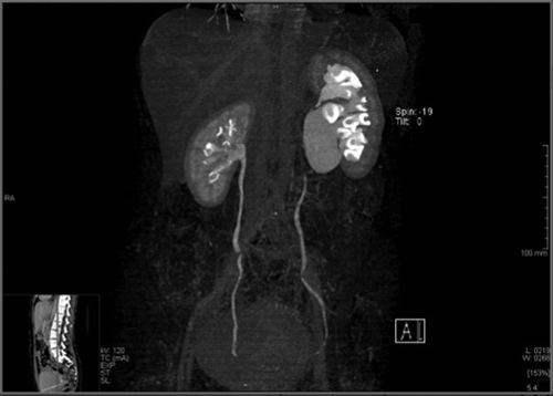
A 29-year-old woman presented to the Emergency Room complaining of dizziness, asthenia, headache and reporting a recent onset of hypertension. The clinical examination was unremarkable except for high blood pressure of 160/110 mmHg. She had a family history of hypertension and no other cardiovascular risk factors. Blood tests were normal and electrocardiogram showed left ventricular hypertrophy. Echocardiography confirmed cardiac damage as identified by septal hypertrophy and showed left atrial enlargement. The patient was treated with clonidine and benzodiazepines, and discharged. A secondary hypertension was suspected and therefore the patient was referred to our hypertension clinic where her blood pressure was still found high (168/110 mmHg). Verapamil and doxazosin were prescribed in order not to affect the renin– angiotensin system (RAS). All tests performed for ruling out a secondary hypertension were normal with the exception of elevated plasma renin activity (PRA) both at baseline and after captopril test (50 mg of captopril): 6.40 and 21.80 μg/l/h, respectively (normal values < 2.62 μg/l/h with a sodium daily income according with a normal Italian diet of 150 mmol). The patient PRA was evaluated 20 days later on the same drug regimen and was found within normal limit. An abdominal ultrasound examination showed a left kidney hydronephrosis and the computerized tomography confirmed a severe dilatation of the renal pelvis and PUJO (). [99mTc] mercaptoacetylglycine (MAG3) dynamic renal scintigraphy (DRS) with diuretic test (furosemide 20 mg i.v.) showed impairment of left kidney drainage with a left renal plasma flow (RPF) of 200 ml/min (44%) ().
Figure 1. Computerized tomography scan revealing a hydronephrosis of the left kidney with a markedly dilated renal pelvis.

Figure 2. [99mTc] mercaptoacetylglycine (MAG3) renal scintigraphy indicating an obstructed pattern on the left kidney (A), which normalized after surgical intervention (B).
![Figure 2. [99mTc] mercaptoacetylglycine (MAG3) renal scintigraphy indicating an obstructed pattern on the left kidney (A), which normalized after surgical intervention (B).](/cms/asset/4947a8b5-90bb-41ed-9de9-a2a9dcf7557e/iblo_a_778005_f0002_b.jpg)
The patient was then treated with ramipril 5 mg/day and amlodipine 5 mg/day with a transient blood pressure reduction and underwent cystoscopy with double-J ureteric stenting. In the following months, the patient's blood pressure levels decreased and antihypertensive medications were tapered accordingly. After 3 months, a 24-h ambulatory blood pressure measurement reported a mean 24-h blood pressure value of 118/74 mmHg, a mean daytime blood pressure of 121/77 mmHg and a mean overnight blood pressure of 112/69 mmHg. Control DRS showed good functional recovery and renal plasma flow of the left kidney was 261 ml/min (49%). (). Six months later, the patient was normotensive with a daytime blood pressure of 118/ 70 mmHg with no antihypertensive medication. Control blood tests showed normal renal function and echocardiogram identified complete reversal to normal of the septal hypertrophy and left atrial enlargement.
Discussion
Hydronephrosis is a common condition and PUJO is the most frequent cause in the paediatric population (Citation1). Approximately 5–10% of children with PUJO are hypertensive (Citation2) and in most cases relief of the obstruction normalizes blood pressure (Citation3).
Few data are available in the literature regarding the adult population in which PUJO is rare and the effect of the correction of the obstruction on blood pressure is still unknown.
In order to comprehend the pathophysiology of hypertension in PUJO, several studies were carried out with conflicting results. However, Carlström and coworkers (Citation4,Citation5) finally demonstrated that animals with hydronephrosis induced by PUJO developed salt-sensitive hypertension, which was directly correlated with the degree of hydronephrosis. Indeed, in hydronephrosis there is a consistent rise in ureteric pressure leading to changes in glomerular filtration, tubular function and renal blood flow. Two major systems involved in the pathogenesis of PUJO-induced hypertension are the RAS and the renal sympathetic nerves (RSN).
Studies performed on experimental models of PUJO have demonstrated that intrarenal RAS is activated throughout the period of obstruction (Citation6). In addition, in animals, the elevated renin level normalized after removal of the hydronephrotic kidney (Citation7). However, hypertension in PUJO cannot be entirely due to the increased plasma renin level, given the negative association found between RAS and blood pressure in experimental animals with PUJO (Citation8).
Enhanced RSN activity can increase plasma renin concentrations and has been shown to lead to sodium retention and salt-sensitive hypertension (Citation9). In the PUJO model of hydronephrosis, renal denervation of the ipsilateral kidney attenuated both hypertension and salt sensitivity in hydronephrotic animals, suggesting that an increased RSN activity contributes to the development of hypertension in hydronephrosis (Citation10). Furthermore, oxidative stress and nitric oxide deficiency in the diseased kidney also appear to significantly contribute to the development of hypertension in animal models of PUJO (Citation11,Citation12).
Currently, pyeloplasty is the standard of care for the surgical correction of PUJO. The indications for surgery in such cases are well defined like a RPF < 40% in unilateral hydronephrosis with a normal contralateral kidney, the presence of symptoms such as abdominal colic or flank pain and rapid worsening of hydronephrosis on serial renal ultrasound, or severe bilateral hydronephrosis (Citation13). Furthermore, as an association between PUJO and hypertension has been shown, we suggest the moderate–severe hypertension with cardiac organ damage could be an additional indication for surgical correction in these patients. Our patient did not complain of PUJO typical clinical manifestations and left RPF was higher than 40%. However, the presence of severe hypertension with cardiac organ damage led us to believe that treating the cause could also prevent the cerebrovascular complications previously described in patients with PUJO-related hypertension (Citation14).
While there are few reports of the long-term outcomes of PUJO correction in adults, no systematic research has been done until now into the effects of PUJO obstruction surgical correction to treat hypertension in affected adults. Kinn (Citation15) followed 71 patients with PUJO, 42 treated with pyeloplasty and 29 without surgical treatment with a prevalence of hypertension of 4.7% in the former group and 6.9% in the latter. No data about blood pressure were available at follow-up. In children, PUJO seems a reversible functional disturbance with no relevant associated anatomic changes and its treatment normalizes blood pressure. In the adult population, PUJO can lead to tubular atrophy and permanent nephron loss as well as secondary hypertension. Of note in our case, we assumed that our patient had a chronic hydronephrosis in which the renal loss of function is irreversible even with correction of the obstruction. Early experiments with dogs showed that if acute unilateral obstruction is corrected within 2 weeks, full recovery of renal function is possible. However, after 6 weeks of obstruction, renal function is irreversibly lost (Citation16).
In conclusion, we reported a case of a 29-year-old woman with PUJO-related hypertension and cardiac organ damage successfully managed by surgical correction of the defect. On the basis of current knowledge regarding the long-term physiological consequences of chronic partial ureteral obstruction, we suggest that the current non-operative management in hydronephrotic patients should be re-evaluated with the aim of preventing renal injury and, most of all, hypertension and its long-term cardiovascular complications such as cardiovascular remodelling and organ damage in later life.
Some questions still remain unanswered: (i) Is there a biomarker that can help to guide treatment decisions? (ii) Should the treatment of PUJO be stratified according to the age of the patient? Our case points out the importance of the renin– angiotensin–aldosterone system in the pathophysiology of hypertension and shows that the development of hypertension due to an increased renin production can be successfully managed by treating the underlying cause of hypertension.
Declaration of interest: The authors report no conflicts of interest. The authors alone are responsible for the content and writing of the paper.
References
- Livera LN, Brookfield DS, Egginton JA, Hawnaur JM. Antenatal ultrasonography to detect fetal renal abnormalities: A prospective screening programme. BMJ. 1989;298:1421–1423.
- Lowe FC, Marshall FF. Ureteropelvic junction obstruction in adults. Urology. 1984;23:331–335.
- de Waard D, Dik P, Lilien MR, Kok, ET, de Jong TPVM. Hypertension is an indication for surgery in children with ureteropelvic junction obstruction. J Urol. 2008;179: 1976–1979.
- Carlström M, Sällström J, Skøtt O, Larsson E, Wåhlin N, Persson AEG. Hydronephrosis causes salt-sensitive hypertension and impaired renal concentrating ability in mice. Acta Physiol (Oxf). 2007;189:293–301.
- Carlström M, Wåhlin N, Sällström J, Skøtt O, Brown R, Persson AE. Hydronephrosis causes salt-sensitive hypertension in rats. J Hypertens. 2006;24:1437–1443.
- Chevalier RL. Molecular and cellular pathophysiology of obstructive nephropathy. Pediatr Nephrol. 1999;13: 612–619.
- Carlström M, Wåhlin N, Skøtt O, Persson AEG. Relief of chronic partial ureteral obstruction attenuates salt-sensitive hypertension in rats. Acta Physiol (Oxf). 2007;189:67–75.
- Tauchi K, Kanehara H. Hypertension and the renin– angiotensin system in the congenital hydronephrosis rat with non-obstructive pelviureteric junction abnormalities. Exp Nephrol. 1996;4:60–64.
- DiBona GF, Kopp UC. Neural control of renal function. Physiol Rev. 1997;77:75–197.
- Carlström M. Causal link between neonatal hydronephrosis and later development of hypertension. Clin Exp Pharmacol Physiol. 2010;37:e14–23.
- Carlström M, Brown RD, Edlund J, Sällström J, Larsson E, Teerlink T, et al. Role of nitric oxide deficiency in the development of hypertension in hydronephrotic animals. Am J Physiol Renal Physiol. 2008;294:F362–370.
- Carlström M, Brown RD, Sällström J, Larsson E, Zilmer M, Zabihi S, et al. SOD1 deficiency causes salt sensitivity and aggravates hypertension in hydronephrosis. Am J Physiol Regul Integr Comp Physiol. 2009;297:R82–92.
- Chertin B, Pollack A, Koulikov D, Rabinowitz R, Hain D, Hadas-Halpren I, et al. Conservative treatment of ureteropelvic junction obstruction in children with antenatal diagnosis of hydronephrosis: Lessons learned after 16 years of follow-up. Eur Urol. 2006;49:734–738.
- Tourchi A, Kajbafzadeh A, Nejat F, Golmohammadi A, Alizadeh F, Mahboobi AH. Bilateral ureteropelvic junction obstruction presenting with hypertension and cerebral vascular accident. J Pediatr Surg. 2010;45:e7–10.
- Kinn AC. Ureteropelvic junction obstruction: Long-term follow-up of adults with and without surgical treatment. J Urol. 2000;164:652–656.
- Leahy AL, Ryan PC, McEntee GM, Nelson AC, Fitzpatrick JM. Renal injury and recovery in partial ureteric obstruction. J Urol. 1989;142:199–203.
