Abstract
Background. Abnormal left ventricular (LV) geometric patterns, particularly concentric LV hypertrophy, are associated with a greater risk of hypertensive complications. The aim of this study was to investigate the association between LV myocardial performance index (LVMPI) and aortic distensibility (AD) with different LV geometric patterns in patients with newly diagnosed hypertension (HT). Methods. We studied 181 patients with newly diagnosed HT (mean age 51.7 ± 5.4 years) and 39 healthy control subjects (mean age 51.2 ± 5.1 years). Echocardiographic examination was performed in all subjects. Four different geometric patterns were determined in hypertensive patients according to LV mass index (LVMI) and relative wall thickness (RWT). AD was calculated from the echocardiographically derived ascending aorta diameters and haemodynamic pressure measurements. LVMPI was calculated from the tissue Doppler-derived ejection time, isovolumic contraction and relaxation times. Results. The highest LVMPI and the lowest AD values were observed in concentric hypertrophy group compared with control, normal geometry, concentric remodelling and eccentric hypertrophy groups (p < 0.05, for all). LVMPI was associated with LVMI (r = 0.497, p < 0.001), RWT (r = 0.270, p < 0.001), AD (r = −0.316, p < 0.001) and E deceleration time (r = 0.171, p = 0.02) in bivariate analysis. In multiple linear regression analysis, LVMPI was independently related to LVMI (β = 0.381, p < 0.001) and AD (β = −0.263, p = 0.001). Conclusions. The LVMPI was highest and AD was lowest in patients with concentric hypertrophy. The LVMPI was independently associated with LVMI and AD in hypertensive patients.
Introduction
Primary hypertension (HT) is a major risk factor for cardiovascular disease. Pressure and volume overload in hypertensive patients leads to left ventricular (LV) hypertrophy (LVH), which is a powerful independent predictor of morbidity and mortality (Citation1,Citation2). There are four different geometric patterns of LV in HT, and these geometric patterns are different in respect to prognosis and LV function (Citation2,Citation3). LV geometric patterns incorporate normal LV structure and concentric remodelling in addition to LVH (eccentric hypertrophy and concentric hypertrophy) (Citation3).
Conventional LV myocardial performance index (LVMPI) is commonly used as a measure of combined systolic and diastolic function (Citation4). Tissue Doppler echocardiography enables us to measure simultaneously both the diastolic and systolic intervals of myocardial motion (Citation5). Determination of LVMPI derived by using pulsed wave tissue Doppler ultrasound scanning requires measurement of only two simple intervals on the same cardiac cycle. Because of this, results are reliable in the presence of heart rate fluctuation (Citation6,Citation7).
Aortic distensibility (AD) is a measurement of vascular elasticity, which reflects the stiffness of aorta (Citation8). Impaired AD influences both LV diastolic and systolic functions (Citation8–11). Previous studies have shown that HT decreases AD (Citation8–11).
In previous studies, there are limited and controversial results related to relationship between LVMPI and LV geometric patterns. Therefore, in the current study, we aimed to investigate tissue Doppler LVMPI and AD in newly diagnosed hypertensive patients with different LV geometries.
Methods
Study populations
Measurements were obtained from 181 patients with newly diagnosed essential HT (mean age = 51.7 ± 5.4 years, male/female = 83/98) and 39 healthy controls (mean age = 51.2 ± 5.1 years; male/female = 18/21). Hypertensive patients had three clinic blood pressure measurements (> 140/90 mmHg) taken at 1-week intervals in the absence of any previous antihypertensive treatment to exclude the pharmacological effects on haemodynamics or ventricular hypertrophy and function. The control group was completely healthy and had multiple blood pressure measurements (< 140/90 mmHg) and were age- and gender-matched in relation to the hypertensive patients. The Local Ethics Committee assessed and approved the study and written informed consent for participation in the study was obtained from all individuals.
Exclusion criteria were secondary or malignant HT, heart failure, positive history or clinical signs of ischaemic heart disease, positive effort test, positive myocardial perfusion scintigraphy, cerebrovascular disease, severe valve disease, atrial fibrillation, receiving any drugs, renal insufficiency (serum creatinine: > 1.5 mg/dl in men and > 1.4 mg/dl in women), major non-cardiovascular diseases and known diabetes or fasting glucose > 126 mg/dl.
Echocardiography
Standard two-dimensional and Doppler echocardiographies were performed using a commercially available echocardiographic machine (Vivid 7R GE Medical System, Horten, Norway) with a 2.0–3.5-MHz transducer. Measurements were made during normal breathing at end expiration. LV end-systolic (LVSd) and end-diastolic diameters (LVDd), end-diastolic interventricular septal thickness (IVSth) and end-diastolic LV posterior wall thickness (PWth) were measured at end-diastole according to established standards of the American Society of Echocardiography (Citation12). LV ejection fraction (EF) was determined by the biplane Simpson's method (Citation13). LV mass (LVM) was calculated using the Devereux formula (Citation14):
Thereafter, LV mass index (LVMI) was obtained by the following formula: LVM/body surface area. LVH was defined according to more stringent criteria as LVMI values exceeding 125 g/m2 in men and 110 g/m2 in women (Citation15). Relative wall thickness (RWT) was measured at end diastole as the ratio of (2 × LV posterior wall thickness)/LVDd. Increased RWT was defined as ≥ 0.45 (Citation3).
Patterns of LV geometry
Geometric patterns were based on the upper normal limits for LVMI and RWT: (i) normal geometry (NG; normal LVMI and normal RWT; (ii) concentric remodelling (CR: normal LVMI and increased RWT); (iii) concentric hypertrophy (CH; increased LVMI and increased RWT); and (iv) eccentric hypertrophy (EH; increased LVMI and normal RWT) (Citation3).
Aortic distensibility
Ascending aorta (Ao) diameters were measured from the same view on the M mode tracing at a level 3 cm above the aortic valve. The systolic diameter (AoS) was measured at the maximum anterior motion of the aorta and the diastolic diameter (AoD) was measured at the peak of the QRS complex on the simultaneously recorded electrocardiogram. Pulse pressure (PP) was obtained simultaneously by cuff sphygmomanometry of the left brachial artery as systolic blood pressure (SBP) minus diastolic blood pressure (DBP). The following indexes of aortic function were calculated:
Standard and tissue Doppler echocardiography
Pulsed Doppler ultrasound scanning recordings of the mitral inflow velocities were obtained from the apical four-chamber view by placing the sample volume between the tips of the mitral leaflets (Citation17). These Doppler parameters were analysed: peak early (E) and late (A) transmitral filling velocities, the ratio of early to late peak velocities (E/A), and deceleration time of E wave (DT). Doppler ultrasound scanning measurements were calculated from an average of five consecutive cardiac cycles.
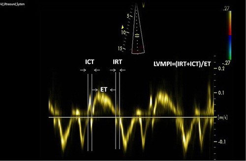
Pulse wave tissue Doppler was performed with a 2-mm sample volume placed at the lateral corner of the mitral annulus from the apical four-chamber view. Filters were set to exclude high-frequency signals and gain minimized. Doppler ultrasound scanning intervals were measured from mitral annular velocity intervals. LVMPI was determined by using this equation (Citation4):
The calculation of tissue Doppler-derived LVMPI was shown in .
Figure 1. The calculation of tissue Doppler-derived left ventricle myocardial performance index (ICT, isovolumic contraction time; ET, ejection time; IRT, isovolumic relaxation time).
Reproducibility
Interobserver variability of the measurements of pulsed wave tissue Doppler parameters and AD measurements was calculated as the difference in two measurements from the same patient by two different observers divided by the mean value. Intraobserver variability was calculated as the difference in two measurements of the same patient by one observer divided by the mean value. Intraobserver and interobserver variabilities were less than 5% for all pulsed wave tissue Doppler parameters and AD measurements.
Statistical analysis
All analyses were conducted using SPSS 17.0 (SPSS for Windows 17.0, Chicago, IL). Distribution of continuous variables was assessed with the one-sample Kolmogorov Smirnov test. Comparison of categorical variables between the groups was performed using the chi-square (χ2) test. Analysis of variance (ANOVA) was used in the analysis of continuous variables. A stratified post hoc analyses of echocardiographic, clinical and laboratory variables were performed according to the LV geometric patterns. The correlations between tissue Doppler LVMPI, AD, haemodynamic and echocardiographic parameters were assessed by the Pearson correlation test. Multiple linear regression analysis was performed to identify the independent associations of tissue Doppler LVMPI and AD. A two-tailed p < 0.05 was considered statistically significant.
Results
Baseline characteristics
Comparison of baseline characteristics in groups is shown in . Body mass index, body surface area, SBP, DBP, PP, total cholesterol, low-density lipoprotein-cholesterol, glucose and creatinine levels were different between the control and patient groups (p < 0.05, for all).
Table I. Comparisons of baseline characteristics among the groups.
Echocardiographic characteristics
Comparisons of echocardiographic characteristics among the groups were reported in the . The lowest LVMPI and highest AD values were found in control group compared with patient groups (p < 0.001, for all). The highest LVMPI and lowest AD values were observed in CH group compared with NG, CR and EH groups (p < 0.001, for all). Also LVMPI value of EH group was higher than NG and CR groups (p = 0.021, p = 0.005, respectively), and its AD value was higher than NG group (p = 0.020).
Table II. Comparisons of echocardiographic characteristics among the groups.
Bivariate and multivariate analysis
Correlation analysis of variables was demonstrated in the . LVMPI value was associated with LVMI (r = 0.497, p < 0.001), RWT (r = 0.270, p < 0.001), AD (r = − 0.316, p < 0.001), DT (r = 0.171, p = 0.02), SBP (r = 0.148, p = 0.047) and DBP (r = 0.178, p = 0.017) in bivariate analysis. LVMPI value was independently related to LVMI (β = 0.381, p < 0.001) and AD (β = − 0.263, p = 0.001) in multiple linear regression analysis. Relationships between LVMPI value, LVMI and AD were shown in and .
Figure 2. Relationship between tissue Doppler-derived left ventricle myocardial performance index and left ventricle mass index.
Figure 3. Relationship between tissue Doppler-derived left ventricle myocardial performance index and aortic distensibility.
Table III. Correlation analysis of variables in patient group.
AD value was independently associated with LVMPI value (β = − 0.186, p = 0.001), LVMI (β = −0.159, p = 0.004) and E/A ratio (β = 0.327, p < 0.001) in multiple linear regression analysis.
Discussion
The main findings of the present study were that: (i) hypertensive patients have impaired LVMPI and AD compared with control subjects; (ii) the major increase in LVMPI values and the major decrease in AD values were observed in CH geometric pattern; (iii) LVMPI value was independently associated with LVMI and AD in patient groups; and (iv) AD was independently related to LVMI and LVMPI.
Previous studies have reported that abnormal LV geometric patterns are associated with a greater risk of hypertensive complications (Citation1–3,Citation18). In particular, hypertensive patients with concentric LV hypertrophy have the highest incidence of cardiovascular events including death (Citation2). Patients with EH or CR have intermediate risk rates. The lowest cardiovascular risk was observed in the group with normal LV geometry (Citation3).
In the present study, tissue Doppler-derived LVMPI in newly diagnosed hypertensive patients was impaired despite having preserved EF. The patients with CH group showed the most deterioration in myocardial performance. There are conflicting reports on the type of association between LVMPI, LV morphology and LVH in essential HT (Citation19–22). Recently, Takasaki et al. reported that LVMPI in CH group was most impaired among all the geometric patterns in untreated hypertensive patients (Citation19). In that study, authors showed that LVMPI was independently related with only LVMI in multiple linear regression analysis. Similar results were also observed in other two studies (Citation20,Citation21). However, current study showed that MPI was not only associated with LVMI but also AD in hypertensive patients. In contrast, Karaye found that LV geometric patterns and LVEF were not associated with MPI in hypertensives, but there was strong association between LV geometric patterns and LV EF (Citation22). In the study of Karaye, 38% of patients had low LV EF unlike to our study. Also, in that study, they found that MPI was related to heart rate, whereas the major advantages of MPI are that it is independent of heart rate (Citation23).
Previous studies have found that concentric and eccentric hypertrophy were associated with a higher degree of cardiovascular events, and systolic and diastolic dysfunction (Citation1–3,Citation7,Citation19–22). In our study, the LVMPI was highest among hypertensive patients with CH, followed by those with EH, with significant difference between the two groups. Also, our study revealed that the LVMPI was significantly higher among hypertensive subjects with EH compared with those with CR. This result suggests that LVMI is a more important parameter than RWT on LVMPI. In current study, LVMPI was increased in hypertensive patients with NG compared with control group. This condition suggests that LV systolic and diastolic functions in hypertensive patients would be impaired even without increased LVMI and RWT. LVH is a measure of preclinical cardiac disease (Citation1). LVH and myocardial fibrosis also lead to diastolic dysfunction. Hypertrophied tissue contains increased amounts of collagen and interstitial fibrosis, and this collagen correlates with impaired Doppler mitral valve inflow patterns (Citation1,Citation24). In present study, relationships between LVMI with E/A ratio and DT, which reflects diastolic functions, were supported by the above-mentioned mechanism. In addition, elevated LV mass is associated with decreased myocardial flow reserve and reduced tolerance of myocardial ischemia (Citation1,Citation25). Increased myocardial ischemia may lead to myocardial scarring and LV dysfunction (Citation26). Additionally, reversing LVH with treatment has been shown to improve systolic and diastolic LV function (Citation27). Therefore, increased LVMPI values, in particularly hypertrophic geometries (CH, EH), reflect the effect of LVH on LV systolic and diastolic function.
In the present study, we found that AD values were impaired in patients with HT compared with control subjects. However, the most significant decrease in AD was observed in CH geometric pattern. This finding may be plausible because of this geometric pattern is the endpoint of HT. We observed an independent association between AD and LVMI. However, AD in EH geometric pattern, which was expressed by increased LVMI, was less impaired when compared with the CH group. This condition may be related to the observation of an eccentric LVH in the early stages of HT according to CH geometric pattern (Citation28). AD is a measurement of vascular elasticity and is a marker for the development or regression of LVH. Reduced AD (reflects to increased aortic stiffness) may participate in the genesis of cardiac hypertrophy in patients with HT. The most important factor in developing cardiac hypertrophy is increased end systolic wall stress (Citation29). End systolic wall stress is influenced not only by the geometric properties of the ventricle but also by increased aortic stiffness (Citation30). Lakatta & Levy showed that increased vascular loading on the heart was a likely cause of the increase in LV wall thickness and wall stress (Citation31).
Finally, the current study showed that LVMPI was independently associated with AD, as well as LVMI. The relationship between MPI and AD in essential HT was reported in only one study (Citation9). However, in that study, LVMPI and AD were not separately investigated according to LV geometric patterns. It is known that AD is an important determinant of LV dysfunction (Citation32). Furthermore, decreased AD influences both left LV diastolic and systolic functions (Citation7–9).
Clinical implications
Our findings may have important clinical implications to the early identification of subclinical LV dysfunction in patients with HT and preserved EF. The early diagnosis of subclinical LV dysfunction with LVMPI may be useful for the appropriate therapeutic approaches in newly diagnosed hypertensive patients.
Study limitations
In the present study, coronary angiography was not performed on the study subjects. However, the patients with a positive effort test, previous coronary artery disease, positive myocardial perfusion scintigraphy, positive history or clinical signs of ischaemic heart disease were excluded from our study.
Conclusions
The present study showed that the LV function assessed with LVMPI and elasticity of aorta assessed with AD were impaired in hypertensive patients compared with the control subjects. These impairments were more prominent in patients with CH than in other LV geometric patterns.
Declaration of interest: The authors report no conflicts of interest. The authors alone are responsible for the content and writing of the paper.
References
- Krauser DG, Devereux RB. Ventricular hypertrophy and hypertension: prognostic elements and implications for management. Herz. 2006;31:305–316.
- Koren M J, Devereux RB, Casale PN, Savage DD, Laragh JH. Relation of left ventricular mass and geometry to morbidity and mortality in uncomplicated essential hypertension. Ann Intern Med. 1991;114:345–352.
- Ganau A, Devereux RB, Roman MJ, de Simone G, Pickering TG, Saba PS, et al. Patterns of left ventricular hypertrophy and geometric remodeling in essential hypertension. J Am Coll Cardiol. 1992;19:1550–1558.
- Tei C, Nishimura RA, Seward JB, Tajik AJ. Noninvasive Doppler-derived myocardial performance index: Correlation with simultaneous measurements of cardiac catheterization measurements. J Am Soc Echocardiogr. 1997;10: 169–178.
- Galiuto L, Ignone G, DeMaria AN. Contraction and relaxation velocities of the normal left ventricle using pulsed-wave tissue Doppler echocardiography. Am J Cardiol. 1998;81:609–614.
- Harada K, Tamura M, Toyono M, Oyama K, Takada G. Assessment of global left ventricular function by tissue Doppler imaging. Am J Cardiol. 2001;88:927–932.
- Ayhan SS, Özdemir K, Kayrak M, Bacaksiz A, Vatankulu MA, Eren Ö, et al. The evaluation of doxorubicin-induced cardiotoxicity: Comparison of Doppler and tissue Doppler-derived myocardial performance index. Cardiol J. 2012;19:363–368.
- Eren M, Gorgulu S, Uslu N, Celik S, Dagdeviren B, Tezel T. Relation between aortic stiffness and left ventricular diastolic function in patients with hypertension, diabetes, or both. Heart. 2004;90:37–43.
- Gur M, Yilmaz R, Demirbag R, Yildiz A, Ozdogru I, Bas MM, et al. Relationship between myocardial performance index and aortic distensibility in patients with essential hypertension. Int J Clin Pract. 2008;62:138–142.
- Dart A, Silagy C, Dewar E, Jennings G, McNeil J. Aortic distensibility and left ventricular structure and function in isolated systolic hypertension. Eur Heart J. 1993;14:1465–1470.
- Celik T, Yuksel UC, Fici F, Celik M, Yaman H, Kilic S, et al. Vascular inflammation and aortic stiffness relate to early left ventricular diastolic dysfunction in prehypertension. Blood Press. 2013;22:94–100.
- Lang RM, Bierig M, Devereux RB, Flachskampf FA, Foster E, Pellikka PA, et al. Chamber Quantification Writing Group. Recommendations for Chamber Quantification: A report from the American Society of Echocardiography's Guidelines and Standards Committee and the Chamber Quantification Writing Group, Developed in Conjunction with the European Association of Echocardiography, a Branch of the European Society of Cardiology. J Am Soc Echocardiogr. 2005;18:1440–1463.
- Schiller NB, Shah PM, Crawford M, DeMaria A, Devereux R, Feigenbaum H, et al. Recommendations for quantitation of the left ventricle by two-dimensional echocardiography. American Society of Echocardiography Committee on Standards, Subcommittee on Quantitation of Two-Dimensional Echocardiograms. J Am Soc Echocardiogr. 1989;2:358–367.
- Devereux RB, Reichek N. Echocardiographic determination of left ventricular mass in man. Anatomic validation of the method. Circulation. 1977;55:613–618.
- Guidelines Committee. 2003 European Society of Hypertension–European Society of Cardiology guidelines for management of arterial hypertension. J Hypertens. 2003;21:1011–1053.
- Stefanadis C, Stratos C, Vlachopoulos C, Marakas S, Boudoulas H, Kallikazaros I, et al. Pressure diameter relation of the human aorta. A new method of determination by the application of a special ultrasonic dimension catheter. Circulation. 1995;92:2210–2219.
- Appleton CP, Hatle LK, Popp RL. Relation of transmitral flow velocity patterns to left ventricular diastolic function: New insights from a combined hemodynamic and Doppler echocardiographic study. J Am Coll Cardiol. 1988;12: 426–440.
- Negri F, Sala C, Re A, Mancia G, Cuspidi C. Left ventricular geometry and diastolic function in the hypertensive heart: Impact of age. Blood Press. 2013;22:1–8.
- Takasaki K, Miyata M, Imamura M, Yuasa T, Kuwahara E, Kubota K, et al. Left ventricular dysfunction assessed by cardiac time interval analysis among different geometric patterns in untreated hypertension. Circ J 2012;76:1409–1414.
- Akintunde AA, Akinwusi PO, Opadijo GO. Relationship between Tei index of myocardial performance and left ventricular geometry in Nigerians with systemic hypertension. Cardiovasc J Afr. 2011;22:124–127.
- Yilmaz R, Seydaliyeva T, Unlü D, Uluçay A. The effect of left ventricular geometry on myocardial performance index in hypertensive patients. Anadolu Kardiyol Derg. 2004;4: 217–222.
- Karaye KM. Relationship between Tei Index and left ventricular geometric patterns in a hypertensive population: A cross–sectional study. Cardiovasc Ultrasound. 2011;9:21.
- Poulsen S, Nielsen J, Andersen H. The influence of heart rate on the Doppler derived myocardial performance index. J Am Soc Echocardiogr. 2000;13:379–384.
- Díez J, Querejeta R, López B, González A, Larman M, Martínez Ubago JL. Losartan-dependent regression of myocardial fibrosis is associated with reduction of left ventricular chamber stiffness in hypertensive patients. Circulation. 2002;105:2512–2517.
- Friehs I, del Nido PJ. Increased susceptibility of hypertrophied hearts to ischemic injury. Ann Thorac Surg, 2003;75:S678–684.
- Kahan T, Bergfeldt L. Left ventricular hypertrophy in hypertension: Its arrhythmogenic potential. Heart, 2005;91: 250–256.
- Wachtell K, Palmieri V, Olsen MH, Gerdts E, Papademetriou V, Nieminen MS, et al. Change in systolic left ventricular performance after 3 years of antihypertensive treatment: The Losartan Intervention For Endpoint (LIFE) study. Circulation. 2002;106:227–232.
- Laufer E, Jennings GL, Korner PI, Dewar E. Prevalence of cardiac structural and functional abnormalities in untreated primary hypertension. Hypertension. 1989;13:151–162.
- Grossman W, Jones D, McLaurin LP. Wall stress and patterns of hypertrophy in the human left ventricle. J Clin Invest. 1975;56:56–64.
- Bouthier JD, De Luca N, Safar ME. Cardiac hypertrophy and arterial distensibility in essential hypertension. Am Heart J 1985;109:1345–1352.
- Lakatta EG, Levy D. Arterial and cardiac aging: Major shareholders in cardiovascular disease enterprises. Circulation. 2003;107:346–354.
- Hundley WG, Kitzman DW, Morgan TM, Hamilton CA, Darty SN, Stewart KP, et al. Cardiac cycle dependent changes in aortic area and distensibility are reduced in older patients with isolated diastolic heart failure and correlate with exercise intolerance. J Am Coll Cardiol. 2001;38:796–802.