Abstract
Background. Postinfectious glomerulonephritis is rare in adults. The characteristics of the disease now differ from what were described decades ago. The goal of this study is to illustrate the clinicopathological spectrum of the disease in the modern era. Methods. Between July 2000 and June 2008, 20 adult cases of postinfectious glomerulonephritis were identified at a medical center in Taiwan. The patients' records were retrospectively reviewed with respect to clinical presentation, microbiology, serology, morphology of renal biopsy, and clinical course. Results. There were 14 males and 6 females. The mean age was 61 years. All patients developed acute renal failure, and the majority (65%) required dialysis support during the disease course. Hypocomplementemia was present in 60% of patients. The most frequently identified infectious agent was Staphylococcus (60%). Histological characteristics showed two distinct patterns of glomerulonephritis: diffuse endocapillary proliferative glomerulonephritis (65%) and focal mesangial proliferative glomerulonephritis (35%). There were no significant differences in the clinical presentation and outcome between the two groups. However, glomerular neutrophil infiltration was more commonly present in diffuse endocapillary proliferative pattern (p = 0.017). The percentage of patients with focal mesangial proliferative pattern significantly increased over time (p < 0.001). At the last follow-up, 6 patients (30%) had died, 6 (30%) were in complete remission, 4 (20%) had partial remission with renal insufficiency, and 4 (20%) were on chronic dialysis. Conclusions. Our data suggested that Staphylococcus had become the leading pathogen in adult postinfectious glomerulonephritis over the past 10 years. Furthermore, atypical histological feature with focal mesangial proliferative pattern was increasingly identified over time. The prognosis was still guarded, carrying a considerable mortality rate and risk for developing chronic renal failure.
INTRODUCTION
Infection has been well documented to cause glomerulonephritis. In the past, the vast majority of cases typically followed streptococcal upper respiratory tract or skin infections, and the disease most frequently affects children. Typical histological findings include diffuse endocapillary proliferative glomerulonephritis accompanied by infiltration with neutrophils within the capillary lumens (exudative features) on light microscopy, C3-dominant granular deposits with or without IgG co-deposition on immunofluorescence, and characteristic subepithelial hump-shaped deposits on electron microscopy. Abrupt symptoms of acute nephritis, hypocomplementemia, and rising anti-streptolysin O (ASLO) titers are diagnostic. Renal biopsy is rarely required. Most children with poststreptococcal glomerulonephritis (PSGN) recover completely. Over recent decades, the pattern of the disease has changed. Not only Streptococcus but also other bacterial, viral, and parasitic agents have been implicated in the pathogenesis of glomerulonephritis.Citation[1] In developed countries, glomerulonephritis associated with non-streptococcal infections is assuming greater important. This is thought to be secondary to a decline in the incidence of group A streptococcal infections in children and a relative increase in the incidence of glomerulonephritis associated with other infections in adults. Furthermore, an increasing number of adult cases have been observed in alcoholics, diabetics, and intravenous drug abusers.Citation[2,3] Atypical clinical presentation often presents complex diagnostic challenges and highlights the important diagnostic role of renal biopsy. The more extensive use of renal biopsy has demonstrated the presence of atypical histological features of the disease.Citation[4,5] While several studies performed before 2000 evaluated the course of adult postinfectious glomerulonephritis,Citation[2,3,6] there have been few reports in the new millennium. The aim of this study was to investigate whether the spectrum of the disease had changed over the past 10 years.
METHODS
Between July 2000 and June 2008, 20 adult cases of postinfectious glomerulonephritis were identified at a medical center in Taiwan, in which there was close temporal relationship between infection and first appearance of renal manifestations, and there was no clinical or laboratory evidence of systemic disease that might cause glomerulonephritis. All patients were submitted to renal biopsy analyzed by light microscopy after hematoxylin-eosin, periodic acid-Schiff, and Masson trichrome stains and by immunofluorescence using antisera against IgG, IgA, IgM, C3, and C1q. The extent of mesangial and endocapillary proliferation was graded as focal (involving <50% of glomeruli) and diffuse (involving ≥50% of glomeruli). Scoring of the chronic tubulointerstitial injury was based on the percentage of tubular atrophy and interstitial fibrosis, and was graded as mild (1+) if involving <25%, moderate (2+) if involving 25–50%, and severe (3+) if involving >50%. Other histological parameters were also scored semiquantitatively on a scale from 0 to 3+. The intensity of immunofluorescence staining was graded on a scale of 0 to 3+. In 11 out of the 20 cases, electron microscopy was performed. Patients' medical records were reviewed for age, sex, type and source of infection, clinical presentation, parameters of renal function, and outcome. Remission was defined as complete if last follow-up serum creatinine was ≤1.2 mg/dL and partial if last follow-up serum creatinine was >1.2 mg/dL.
Statistical analyses were performed using SPSS version 12 (SPSS Inc., Chicago, Illinois, USA) for Windows and produced by simple non-parametric test (Mann-Whitney U test and Fisher's exact test). A p value < 0.05 was considered significant.
RESULTS
There were 14 males and 6 females, with mean age of 61 ± 15 years (range 30–80 years). Clinical characteristics at presentation are summarized in . All patients developed acute renal failure. The mean 24-hour urinary protein excretion was 3.2 g, and 41.1% of patients had nephrotic range proteinuria. Hematuria was present in all patients. Leukocyturia was seen in the majority of patients. The mean peak serum creatinine was 6.65 mg/dL. Hypocomplementemia was present in 60% of patients. Thirteen out of 20 patients (65%) required dialysis support during the disease course.
Table 1 Clinical details of 20 adult patients with postinfectious glomerulonephritis
All but one patient (case 9) had renal manifestations during the course of intercurrent infections. In case 9, there was no clinical evidence of infection at presentation, and the diagnosis was made according to rising ASLO titers and classic histological criteria. The sites of infection included skin (20%), heart/endocarditis (20%), lung (15%), bone/joint (15%), urinary tract (15%), and deep-seated abscess (10%). The two most frequently identified infectious agents were Staphylococcus (60%) and Streptococcus (15%). In a total of 12 patients with staphylococcal infection, 8 (67%) had methicillin-resistant Staphylococcus aureus (MRSA) and 4 (33%) had methicillin-sensitive Staphylococcus aureus (MSSA).
Histological characteristics are summarized in . Two distinct patterns of glomerulonephritis were identified: diffuse endocapillary proliferative glomerulonephritis (65%) and focal mesangial proliferative glomerulonephritis (35%). Glomerular crescents were present in 45% of cases (affected ≥50% of glomeruli in 20% of cases). Glomerular neutrophil infiltration was seen in 55% of cases. Segmental glomerular necrosis was seen in one case. Acute tubular injury with luminal ectasia, epithelial simplification, loss of brush border, and nuclear enlargement was a common feature, identified in 65% of cases. All but two cases had interstitial infiltration with inflammatory cells. Only four biopsies showed characteristic subepithelial hump-shaped deposits.
Table 2 Histological features of 20 adult patients with postinfectious glomerulonephritis
All but six patients (30%) who died within three months after diagnosis were followed for a mean of 27 ± 23 months. Six patients (30%) entered complete remission. Four patient (20%) had partial remission, with serum creatinine levels ranging from 1.9 to 4.1 mg/dL, and four patients (20%) had to be submitted to chronic dialysis.
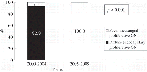
By comparison, there were no significant differences in the clinical presentation and outcome between patients with diffuse endocapillary proliferative pattern and focal mesangial proliferative pattern. However, glomerular neutrophil infiltration was more commonly present in diffuse endocapillary proliferative pattern (p = 0.017; see ). Our data suggested that the percentage of patients with focal mesangial proliferative pattern significantly increased over time (p < 0.001; see ).
Table 3 Comparison of clinicopathological features between diffuse endocapillary and focal mesangial proliferative glomerulonephritis associated with infection in adults
DISCUSSION
Classically, postinfectious glomerulonephritis occurs after streptococcal infections, typically involving upper respiratory tract or skin. The disease most frequently affects children and is relatively uncommon in adults. In developed countries, the incidence of typical PSGN has declined sharply over the last 50 years, probably due to the improved environmental sanitation and better public health service. On the contrary, adult cases of glomerulonephritis associated with non-streptococcal infections are increasingly recognized in the recent years. Adults with an immunocompromised background are particularly at risk for developing the disease.Citation[2,3] More diverse sites of infection and microorganisms have been linked to adult postinfectious glomerulonephritis. Previous studies have demonstrated that Staphylococcus was responsible for an increasing number of cases.Citation[3,7] Our series also showed that Staphylococcus was the most commonly identified infectious agent. Moreover, MRSA accounted for the majority of cases (see ). In our series, the site of infection could be indentified in the majority of cases; the most common sites were skin (20%), heart/endocarditis (20%), lung (15%), bone/joint (15%), and urinary tract (15%). Considering that all 242 patients compiled from four modern studies,Citation[2,3,6,7] the most common sites of infection included upper respiratory tract (24%), skin (16%), lung (14%), heart/endocarditis (9%), and teeth (7%). In these studies, 7–16% of patients had no clinical evidence of infection preceding the renal disease, and in 24–59% of patients, the offending microorganism could not be identified. These data suggested that postinfectious glomerulonephritis should be included in the differential diagnosis of nephritic/nephrotic syndrome in adults, even in the absence of a history of infection.
It has been demonstrated that there is a broad spectrum of glomerular histological findings in adult postinfectious glomerulonephritis.Citation[4,5] The classic glomerular pattern is diffuse endocapillary proliferative glomerulonephritis. This pattern was present in approximately two-thirds of patients in our series. Focal mesangial proliferative glomerulonephritis was present in the remaining seven cases. Importantly, our data suggested a significant escalation of focal mesangial proliferative pattern over time (see ). The cause was probably also related to a growing number of cases of glomerulonephritis associated with staphylococcal infection, particularly MRSA infection. Over the decades, two histological patterns of glomerulonephritis associated with staphylococcal infection are well-defined. One exhibits a diffuse endocapillary and exudative pattern, resembling classic PSGN, in patients with Staphylococcus aureus infection; the other displays a pattern identical to membranoproliferative glomerulonephritis in patients with Staphylococcus epidermidis infection secondary to ventriculovascular shunts. In recent years, however, a third form of Staphylococcus-associated glomerulonephritis characterized by mesangial proliferation with IgA-dominant or co-dominant deposits has been increasingly recognized, which generally occurs in patients with infections caused by MRSA.Citation[8–10] It is speculated that enterotoxins produced by MRSA may serve as superantigens that contribute to this type of glomerulonephritis.Citation[11] In our series, all seven patients (cases 14–20, 1 identified within 2000–2004 and 6 identified within 2005–2008) with mesangial proliferative glomerulonephritis had IgA mesangial staining (see ). Among them, five patients had MRSA and one patient had MSSA (see ). The remaining one patient (case 17) had unusual pathogen of Mycobacterium tuberculosis.
The prognosis of adult postinfectious glomerulonephritis has not been well defined, but there is general agreement that the prognosis is less favorable than PSGN in children. Before the 1990s, complete remission was reported in 60–80% of adults.Citation[12–15] More recent studies found complete remission in only 26–56% of adults.Citation[3,6,7] In our series, complete recovery was found in 30% of patients. Moreover, short-term mortality was found in 30% of patients. All these data strongly suggest that the prognosis of postinfectious glomerulonephritis is worsening in adults. This is probably due to typical PSGN becoming rarer and the number of patients with severe underlying diseases progressively increasing. This study also showed that patients with less severe glomerular change with focal mesangial proliferative glomerulonephritis had no better outcome than patients with more severe glomerular change with diffuse endocapillary proliferative glomerulonephritis (see ). Previous reports of Staphylococcus-associated mesangial glomerulonephritis had the same results.Citation[10,11] This was probably due to other histological findings, such as the severity of tubulointerstitial damage, and the clinical underlying diseases were also found to affect the prognosis.Citation[6]
In conclusion, this study suggested that the clinicopathological spectrum of postinfectious glomerulonephritis had changed over the past 10 years. Atypical clinical and histological features may present diagnostic challenges. Staphylococcus had become the leading infectious agent. Furthermore, atypical histological feature with mesangial proliferative pattern was increasingly identified over time. This was probably due to a growing number of cases of glomerulonephritis associated with MRSA infection, which is now endemic in most hospitals and is increasingly identified from community-acquired infections.Citation[16] Our data showed an unfavorable prognosis in adult postinfectious glomerulonephritis. However, this result was probably influenced by the fact that only patients with severe disease received renal biopsy and were enrolled into the study.
ACKNOWLEDGMENTS
The authors report no conflicts of interest. The authors alone are responsible for the content and writing of the paper.
REFERENCES
- Sotsiou F. Postinfectious glomerulonephritis. Nephrol Dial Transplant. 2001;16(Suppl. 6):68–70.
- Keller CK, Andrassy K, Waldherr R, Ritz E. Postinfectious glomerulonephritis—is there a link to alcoholism?. Q J Med. 1994;87:97–102.
- Montseny JJ, Meyrier A, Kleinknecht D, Callard P. The current spectrum of infectious glomerulonephritis: Experience with 76 patients and review of the literature. Medicine (Baltimore). 1995;74:63–73.
- Edelstein CL, Bates WD. Subtypes of acute postinfectious glomerulonephritis: A clinico-pathological correlation. Clin Nephrol. 1992;38:311–317.
- Sotsiou F, Dimitriadis G, Liapis H. Diagnostic dilemmas in atypical postinfectious glomerulonephritis. Semin Diagn Pathol. 2002;19:146–159.
- Moroni G, Pozzi C, Quaglini S, Long-term prognosis of diffuse proliferative glomerulonephritis associated with infection in adults. Nephrol Dial Transplant. 2002;17:1204–1211.
- Nasr SH, Markowitz GS, Stokes MB, Said SM, Valeri AM, D'Agati VD. Acute postinfectious glomerulonephritis in the modern era: Experience with 86 adults and review of the literature. Medicine (Baltimore). 2008;87:21–32.
- Nasr SH, Markowitz GS, Whelan JD, IgA-dominant acute poststaphylococcal glomerulonephritis complicating diabetic nephropathy. Hum Pathol. 2003;34:1235–1241.
- Satoskar AA, Nadasdy G, Plaza JA, Staphylococcus infection-associated glomerulonephritis mimicking IgA nephropathy. Clin J Am Soc Nephrol. 2006;1:1179–1186.
- Nasr SH, Share DS, Vargas MT, D'Agati VD, Markowitz GS. Acute poststaphylococcal glomerulonephritis superimposed on diabetic glomerulosclerosis. Kidney Int. 2007;71:1317–1321.
- Koyama A, Kobayashi M, Yamaguchi N, Glomerulonephritis associated with MRSA infection: A possible role of bacterial superantigen. Kidney Int. 1995;47:207–216.
- Hinglais N, Garcia-Torres R, Kleinknecht D. Long-term prognosis in acute glomerulonephritis: The predictive value of early clinical and pathological features observed in 65 patients. Am J Med. 1974;56:52–60.
- Lien JW, Mathew TH, Meadows R. Acute post-streptococcal glomerulonephritis in adults: A long-term study. Q J Med. 1979;48:99–111.
- Vogl W, Renke M, Mayer-Eichberger D, Schmitt H, Bohle A. Long-term prognosis for endocapillary glomerulonephritis of poststreptococcal type in children and adults. Nephron. 1986;44:58–65.
- Chugh KS, Malhotra HS, Sakhuja V, Progression to end stage renal disease in post-streptococcal glomerulonephritis (PSGN)—Chandigarh Study. Int J Artif Organs. 1987;10:189–194.
- Fridkin SK, Hageman JC, Morrison M, Methicillin-resistant Staphylococcus aureus disease in three communities. N Engl J Med. 2005;352:1436–1444.

