Abstract
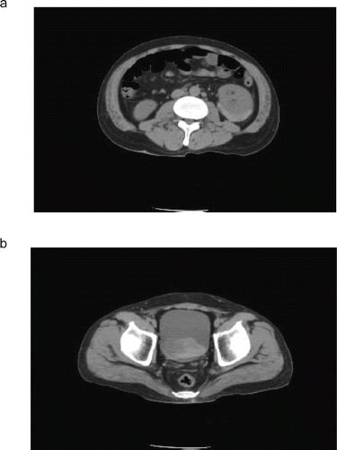
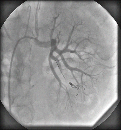
Renal artery pseudoaneurysm is a rare clinical entity that has been reported after renal biopsy, percutaneous renal surgery, penetrating trauma, and rarely blunt renal trauma. We present the case of a 37-year-old man with ruptured renal artery pseudoaneurysm accompanied by massive gross hematuria, urinary clot retention, and bladder tamponade, which were the presenting signs seven hours after renal biopsy. Abdominal CT scan showed a large perinephric, intracapsular hematoma of left kidney. His angiogram revealed a left renal segmental artery pseudoaneurysm that measured 1 cm × 1 cm. He was successfully treated by selective embolization of the arterial branch supplying the pseudoaneurysm.
INTRODUCTION
Renal artery pseudoaneurysm is a rare clinical entity; however, it has great clinical significance when encountered because of its propensity for rupture.Citation[4] It is a rare complication of renal biopsy percutaneous renal procedures, penetrating trauma and, rarely, blunt renal trauma.
The clinical manifestations of renal artery pseudoaneurysms may vary from incidental to causing hypertension, flank pain, hematuria, and rupture. The risk of rupture is estimated low, but it is associated with a mortality rate as high as 80%.Citation[3] Aneurysms larger than 2 cm in diameter are considered to have a high risk of rupture, although ruptures have also been reported in smaller aneurysm.
Angiographic embolization is now considered a safe and effective treatment in patients with renal artery pseudoaneurysm. This treatment should be the procedure of choice due to its advantage of being minimally invasive and its selective treatment with maximal preservation of renal parenchyma.
CASE REPORT
A 37-year-old was admitted to our rheumatology department of our hospital because of dyspnea and arthralgia. Systemic sclerosis was diagnosed on proximal scleroderma, Raynaud's phenomenon, and the lung abnormality of computed tomography as well as positive antinuclear antibody. Because of proteinuria and microscopic hematuria, he was moved to our department and had renal biopsy performed. After seven hours of biopsy, flank pain, severe gross hematuria, urinary clot retention, and bladder tamponade were recognized, and abdominal CT showed a large perinephric, intracapsular hematoma of the left kidney (see and ). Gross hematuria persisted the next day, so we decided to perform angiography. His angiogram revealed a left renal segmental artery pseudoaneurysm that measured 1 cm × 1 cm and its rupture, and then the subsequent coiling of this spontaneously ruptured left renal segmental artery pseudoaneurysm (see ).
DISCUSSION
Renal artery pseudoaneurysm is a rare complication of renal biopsy percutaneous renal procedures,Citation[6] but when encountered, it is of great clinical significance because of propensity for rupture.Citation[4] A pseudoaneurysm is the presence of arterial blood entering into adjoining tissue with continuous blood flow within this space. Symptoms may include abdominal tenderness, abdominal mass, hematuria, hypertension, and shockCitation[8]; however, incidence for ruptured renal pseudoaneurysm and the mortality rate of pseudoaneurysm are difficult to establish due to the lack of reported cases in the literature.Citation[1] When there is rupture, there are four spaces the blood can be redistributed (viz., retroperitoneal, intraperitoneal, intrarenal, and intrapelvic). Most intraparenchymal renal artery pseudoaneurysm ruptures are self-contained, leading to increased probability of tamponade and improved mortality.Citation[7] The same principle may apply to extraparenchymal aneurysms that are contained in the retroperitoneum with concomitant tamponade.Citation[5]
Early detection and embolization are important in treating this life-threatening injury with reported high success rates.Citation[2] Treatment of renal pseudoaneurysm consists of nephrectomy, open vascular surgery, or angiographic embolization, depending on the patient's clinical condition. Angiographic embolization should be the procedure of choice due to its advantage of being minimally invasive and its selective treatment with maximal preservation of renal parenchyma. Surgical indications for repair include overt ruptures, aneurysm greater than 2 cm, renovascular hypertension, expansion of the aneurysm, and evidence of renal damage.Citation[3]
We herein showed a case in which massive microhematuria, urinary clot retention, and bladder tamponade seven hours after renal biopsy were the presenting signs of renal pseudoaneurysm rupture, and demonstrated that it can be managed successfully with angiographic embolization.
ACKNOWLEDGMENTS
We thank Takako Pezzotti and Ayumi Hosotani (Kyoto University) for excellent technical assistance. The authors report no conflict of interest.
REFERENCES
- Albani JM, Novick AC. Renal artery pseudoaneurysm after partial nephrectomy: Three case reports and a literature review. Urology. 2003;62:227–231.
- Breyer BN, McAninch JW, Elliott SP, Minimally invasive endovascular techniques to treat acute renal hemorrhage. J Urol. 2008;179:2248–2252.
- Cohenpour M, Strauss S, Gottlieb P, Pseudoaneurysm of the renal artery following partial nephrectomy: Imaging findings and coil embolization. Clin Radiol. 2007;62:1104–1109.
- Halachmi S, Chait P, Hodapp J, Renal pseudoaneurysm after blunt renal trauma in a pediatric patient: Management by angiographic embolization. Urology. 2003;61:224.
- Jeanmonod R, Lewis C. Bilateral renal artery aneurysm rupture in a man with leukemia: Report of a case. J Vasc Surg. 2008;47:871–873.
- Lee RS, Porter JR. Traumatic renal artery pseudoaneurysm: Diagnosis and management techniques. J Trauma. 2003;55:972–978.
- Porcaro AB, Migliorini F, Pianon R, Intraparenchymal renal artery aneurysms. Case report with review and update of the literature. Int Urol Nephrol. 2004;36:409–416.
- Simonetti G, Rossi P, Passariello R, Iliac artery rupture: A complication of transluminal angioplasty. AJR Am J Roentgenol. 1983;140:989–990.


