Abstract
Human parvovirus B19 infection causes several clinical syndromes. Systemic complications due to this illness are rare. Parvovirus B19 associated renal disease is being increasingly recognized. We report two cases with characteristic presentation with a nephrotic syndrome due to parvovirus B19. We discuss the importance of identifying this infection as a cause of endocapillary glomerulonephritis.
INTRODUCTION
Human parvovirus B19 infection is a common childhood exanthematous illness. This infection is usually asymptomatic or mild in adults and systemic complications are rare. Major clinical manifestations include erythema infectiosum, arthritis, arthralgia, transient aplastic crisis, and fetal hydrops. Parvovirus B19-associated renal disease is not well documented and the characteristic presentation with a nephrotic syndrome has previously been described in the United Kingdom only once before in the clinical setting of patients with sickle-cell disease.Citation1 We describe two previously healthy individuals who developed an acute and severe nephrotic syndrome following parvovirus B19 infection. We discuss the importance of identifying parvovirus infection as the cause of an endocapillary glomerulonephritis.
CASE-1
A 41-year-old woman was admitted with 3 days of generalized edema. She described an illness associated with fever, arthralgia, and a rash on her lower limbs 2 weeks before admission. Her two children had a similar illness associated with a viral exanthem a few weeks previously. There were no other systemic symptoms and on systematic inquiry there were no abnormalities in the cardiovascular, upper and lower respiratory, gastrointestinal, or genitourinary tracts. She had an unremarkable past medical and family history, and was on no medication.
On examination, she had an increased jugular venous pressure at 5 cm and generalized pitting edema. She had no systemic signs. Her pulse was 70 beats/min and her blood pressure was 160/90 mmHg. Her cardiovascular system, respiratory system, and abdominal examinations were clinically normal. Urinalysis showed 3+ protein and 2+ blood. Initial investigations are shown in and .
TABLE 1. Initial biochemical, hematological, and radiological investigations of both cases
TABLE 2. Initial microbiological and immunological investigations of both patients
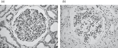
A diagnosis of post-infectious glomerulonephritis was made and renal histology was obtained. Light microscopy showed 10 glomeruli all of which showed exudation and proliferation of mesangial and endothelial cells with neutrophil infiltration (a). The interstitium and vessels were normal. The tubules showed mild dilatation and cellular atrophy. Immunostaining showed strong granular reactivity for IgG, IgM, C3, and C1q. These changes are consistent with an endocapillary glomerulonephritis secondary to parvovirus B19 infection.
FIGURE 1. (a) Patient 1: Light microscopy. (b) Patient 2: Immunostaining highlighted granular capillary loops with IgG, IgM, C3, and C1q.
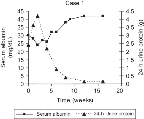
Initially, increasing peripheral edema and eventually pulmonary edema had accumulated in the patient. She was treated with diuretics and salt restriction, and progressively improved. She was discharged after 28 days. Over a period of 4 weeks, her edema resolved. Renal function, 24-h urine protein, serum albumin, and the complement levels normalized within 8 weeks. Serial 24-h urine protein and serum albumin results are shown in . She remained well without any evidence of recurrence of proteinuria on follow-up over 2 years and was discharged from clinic.
FIGURE 2. A graphical representation of the resolution of proteinuria and the serum albumin levels of Case-1.
CASE-2
A 47-year-old woman was admitted for investigation of generalized edema. Two weeks before admission, she developed an erythematous rash involving her lower limbs and abdomen, and had experienced a large joint enthesopathy. These symptoms resolved without specific intervention. Three days before admission, she presented to her GP with a 1-week history of increasing edema. There were no systemic symptoms. There were no cardiovascular, upper or lower respiratory tract, gastrointestinal, or genitourinary tract symptoms. There was neither relevant past medical nor family history, and she was on no medication.
On admission, the jugular venous pressure was increased at 5 cm. There was generalized pitting edema. She was apyrexial and there were no systemic signs. Blood pressure was 160/85 mmHg, heartbeats were normal, and there was no abnormality in the respiratory system and gastrointestinal tracts. Urinalysis showed 3+ protein and 2+ blood. Initial investigations are shown in and .
A diagnosis of post-infectious glomerulonephritis was made and renal histology was obtained. Light microscopy showed 20 glomeruli all of which showed an increase in mesangial matrix and cellularity, leukocyte infiltration, and endothelial cell swelling. There was no necrosis, sclerosis, or extracapillary proliferation. The interstitium and tubules were normal. The vessels were normal without evidence of vasculitis. Immunostaining highlighted granular capillary loops with C3, C1q, IgM, and IgG (b). Mesangial deposition was scanty. These changes are consistent with an endocapillary glomerulonephritis secondary to parvovirus B19 infection.
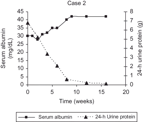
Management was supportive and expectant. She was treated with salt restriction and furosemide. Edema and hypertension resolved within 8 weeks, paralleling a reduction in proteinuria. Complement levels normalized within 4 weeks. Her progression of proteinuria and serum albumin levels are graphically shown in . She has remained in remission without recurrence of proteinuria on follow-up over a 2-year period.
FIGURE 3. A graphical representation of the resolution of the proteinuria and serum albumin levels of Case-2.
DISCUSSION
We report two patients who presented with an acute nephrotic syndrome secondary to an endocapillary glomerulonephritis. Both patients had a viral-type prodrome approximately 2 weeks before presentation. An autoimmune process was serologically excluded. An acute parvovirus B19 infection was confirmed by IgM positivity at presentation and disappearance of IgM antibodies on convalescent sera.
Post-streptococcal glomerulonephritis is the paradigm of post-infectious acute endocapillary glomerulonephritis. This is rare in developed countries and other infections, including varicella, mumps, measles, Epstein–Barr virus (EBV), hepatitis B, Hantaan virus, cytomegalovirus (CMV), and Coxsackie virus, must be considered as alternative causes.Citation2–4 Measles, varicella, EBV, and CMV antigens have been demonstrated in the glomeruli by a variety of techniques including PCR, in situ hybridization, and direct immunofluorescence.Citation2–4
Parvovirus infection classically causes erythema infectiosum in children. The sobriquet, ‘Fifth Disease,’ originates from its ranking among childhood infections after measles, mumps, rubella, and chicken pox. It appears to be a standard serum sickness, which may cause an acute arthropathy, transient heart failure, meningitis, cutaneous vasculitis, and hepatitis. Markenson et al. reported two patients with sickle-cell disease who developed a nephrotic syndrome.Citation5 Wierenga et al. described a further seven patients who developed a nephrotic syndrome in the context of an aplastic crisis induced by parvovirus B19.Citation6 Collapsing glomerulopathy has also been associated with parvovirus B19 infection in a retrospective analysis of archived renal biopsy material: this may not prove causality.Citation7 Only recently has renal involvement due to parvovirus B19 infection in healthy individuals been described and overall 16 cases with an endocapillary proliferative glomerulonephritis and, usually, a nephrotic syndrome have been described since 1999.Citation8–11 A single case of cutaneous vasculitis with proteinuria and a proliferative glomerulonephritis in association with parvovirus B19 infection is reported in the UK literature, but without complete clinical, biochemical, immunological, serological, and immunohistochemical data, to support a diagnosis of post-infectious endocapillary glomerulonephritis.Citation11 More recently, Quek et al. described three patients in United Kingdom who developed nephrotic syndrome following parvovirus B19 infection in patients with sickle-cell disease.Citation1
Our two cases support the good prognosis of parvovirus B19-induced endocapillary glomerulonephritis in previously healthy adults notwithstanding relatively dramatic acute clinical manifestations. Confirmation of the diagnosis and the excellent prognosis will prevent speculative and potentially harmful immunosuppression. Clinical suspicion may be raised by a prodrome of a viral exanthem and arthralgia, especially where the other family members are affected. Hypocomplementemia is a common feature of many post-infectious and some autoimmune [systemic lupus erythematosus (SLE)] causes of glomerulonephritis, and has no discriminatory value. Acute and convalescent serology for parvovirus B19 should now be a routine part of the assessment of patients presenting with an acute nephrotic syndrome.
Declaration of interest: This is to confirm that none of the authors have received financial support or funding to write this paper. We also can confirm none of the authors have any commercial relationships relavent to the article's subject matter. All three authors have no conflict of interest to declare.
REFERENCES
- Quek LL, Sharpe C, Dutt N, Acute human parvovirus B19 infection and nephrotic syndrome. Br J Haematol. 2010;149: 289–291.
- Lin CY, Hsu HC. Measles and acute glomerulonephritis. Pediatrics. 1983;71:398–401.
- Lin CY, Hsu HC, Hung HY. Nephrotic syndrome associated with varicella infection. Pediatrics. 1985;75:1127–1131.
- Ozawa T, Srewart JA. Immune-complex glomerulonephritis associated with Cytomegalovirus infection. Am J clin Pathol. 1979;72:103–107.
- Markenson AL, Chandra M, Lewy JE, Miller DR. Sickle cell anaemia, the nephrotic syndrome and hypoplastic crisis in a sibship. Am J Med. 1978;64:719–723.
- Wierenga KJJ, Pattison JR, Brink N, Glomerulonephritis after human Parvovirus infection in homozygous sickle-cell disease. Lancet. 1995;346:475–476.
- Moudgil A, Nast CC, Bagga A, Association of parvovirus B19 infection with idiopathic collapsing glomerulopathy. Kidney Int. 2001;59:2126–2133.
- Komatsuda A, Ohtani H, Nimura T, Endocapillary proliferative glomerulonephritis in a patient with Parvovirus B19 infection. Am J Kidney Dis. 2000;36:851–854.
- Mori Y, Yamashita H, Umed Y, Association of Parvovirus B19 infection with acute glomerulonephritis in healthy adults: Case report and review of the literature. Clin Nephrol. 2002;57:69–73.
- Takeda S, Takaeda C, Takazakura E, Haratake J. Renal involvement induced by human Parvovirus B19 infection. Nephron. 2001;89:280–285.
- Chakravarty K, Merry P. Systemic vasculitis and atypical infections: Report of two cases. Postgrad Med J. 1999; 75: 544–546.