Abstract
Under common practice, treatment of diabetic nephropathy (DN) is usually initiated at late stage of CKD due to the insensitiveness of the available diagnostic markers. Such treatment fails to restore renal perfusion and function. This is due to the defective mechanism of vascular homeostasis and impaired nitric oxide production observed in late stage of DN. In contrast, the mechanism of vascular repair is adequately functional in early stage of DN (normoalbuminuria). In this study, we treated 50 normoalbuminuric diabetic patients with multidrug vasodilators, namely ACE inhibitor, angiotensin receptor blocker, ± calcium channel blocker in conjunction with correction of metabolic disorders for 24–36 months. Following the treatment, increment in peritubular capillary flow in response to vasodilators was observed, and thus supports the adequate role of vascular repair. In addition, increase in renal function documented in this study also implies that an effective preventive strategy to minimize end-stage renal disease can be accomplished in normoalbuminuric DN.
INTRODUCTION
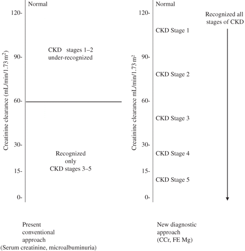
Diabetic nephropathy (DN) is reflected by microalbuminuria (30–300 µg of urinary albumin/gram creatinine) or serum creatinine concentration greater than 1 mg/dL. However, serum creatinine is usually within normal limit in the early stage of CKD, and the abnormally elevated level of serum creatinine becomes apparent only when the measured creatinine clearance drops below 50% level. Similarly, microalbuminuria becomes apparent only when the measured creatinine clearance is approaching the 50% level. This implies that under current practice, such diagnostic markers fail to recognize early stage of DN (CKD stages 1–2).Citation1,Citation2 Treatment, therefore is usually initiated at a rather late stage, and fails to restore renal perfusion and function. Such therapeutic failure is due to the impaired vascular homeostasis and impaired nitric oxide (NO) production in response to vasodilator treatment. In this regard, HohensteinCitation3 has recently demonstrated that receptor-bound vascular endothelial growth factor (VEGF) or VEGF activation was confined to the glomerular endothelium and was increased predominantly in the endothelium of only mildly injured glomeruli, but significantly decreased in more severely injured glomeruli. This implies that receptor-bound VEGF may be lost into the circulation in severely injured glomeruli with advanced renal microvascular disease. To support this view, we demonstrated an increased number of circulating endothelial cell receptor-bound VEGF that is presumably lost into the circulation during vascular injury.Citation4 Interestingly, during renal disease progression in late stage of DN characterized by the progressive reduction in peritubular capillary flow, the components of this receptor-bound reflect a predominant VEGFR2 expression, whereas the VEGFR1 was defective.Citation5 This would imply that VEGFR2 is antiangiogenic and VEGFR1 is angiogenic. Altered vascular homeostasis observed in late stage of DN is characterized by defective angiogenic factors, namely VEGFR1, angiopoietin 1, endothelial progenitor cell, in conjunction with abnormally elevated antiangiogenic factors, namely VEGFR2, angiotensin II, and angiopoietin 2.Citation5–10 With respect to defective angiogenic factor, it would physiologically impair Akt phosphorylation and uncoupling of eNOS and impair NO production.Citation11,Citation12 This would lead to an impaired stimulation of endothelial cell proliferation and maturation inadequately replacing for the endothelial cell loss from the vascular wall into the circulation, resulting in an insufficient (physiologic) vasculogenesis and vascular repair. Simultaneously, the abnormally elevated antiangiogenic factors would activate through the Akt-NO-independent pathway, inducing pathologic proliferations of abnormally immature endothelial cells and vascular smooth muscle cells (VSMC). The abnormal endothelial cell would be consistent with the endothelial-myofibroblast transition cell documented by Li in diabetic kidney.Citation13 The VSMC proliferation would induce progressive thickening of vascular wall, a narrowing of vascular lumen, and eventually a progressive reduction in vascular perfusion inducing chronic ischemic injury to the tubulointerstitium. Collectively, the altered vascular homeostasis observed in late stage of DN would incriminate in mixed pictures of insufficient (physiologic) vasculogenesis and vascular repair, and of pathologic neoangiogenesis and progressive macro- and microvascular diseases. These pathologic vascular diseases correlate with the hemodynamic alteration characterized by a progressive reduction in peritubular capillary flow observed in late stage of DN.
In contrast to the preceding information, we have recently demonstrated that the mechanism of vascular repair appears to be adequately functional in early stage of DN (normoalbuminuria). All the angiogenic and antiangiogenic factors were not significantly different from the normal control.Citation14 Theoretically, in this normoalbuminuric stage, a normal level of VEGF would activate through the classical pathway (VEGF → VEGFR1), by which it would physiologically induce Akt phosphorylation, coupling of eNOS, and enhance NO production. Collectively they would integrate in the physiologic stimulation of endothelial cell proliferation resulting in an adequate process of angiogenesis and vascular repair. This implies that the environment of adequate vascular repair in early stage of DN would be favorable for enhancing the renal perfusion to correct the chronic ischemic state, eventually restoring the renal function. We, therefore, planned to treat 50 type 2 DN patients associated with mildly impaired renal perfusion and function with multidrug vasodilators.
MATERIAL AND METHODS
Fifty diabetic nephropathic patients aged 24–72 years (26 males and 24 females) were included in this prospective uncontrolled trial study. This study was approved by the Medical Ethics Committee, Faculty of Medicine, King Chulalongkorn Memorial Hospital. DN is reflected by (1) an abnormally elevated level of fractional excretion of magnesium (FE Mg)—an index of chronic kidney disease, as FE Mg has been demonstrated earlier to correlate directly with the magnitude of tubulointerstitial fibrosisCitation15,Citation16 and (2) a decline in measured creatinine clearance. It is noted that all these diabetic patients had normal values of serum creatinine and normoalbuminuria. The therapeutic strategy included vasodilators such as ACE inhibitor (Enaril 5–20 mg/day), angiotensin receptor blocker (Telmisartan 40–80 mg/day or Losartan 50–100 mg/day) ± calcium channel blocker 5–10 mg/day, and antioxidants such as vitamin C (1000–2000 mg/day), vitamin E (400–800 units/day), in addition to correction of metabolic disorders. With respect to vasodilators, a low dose of ACE inhibitor and angiotensin receptor blocker are used in mild CKD (CKD stage 1) patients. Moderate dose of ACE inhibitor and angiotensin receptor blocker are used in moderate CKD (CKD stage 2–3) patients. Calcium channel blocker is added when necessary. Improvement in creatinine clearance is the therapeutic target by adjusting the dose of vasodilators. Metabolic disorders are hyperlipidemia which is treated with lipid-lowering substances such as lipitor. High sugar is controlled with metformin and Actos. All patients were advised to drink water ad lib (3 L/day). These patients complied well with the study and recommendation.
Renal Function Study
Renal function study was performed under 10 h urinary collection. No diuretic was administered during or within 24 h before the test. Briefly, after a regular supper, no additional food except drinking water ad lib was allowed. The patients were instructed to void at 7 pm, and the urine was totally collected from 7 pm to 5 am. Clotted blood from venipuncture was drawn at the end of the test for analysis of creatinine and magnesium levels. Urine samples were analyzed as blood samples by the Renal Metabolic Laboratory Unit. For analysis of (1) creatinine and (2) magnesium, (1) the methods described by Faulkner and King and (2) atomic absorption spectrophotometer (model 1100 G; Perkin Elmer, Norwalk, CT, USA) were used, respectively. A reflection of tubulointerstitial fibrosis and chronic kidney disease was derived from the determination of FE Mg, which was calculated through the formula:
Renal Hemodynamic Study
Simultaneously, effective renal plasma flow using 131I-labeled o-iodohippuric acid (hippuran) and of glomerular filtration rate (GFR) using 99mTc-labeled diethylene triamine pentaacetic acid were assessed by previously described method.Citation17 The peritubular capillary flow is derived from the subtraction of GFR from renal plasma flow and is in mL/min/1.73 m2.
Statistics
Comparison of the sample mean of two quantitative variables was determined by the paired student t-test. p-Values below 0.05 were considered significantly different.
RESULTS
A significant decline in fasting blood sugar and Hb A1c was observed following treatment with antidiabetic agents. With respect to renal function study, improvements in measured creatinine clearance, declines in FE Mg, and microalbumin/creatinine ratio were observed following vasodilators treatment. Coinciding with this, a restoration of renal perfusion was also documented, as depicted in .
Table 1. Pre-treatment and post-treatment values of blood chemistries, renal function, and hemodynamics
DISCUSSION
These 50 normoalbuminuric type 2 diabetic patients associated with normal serum creatinine value and normoalbuminuria cannot be differentiated from the normal population by the currently available diagnostic markers. However, under new diagnostic approach, it would easily be screened by (1) measurement of creatinine clearance, which was significantly defective; (2) FE Mg, which was doubling the normal value reflecting the presence of tubulointerstitial fibrosis; and (3) evidence of renal microvascular disease and renal ischemic environment in these patients which is reflected by the decline in peritubular capillary flow which directly supplies the tubulointerstitium. In general, these patients would usually be left unattended, and allowed the inflammatory process and the ischemic environment to continue on, until the progressive impairment in renal function drops to 50% level, or the microalbuminuria emerges. Treatment of DN is therefore initiated at a rather late stage, and the treatment with vasodilators in such stage fails to restore renal perfusion or function. Vasodilators treatment in this late stage is ineffective due to impaired NO production secondary to the altered vascular homeostasis associated with the defective renal microvascular disease ().
Figure 1. Comparison between current diagnostic markers versus new diagnostic approach to screen for staging of chronic kidney disease.

To overcome the present unsolved clinical problem, we have implemented to treat the diabetic patients at the early stage under the environment favorable for renal angiogenesis and regeneration. In fact, the result of our study has supported the above conceptual view. Improvement in peritubular capillary flow is substantiated following the treatment with vasodilators in normoalbuminuric DN, which reflects an adequate production of NO in response to vasodilators. Improvement in peritubular capillary flow would inhibit the degenerative process toward tubulointerstitial fibrosis reflected by the decline in the magnitude of FE Mg following vasodilators treatment—an implication of renal regeneration. In addition, increment in measured creatinine clearance or GFR is accomplished following enhanced renal perfusion. With respect to the GFR or creatinine clearance, the initial value of GFR or creatinine clearance in type 2 DN is usually below the value of normal control. This finding is quite contrasting to the GFR or creatinine clearance observed in type 1 DN, which shows an elevated value above normal control. Following the treatment with vasodilators, there is usually a transient drop in creatinine clearance in the initial stage due to the correction of hyperfiltration phenomenon. Following this, an increase in creatinine clearance has been persistently observed. Improvements in peritubular capillary flow and creatinine clearance or GFR are probably due to both effects of vasodilators and control of diabetes. Recently, a similar benefit of vasodilators has been reported in normoalbuminuric type 2 DN by Ritt et al.Citation18 In conclusion, this therapeutic benefit renders support that an effective preventive strategy to minimize the development of end-stage renal disease in type 2 DN would be plausibly accomplished by implementing the treatment at the early stage of DN.
ACKNOWLEDGMENTS
We are grateful to the supports of Thailand Research Fund, National Research Council Fund of Thailand, and the Royal Institute of Thailand.
Declaration of interest: The authors report no conflicts of interest. The authors alone are responsible for the content and writing of the paper.
REFERENCES
- Futrakul N, Sila-asna M, Futrakul P. Therapeutic strategy towards renal restoration in chronic kidney disease. Asian Biomed. 2007;1:33–44.
- Futrakul N, Vongthavarawat V, Sirisalipotch S, Chairatanarat T, Futrakul P, Suwanwalaikorn S. Tubular dysfunction and hemodynamic alteration in normoalbuminutic type 2 diabetes. Clin Hemorheo Microcirc. 2005;32:57–65.
- Hohenstein B, Hansknicht B, Bochmer K, Local VEGF activity but not VEGF expression is tightly regulated during diabetic nephropathy in man. Kidney Int. 2006;69:1654–1661.
- Futrakul N, Butthep P, Futrakul P, Sitprija V. Glomerular endothelial dysfunction in type 2 diabetes mellitus. Ren Fail. 2006;28:523–524.
- Futrakul N, Butthep P, Futrakul P. Altered vascular homeostasis in type 2 diabetic nephropathy. Ren Fail. 2009;31:207–210.
- Natarajan R, Bai W, Lanting L, Effects of high glucose on vascular endothelial growth factor expression in vascular smooth muscle cell. Am J Physiol. 1992;273:H2224–H2231.
- Bortoloso E, Dol Prete D, Vestra MD, Quantitation and qualitative changes in vascular endothelial growth factor gene expression in glomeruli of patients with type 2 diabetes. Eur J Endocrinol. 2004;150:799–807.
- Cooper ME, Vranes D, Youssef S, Increased renal expression of vascular endothelial growth factor (VEGF) and its receptor VEGFR2 in experimental diabetes. Diabetes. 1999;48:2229–2239.
- Loomans CJ, de Koning EJ, Staal FJ, Endothelial progenitor cell dysfunction a novel concept in the pathogenesis of vascular complications of type 1 diabetes. Diabetes. 2004;53:195–199.
- Williams B, Baker AQ, Gallacher B, Angiotensin II increases vascular permeability factor gene expression by human vascular smooth muscle cells. Hypertension. 1995;25:913–917.
- Miyauchi H, Minamino T, Tatono K, Akt negatively regulates the in vitro life span of human endothelial cells via a p53/p21-dependent pathway. EMBO J. 2004;23:212–220.
- Nakagawa T, Sato W, Suertin YY, Uncoupling of a vascular endothelial growth factor with nitric oxide as a mechanism for diabetic vasculopathy. J Am Soc Nephrol. 2006;17:736–745.
- Li J, Bertram JF. Review: Endothelial-myofibroblast transition, a new player in diabetic renal fibrosis. Nephrology. 2010;15:507–512.
- Futrakul N, Futrakul P. Vascular repair is adequately functional in early stage of diabetic nephropathy. Asian Biomed. 2010; in press.
- Futrakul P, Yenrudi S, Futrakul N, Tubular function and tubulointerstitial disease. Am J Kidney Dis. 1999;33:886–891.
- Deekajorndech T. Fractional excretion of magnesium (FE Mg) in systemic lupus erythematosus. J Med Assoc Thai. 2005;88:743–745.
- Futrakul N, Futrakul P, Siriviriyakul P. Correction of peritubular capillary flow reduction with vasodilators restores function in focal segmental glomerulosclerotic nephrosis. Clin Hemorheo Microcirc. 2004;31:197–205.
- Ritt M, Ott C, Raff U, Renal vascular endothelial function in hypertensivepatients with type 2 diabetes mellitus. Am J Kidney Dis. 2009;53:281–289.
