Abstract
Aim: To investigate and describe cardiac troponins in subjects with acute kidney injury (AKI). Methods: A prospective observational study of troponin in subjects presenting with AKI in a tertiary hospital. Creatine kinase-MB (CKMB), troponin I (Abbott Laboratories), and troponin T (Roche 4th generation) were measured. Patients with conditions known to cause elevated troponin levels were excluded. Results: Nineteen subjects were enrolled in the study. Six subjects had troponin I and T concentrations above the 99th percentile of a reference population. There was high concordance of result between troponin I and troponin T. However, the concordance of elevated troponin levels with CKMB was less marked at 45%. Statistically significant factors associated with elevated troponin levels were age over 60 years, abnormal electrocardiogram, and history of previous ischemic heart disease. Conclusion: This is the first study able to demonstrate impaired renal function occurring acutely, without known confounders, results in elevated troponin levels. This suggests that impaired renal function disease influences plasma troponin levels in AKI as well as in chronic kidney failure.
INTRODUCTION
Cardiac troponins are often tested routinely in the clinical context of chest pain, but comorbid conditions can result in elevated levels in pathologies other than acute coronary syndrome.Citation1 Elevated troponins in end-stage renal disease (ESRD) have been well documented.Citation2 The cause of elevated troponins in ESRD is unclear but possibilities include cardiac dysfunction, left ventricular hypertrophy, and subclinical myocardial infarction.Citation3 Although elevated troponins in ESRD can cause false-positive results for acute coronary syndrome, elevated levels have been shown to have prognostic significance.Citation4,5 The largest study of 733 patients had demonstrated that elevated troponins T and I were predictive of increased mortality.Citation6
In contrast, cardiac troponins in acute kidney injury (AKI) patients have been sparsely investigated. Patients presenting with AKI and an elevated cardiac troponin are not an uncommon scenario. One study had shown that “acute renal failure” had the highest troponin T and I levels when compared to patients with chronic renal impairment, with chronic renal failure, and on hemodialysis.Citation7 However, this study included patients with “acute renal failure” due to multi-organ failure. Multi-organ failure has been described as a cause of elevated cardiac troponins and these results have been confounded.Citation8 To date, there have been no studies that have solely investigated cardiac troponins and AKI.
We present a prospective observational study: troponins in AKI (TAKI) describing troponin T and I levels in AKI.
SUBJECTS
This study was approved by the New Zealand Health and Disability Northern X Regional Ethics Committee. After informed written consent was obtained, 19 subjects were enrolled over a period between 1 April 2008 and 31 November 2008. Patients over the age of 18 years presenting with AKI were considered for the study. AKI was defined as an increase of serum creatinine ×2 or glomerular filtration rate (GFR) decrease of >50% from baseline (where a baseline creatinine was not available, a theoretical baseline creatinine was used based on age, weight, and sex). These criteria are consistent with the second international consensus of the Acute Dialysis Quality Initiative group.Citation9 Subjects were excluded if there were diagnoses of multi-organ failure, acute myocardial infarction, myocarditis, pericarditis, infiltrative cardiac disease, defined arrhythmias (atrial fibrillation, atrial flutter, ventricular tachycardias), pulmonary embolus, congestive heart failure, and sepsis.
METHODS AND MATERIALS
Heparin plasma samples on admission, at peak plasma creatinine, and pre-discharge from subjects were sought. The samples were centrifuged at 2400 × g for 5 min. The supernatant was stored at −20°C for up to 3 months. Plasma samples were batched and tested after further centrifugation for creatine kinase-MB (CKMB), cardiac troponin T, and troponin I. CKMB and troponin T (Roche Diagnostics GmbH, Mannheim, Germany) were tested on the Roche modular automated clinical chemistry analyzer. Troponin I (Abbott Laboratories, Abbott Park, IL, USA) was measured with Abbott CI8200 automated clinical chemistry analyzer. Cardiac troponin I (Abbott Laboratories) has an observed 99th percentile of 0.028 μg/L. The troponin I concentration that meets the ≤10% total coefficient of variation (CV) stated in the package insert is 0.032 μg/L.Citation10 Local quality control data showed a 10% CV performance was attained at a troponin I concentration of 0.04 μg/L. Cardiac troponin T by Roche has an observed 99th percentile at <0.01 μg/L and a precision of ≤10% total CV for samples ≥0.035 μg/L.Citation11 At a troponin T level of 0.014 μg/L, it had a CV of 20%. For the purposes of this study, an elevated troponin T level was defined as ≥0.01 μg/L, and an elevated troponin I was defined as ≥0.032 μg/L.
The investigators were blinded to the troponin I, troponin T, and CKMB results.
Subjects enrolled in the study had their clinical records reviewed for relevant demographic characteristics and clinical investigations including urine output, blood pressure, renal biopsy, renal imaging, and final discharge diagnosis. The etiology of AKI was based on clinical grounds as detailed in the final hospital documentation notes.
Statistical Analysis
p-Values were based on unpaired Student’s t-tests for parametric values (Prism 3.0) and Fisher’s exact one-tailed test for nonparametric values (www.openepi.com).
RESULTS
Nineteen patients were enrolled over a period between 1 April 2008 and 31 November 2008. Thirty-nine samples were collected from the 19 patients.
Admission, peak creatinine, and discharge specimens were sought, but due to late referrals to the renal service many of these patients only two or one sample(s) were tested.
outlines the baseline clinical characteristics and laboratory variables of the cohort as compared to subjects with elevated troponin levels. The cohort’s mean age was 57 ± 19.7 years and 79% were men; 11% had diabetes mellitus and 16% had a previous history of ischemic heart disease (IHD). We excluded patients with defined arrhythmias: atrial fibrillation, atrial flutter, and ventricular tachycardias. However, 50% of our cohort had an abnormal electrocardiogram (ECG) which did not meet our exclusion criteria; these included ventricular ectopics, left ventricular hypertrophy, first degree heart blockage, T-wave inversion, and pathological q waves. As compared to the overall cohort, subjects with elevated troponins were statistically significantly (p < 0.05) older: mean age 74 ± 6.4 years; have abnormal ECGs (83%), and have a previous history of IHD (50%). They also had higher peak urea and creatinine levels but this did not reach statistical significance.
Table 1. Clinical characteristics of all subjects and subjects with elevated troponin T or I.
describes the most common etiology for acute injury for the overall cohort. The most common in descending order were medication (26%), glomerulonephritis (21%), post-renal obstruction (prostate cancer, 11%), pre-renal (dehydration, 11%), acute tubular necrosis (11%), cast nephropathy (5%), tumor lysis (5%), and pyelonephritis (5%).
Table 2. Etiologies of acute kidney injury.
outlines the etiological causes of AKI and clinical characteristics of those with elevated troponins. Six patients had either elevated troponin I (>0.03 μg/L) or elevated troponin T (≥0.01 μg/L). Those with elevated troponin I had mean measured peak value of 0.08 μg/L with a standard deviation of 0.079; the range was from 0.02 to 0.24 μg/L. Those with elevated troponin T had mean measured peak values of 0.03 μg/L with a standard deviation of 0.012; the range was from 0.02 to 0.05 μg/L. All subjects except one had both troponins I and T above our designated cut-offs. The causes of AKI in this cohort were drugs (gentamicin), cast nephropathies, pre-renal (dehydration), anti-neutrophil cytoplasmic antibody (ANCA) vasculitis, and Goodpasture’s glomerulonephritis. The ECG abnormalities in this cohort were left ventricular hypertrophies, first degree heart blockage, and T-wave inversion in the lateral leads. All those with a history of IHD also had abnormal ECGs.
Table 3. Clinical characteristics of subjects with elevated troponins.
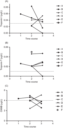
demonstrates the changes in troponin I, troponin T, and CKMB during hospital admission, at peak creatinine, and at discharge. The relative change of troponin I was more marked during the hospital stay as compared to troponin T.
Figure 1. Time course for cardiac markers (troponin I, troponin T, and CKMB) for subjects with elevated troponin concentrations. 1, admission; 2, peak creatinine; 3, discharge. A, B, C, D, E, and F reflect the same patients as in Dashed line = 0.03 μg/L (99th percentile). One value was omitted for visual clarity; it was a value of 0.24 μg/L for patient F.
DISCUSSION
This study is the first to investigate the effects of AKI in isolation on measured plasma cardiac troponin levels. Our exclusion criteria excluded patients with conditions that are known to cause elevated troponin levels.
This study has demonstrated that cardiac troponins are elevated in a significant proportion (32%) of patients with AKI alone. There was high concordance of results of troponin I and troponin T. In subjects with elevated troponin concentrations, all except one had both troponins I and T above the 99th percentile during admission to hospital. There was less concordance between CKMB and cardiac troponins levels, with only 45% of subjects with elevated troponin having elevated CKMB levels (>4 g/L). This is expected as troponins T and I are more sensitive and specific for myocardial injury.
Earlier studies of cardiac troponins and ESRD had shown that troponin T levels were more likely to be elevated compared to troponin I levels but our study demonstrated similar performance which supports a report by Hickman et al.Citation12 that demonstrated newer cardiac troponin I assays have similar performance in detecting elevated troponin concentrations in patients with ESRD.
The limitation in this study is the limited subject numbers; therefore one should be cautious in extrapolating these results. The diagnosis of AKI was based on clinical grounds and many patients did not have baseline creatinine concentrations to compare against.
We are aware that the Abbott Architect troponin I assay and the Roche 4th generation troponin T assays did not meet the 10% CV at the 99th percentile as desired by guidelines from the European Society of Cardiology and the American College of Cardiology.Citation13 Many laboratories have previously instituted a diagnostic cut-off meeting the 10% CV, which is invariably above the 99th percentile to avoid false positives. However, a study by Apple et al.Citation14 noted that irrespective of the total imprecision of the assay at the 99th percentile limit, only the 99th percentile cut-off value should be used for cardiac troponins in clinical practice as the patients misclassified on serial troponin measurement for assays with CV < 25% would be insignificant. Thus, we believe that using the 99th percentile for the Abbott troponin I and Roche troponin T is the valid cut-off to use in this study.
In this study, subject factors such as advanced age, abnormal ECG, and past history of heart disease were significantly associated with elevated troponin levels. The cause of elevated cardiac TAKI is unclear but postulated causes of elevated troponins in ESRD may be relevant. Factors associated with elevated cardiac troponins in ESRD described in the literature have included the transthoracic echocardiographic parameters: left atrial size, left ventricular mass index, the ratio of transmitral early left ventricular filling velocity to early diastolic Doppler tissue imaging velocity of the mitral annulus, and the prevalence of left ventricular dysfunction as well as age, diabetes, and being on dialysis.Citation15 In patients with congestive heart failure, renal function (GFR) correlated significantly and more strongly than cardiac function with serum troponin T levels. And the authors postulated that it is the impaired renal function that causes the accumulation of troponin.Citation16,17
Factors associated with elevated TAKI such as previous heart disease and advanced age suggest that these patients were more likely to have underlying heart disease and therefore possibly higher basal serum troponin concentrations. A study investigating the plasma elimination of cardiac troponin I after acute myocardial infarction in ESRD showed increased half-life in those patients with ESRD compared with normal patients.Citation18
Recent studies involving highly sensitive troponin assays have demonstrated that in apparently healthy populations, the elderly were more likely to have cardiac troponin concentrations above the 99th percentile, perhaps indicating underlying subclinical cardiac disease.Citation19
This study is the first to investigate the effects of AKI in isolation on measured plasma cardiac troponin levels. The short timeframe of AKI suggests factors resulting from chronic fluid overload and ESRD, such as left ventricular hypertrophy and left ventricular dysfunction, are not the primary cause of elevated troponins. Instead, it supports the notion that renal function by itself may have effects on observed troponin levels. Compared to a study by Collinson et al.,Citation7 which included patients with multi-organ failure, our data showed relatively mild elevations of troponin T and I in patients with AKI.
Diris et al.Citation20 have postulated that there is a constant micro loss of cardiomyocytes during normal life and clearance of troponin T normally happens with such speed that serum concentrations are below the current detection limit of 0.01 μg/L. By definition, 99% of healthy subjects without renal impairment have troponin levels within the reference intervals. But perhaps among patients with subclinical cardiac disease, AKI is sufficient to raise basal troponin levels due to the increased troponin half-life and reduced elimination, resulting in higher probabilities of measured troponin levels being above the 99th percentile.
ACKNOWLEDGMENT
We would like to thank Roche New Zealand and Abbott Laboratories for supplying reagents free of charge for this study and the A+ Trust for funding.
Declaration of interest: The authors report no conflicts of interest. The authors alone are responsible for the content and writing of the paper.
REFERENCES
- Korff S, Katus HA, Giannitsis E. Differential diagnosis of elevated troponins. Heart. 2006;92:987–993.
- Li D, Keffer J, Corry K, Vazquez M, Jialal I. Nonspecific elevation of troponin T levels in patients with chronic renal failure. Clin Biochem. 1995;28:474–477.
- De Zoysa JR. Cardiac troponins and renal disease. Nephrology. 2004;9:83–88.
- Ooi DS, Veinot JP, Wells GA, House AA. Increased mortality in hemodialyzed patients with elevated serum troponin T: One-year outcome study. Clin Biochem. 1999;32:647–652.
- Dierkes J, Domrose U, Westphal S, . Cardiac troponins predicts mortality in patients with end-stage renal disease. Circulation. 2000;102:1964–1969.
- Apple FS, Marakami MM, Pearce LA, Herzog CA. Predictive value of cardiac troponin I and T for subsequent death in end-stage renal disease. Circulation. 2002;106:2941–2945.
- Collinson PO, Hadcocks L, Foo Y, . Cardiac troponins in patients with renal dysfunction. Ann Clin Biochem. 1998;35:380–386.
- Collison PO, Stubbs PJ. Are troponins confusing? Heart. 2003;89:1285–1287.
- Bellomo R, Ronco C, Kellum JA, Mehta RL, Palevsky P. Acute renal failure-definition, outcome measures, animal models, fluid therapy and information technology needs: The second international consensus conference of the acute dialysis quality initiative (ADQI) group. Crit Care. 2004;8:R204–R212.
- Architect STAT troponin-I. Package insert, Cat. Log no. 840549/R6. Chicago, IL: Abbott Laboratories; 2008.
- Troponin T Roche diagnostic package insert, 2007-09 V4 English. Mannhiem: Roche Diagnostics GmbH, 2007–2009.
- Hickman PE, Koerbin G, Southcott E, Tate J. Newer cardiac troponin I assays have similar performance to troponin T in patients with end-stage renal disease. Ann Clin Biochem. 2007;44:285–289.
- Apple FS, Wu AH, Jaffe AS. European society of cardiology and American college of cardiology guideline for redefinition of myocardial infarction: How to use existing assays clinically and for clinical trials. Am Heart J. 2002;144:981–986.
- Apple FS, Parvin CA, Buechler KF, Christenson RH, Wu AH, Jaffe AS. Validation of the 99th percentile cutoff independent of assay imprecision (CV) for cardiac troponin monitoring for ruling out myocardial infarction. Clin Chem. 2005;51:2198–2200.
- Jeon DS, Lee MY, Kim CJ, . Clinical findings in patients with cardiac troponin T elevation and end-stage renal disease without acute coronary syndrome. Am J Cardiol. 2004;94:831–834.
- Aksoy N, Ozer O, Sari I, Sucu M, Aksoy M, Geyikli I. Contribution of renal function impairment to unexplained troponin T elevation in congestive heart failure. Ren Fail. 2009;31:272–277.
- Tsutamoto T, Kawahara C, Yamaji M, . Relationship between renal function and serum cardiac troponin T in patients with chronic heart failure. Eur J Heart Fail. 2009;11:653–658.
- Ellis K, Dreisbach AW, Lertora JJ. Plasma elimination of cardiac troponin in end-stage renal disease. South Med J. 2001;94:993–996.
- Venge P, Johnston N, Lindahl B, James S. Normal plasma levels of cardiac troponin I measured by the high-sensitivity cardiac troponin I access prototype assay and the impact on the diagnosis of myocardial infarction. J Am Coll Cardiol. 2009;54:1165–1172.
- Diris JH, Hackeng CM, Kooman JP, Pinto YM, Hermens WT, van Dieijen-Visser MP. Impaired renal clearance explains troponin T fragments in hemodialysis patients. Circulation. 2004;109:23–25.