Abstract
Electrolyte disorders can alter cardiac ionic currents and depending on the changes can promote proarrhythmic effects. Potassium (K+) is the most common intracellular cation related to arrhythmic disorders. Hyperkalemia is mainly seen in the setting of impaired renal function. Severe hyperkalemia may lead to rhythm disorders. Herein, we report a patient with accelerated idioventricular rhythm (AIVR) due to hyperkalemia, which was successfully treated with glucose-insulin (GI) infusion.
INTRODUCTION
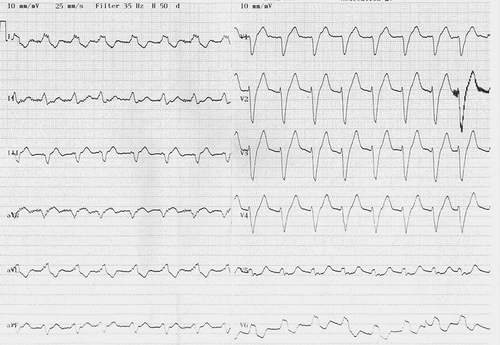
A 68-year-old man was admitted to the emergency department with a syncope attack. Twelve-lead electrocardiography (ECG) revealed atrial standstill with absence of P waves and accelerated idioventricular rhythm (AIVR) (). On examination, the blood pressure was 120/70 mmHg, and the patient was in mild respiratory distress. The patient had a history of coronary artery disease (CAD). Bedside echocardiography revealed mild diffuse (global) hypokinesia of the left ventricle with an ejection fraction (EF) of 50%. Because of previous history of CAD and AIVR on ECG, coronary angiography was performed. A previously implanted bare metal stent in the left anterior descending coronary artery did not show signs of restenosis. There were no significant (≥50%) lesions in the other coronary arteries. The plasma levels of both troponin I and creatine kinase-MB (CK-MB) isoform were normal on admission and serial cardiac enzymes were also within normal limits. Biochemical investigations were as follows: serum sodium, 128 mEq/L (136–141 mEq/L); potassium, 6.4 mEq/L (3.6–5.1 mEq/L); urea, 135 mg/dL (17–43 mg/dL); and creatinine, 2.4 mg/dL (0.7–1.2 mg/dL). There was no metabolic acidosis in the arterial blood gas sample. The patient was not anuric. Therefore, immediate hemodialysis was not performed. Glucose-insulin (GI) infusion was immediately started to reduce plasma potassium levels. After GI infusion, serum potassium level was decreased to 5.1 mEq/L, sinus rhythm with normal PQ interval and normal QRS duration was restored, and AIVR disappeared. We related the occurrence of AIVR to hyperkalemia caused by renal failure. After 4 days of hospitalization, the patient was discharged with full recovery.
Figure 1. Twelve-lead ECG showing accelerated idioventricular rhythm in a patient with hyperkalemia.

The electrocardiographic manifestations of hyperkalemia are related to the serum potassium level (.Citation1 Severe hyperkalemia may cause idioventricular rhythm.Citation2 AIVR is a ventricular rhythm consisting of three or more consecutive monomorphic beats, with gradual onset and gradual termination. Less commonly, AIVR is polymorphic.Citation3 The discharge rate of the ectopic ventricular focus is similar to the sinus rate (isorhythmic) between 50 and 120 bpm. AIVR is usually a benign and well-tolerated arrhythmia. Most of the cases will require no treatment. In rare situations, such as sustained or incessant AIVR or when atrioventricular (AV) dissociation induces syncope, the risk of sudden death is higher, and the arrhythmia should be treated. Different terminologies were used to describe AIVR: non-paroxysmal ventricular tachycardias (VT), isorhythmic slow VT, and, the curious term, benevolent tachycardia.Citation3 AIVR is often a clue to certain underlying conditions, like myocardial ischemia – reperfusion, digoxin toxicity, and cardiomyopathies.Citation4–6 AIVR induced by hyperkalemia has been previously described in the literature.Citation7 To the best of our knowledge, hyperkalemia-induced AIVR is very rare condition and this is the second reported case. Of importance is the fact that hyperkalemia can produce AIVR, and emergency physicians, cardiologists, and internists should be aware of this arrhythmia in patients with renal failure. In addition, hyperkalemia-induced AIVR may resolve after treatment of hyperkalemia.
Table 1. Electrocardiographic manifestations of serum hyperkalemia relative to serum potassium level.
Declaration of interest: The authors report no conflicts of interest. The authors alone are responsible for the content and writing of the paper.
REFERENCES
- El-Sherif N, Turitto G. Electrolyte disorders and arrhythmogenesis. Cardiol J. 2011;18:233–245.
- Diercks DB, Shumaik GM, Harrigan RA, Brady WJ, Chan TC. Electrocardiographic manifestations: Electrolyte abnormalities. J Emerg Med. 2004;27:153–160.
- Grimm W, Marchlinski FE. Accelerated idioventricular rhythm and bidirectional ventricular tachycardia. In: Zipes D, Jalife J, eds. Cardiac Electrophysiology: From Cell to Bedside. 4th ed., Philadelphia, PA: Saunders; 2004:700–704.
- Goldberg S, Greenspon AJ, Urban PL, . Reperfusion arrhythmia: A marker of restoration of antegrade flow during intracoronary thrombolysis for acute myocardial infarction. Am Heart J. 1983;105(1):26–32.
- Castellanos A, Azan L, Bierfield J, Myerburg RJ. Digitalis-induced accelerated idioventricular rhythms: Revisited. Heart Lung. 1975;4(1):104–110.
- Grimm W, Hoffmann J, Menz V, Schmidt C, Müller HH, Maisch B. Significance of accelerated idioventricular rhythm in idiopathic dilated cardiomyopathy. Am J Cardiol. 2000;85(7):899–904, A10.
- Turner PP. Repetitive atrial standstill with idioventricular rhythm from hyperkalemia. Br Heart J. 1962;24:389–392.
