Abstract
Vancomycin causing acute kidney injury has traditionally been associated with acute interstitial nephritis. There have been a few case reports of biopsy-proven acute tubular necrosis (ATN) from vancomycin in the pediatric literature and only one previous report in the adult population. Here, we report a second case of biopsy-proven ATN resulting from vancomycin toxicity.
INTRODUCTION
The original indication for the use of vancomycin was penicillin-resistant Staphylococcus aureus, as approved for the intravenous administration by the FDA in 1958 by fast-tracked process.Citation1 Vancomycin is a glycopeptide antibiotic that inhibits cell wall synthesis by preventing incorporation of two key peptide subunits into peptiglycan matrix, the major component of Gram-positive bacteria wall.Citation2 Vancomycin has been primarily indicated for life-threatening infections with Gram-positive bacteria that are unresponsive to other less toxic antibiotics.Citation3 In principle, it should not be used in methicillin-sensitive S. aureus (MSSA) because vancomycin is inferior in its efficacy to beta-lactamase-resistant penicillins such as Nafcillin. Thus, in this context, vancomycin has been reserved as drug of last resort and has not been used as a first-line antibiotic.Citation4,5
The use of vancomycin has increased dramatically in recent years with the emergence of MSSA as an important hospital pathogen. Thus, vancomycin is often prescribed as the first-line antibiotic for hospital-acquired infection with proven or even suspected resistant Gram-positive organisms. Because of poor tissue penetration and concomitant decrease in bacteria killing, achieving the minimum inhibitory concentration has required scaling up the dose of vancomycin, leading to a parallel rise in the incidence of drug toxicity.Citation6,7 In particular, acute kidney injury has become a significant clinical concern with the use of vancomycin. As vancomycin is primarily cleared by glomerular filtration, failing kidney function leads to retention and build up of highly toxic levels of antibiotic, perpetuating a vicious cycle of renal injury.Citation8
The most common vancomycin-associated nephrotoxicity is acute interstitial nephritis, although other lesions have been rarely described.Citation9–11 The present report highlights the complexity in establishing precise diagnosis of acute kidney injury in patients who are infected with MSSA and who are treated with vancomycin. It also underscores the crucial role of kidney biopsy as a guide to selecting a rational therapeutic approach.
CASE REPORT
G. O., a 45-year-old male with poorly controlled insulin-dependent diabetes mellitus, was admitted to the hospital for a planned excision of his first toe secondary to a non-healing wound and possibility of osteomyelitis. His most recent glycosylated hemoglobin A1c was 10. The patient underwent a right hallux amputation and subsequently was found to have MSSA infection. He was then started on vancomycin with initial dose of 1500 mg every 6 hours. After 4 days of treatment the patient was noted to have an acute rise in serum creatinine to 3.4 mg/dL (baseline 0.6 mg/dL).
A renal ultrasound was performed which showed no evidence of hydronephrosis. There were no episodes of hypotension noted. The patient appeared euvolemic on physical exam. A vancomycin trough was drawn and found to be 101.4 μg/mL. The vancomycin was subsequently discontinued and he was started on clindamycin. There were no other ischemic or toxic insults that could account for the acute kidney injury.
After discontinuing the drug, the vancomycin troughs decreased from 101.4 to 91.6 then 65.6 μg/mL. However, the serum creatinine continued to rise from 3.4 to 6.1 mg/dL. Because a spot urine protein to creatinine ratio showed 3 gm of proteinuria further work up was ordered. The patient was empirically started on Solu-Medrol (Mckesson Drug Co., Tolleson, AZ, USA) and a biopsy was performed. Meanwhile, a serological work up including complements, antinuclear antibody, antinuclear cytoplasmic antibody, antiglomerular basement membrane antibody, and protein electrophoresis was normal.
RENAL BIOPSY FINDINGS
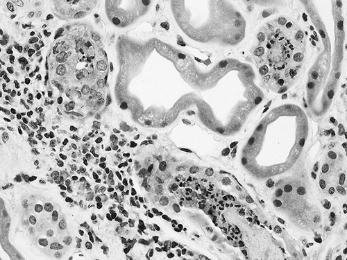
Light microscopic evaluation of the renal biopsy specimen demonstrated renal cortex with 29 glomeruli, of which 2 were globally sclerosed. A few glomeruli contained a mild increase in mesangial matrix compatible with diabetic injury, although nodular sclerosis characteristic of advanced diabetic nephropathy was not present. The glomerular basement membranes appeared normal, and no glomerular proliferation, inflammation, or other abnormalities were seen. Most of the changes involved the renal tubules and interstitium. The interstitium was diffusely expanded by edema. Tubules throughout the biopsy core showed evidence of acute injury, including flattening of the epithelium, sloughing of cells into the luminal space and focal nuclear dropout (). Small patches of tubular and interstitial inflammation were identified, although most of the biopsy was free of inflammation. Where present, the inflammatory infiltrates were composed of a mixed population of lymphocytes, macrophages, and neutrophils with occasional plasma cells. Only rare eosinophils were seen. Within the inflammatory foci, a few tubules contained neutrophils as well as cellular debris (). No significant interstitial fibrosis or tubular atrophy was present. Arterioles showed moderate to severe hyalinosis, although muscular arteries appeared unremarkable.
Figure 1. The tubular epithelium is flattened, with loss of brush borders and decreased numbers of nuclei. Several tubules show reactive nuclear enlargement with sloughing of cell debris into the tubular lumens.
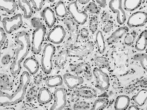
Figure 2. Small foci of interstitial inflammation are present, containing lymphocytes, macrophages, and occasional plasma cells and only rare eosinophils. Several tubules contain a mixture of neutrophils and cell debris (neutrophil casts).
Biopsy tissue examined by immunofluorescence microscopy contained renal cortex with 14 glomeruli, of which one was globally sclerosed. The glomerular and tubular basement membranes demonstrated pseudo-linear staining for immunoglobulin G (IgG) and albumin, consistent with the patient’s history of diabetes. No immune complex deposition was identified. Tissue submitted for electron microscopy contained tubules and interstitium only; ultrastructural examination was not performed.
The biopsy diagnosis was that of acute tubular injury/acute tubular necrosis (ATN), with minimal patchy tubular interstitial inflammation. Allergic interstitial nephritis was not favored, given the sparse nature of the infiltrates and lack of prominent eosinophils. The presence of neutrophil tubular casts did raise the possibility of intrarenal infection, although no clinical evidence of infection was identified. The glomerular mesangial increase and immunofluorescence staining pattern indicated early diabetic injury, without features of advanced diabetic nephropathy.
CASE CONTINUED
There was no other likely alternative explanation for the ischemic or toxic injury noted on the renal biopsy. Following the renal biopsy, the patient’s creatinine continued to rise, his urine output decreased to an oliguric range, and he was started on hemodialysis. Because the biopsy showed no suggestions of acute interstitial nephritis the steroids were discontinued. Although the patient did require hemodialysis for over 2 months, he eventually made a full recovery and subsequently regained renal function.
DISCUSSION
This case highlights the difficult therapeutic dilemmas commonly encountered in hospitalized patients treated with vancomycin. The dramatic increase in vancomycin use as the mainstay in combating life-threatening infections with MSSA has increased the occurrence of acute kidney injury. The most commonly reported vancomycin-induced nephrotoxicity is acute interstitial nephritis. ATN as a complication of vancomycin has been primarily reported in the pediatric literature and one case of biopsy-proven ATN has been reported in an adult.Citation12,13 Although the efficacy of corticosteroids and other immunosuppressive agents in drug-induced acute interstitial nephritis has not been established by randomized controlled clinical trials, they are still used as therapeutic agents if improvement of renal function is not evident after withdrawal of the offending drug. In the case of drugs which are cleared by glomerular filtration such as vancomycin, withdrawal of the antibiotic in the presence of impaired kidney function may result in highly nephrotoxic drug levels, which extend the renal injury. In this instance early hemodialysis may be the most rational therapeutic approach. Appropriate dose of corticosteroids should be maintained in the patient until the vancomycin drug levels are undetectable. The timing of steroid administration is the most important factor. Unnecessary delay in starting the steroids may lead to fibrogenesis with development of interstitial scarring and irreversible renal insufficiency.
Establishment of the histopathologic diagnosis on renal biopsy is critical in determining the mechanism of renal injury and deciding whether corticosteroids or other immunosuppressive agents are indicated. Unwarranted use of high-dose steroids is not only redundant but may be detrimental to the outcome of the underlying infection. Other biopsy-proven kidney injuries such as ATN or post-infectious glomerulonephritis do not justify immunosuppressive treatment.
This case underscores the need to consider ATN in the differential diagnosis of vancomycin-induced kidney injury. The mechanism of tubular injury in the setting of vancomycin therapy is not readily understood. However, experimental studies in rats have shown that vancomycin may induce oxidative stress resulting in renal tubular injury. In rats, vancomycin has been shown to increase malondialdehyde levels, a marker of oxidative stress, and decrease the levels of antioxidative enzymes such as superoxide dismutase and catalase. Examination of rat kidney tissue demonstrated epithelial necrosis, vacuolization of epithelial cells, and interstitial edema. Administration of a strong antioxidative agent significantly ameliorated the tubular damage.Citation14 In that study the trough levels of vancomycin correlated with the severity of tubular injury. As vancomycin nephrotoxicity remains a serious medical concern, additional studies, including clinical investigations are warranted.
Declaration of interest: The authors report no conflicts of interest. The authors alone are responsible for the content and writing of the paper.
REFERENCES
- Chua K, Howden BP. Treating Gram-positive infections: Vancomycin update and the whys, wherefores and evidence base for continuous infusion of anti-Gram-positive antibiotics. Curr Opin Infect Dis. 2009;22:525–534.
- Ahmida MH. Protective role of curcumin in nephrotoxic oxidative damage induced by vancomycin in rats. Exp Toxicol Pathol. 2010; DOI:10.1016/j.etp.2010.07.010.
- Shen WC, Chiang YC, Chen HY, . Nephrotoxicity of vancomycin in patients with methicillin-resistant Staphylococcus aureus bacteremia. Nephrology (Carlton). 2011; DOI: 10.1111/j.1440-1797.2011.01488.x.
- Gonzales C, Rubio M, Romero-Vivas J, Gonzales M, Picazzo JJ. Bacteremic pneumonia due to Staphylococcus aureus: A comparison of disease caused by methicillin-resistant and methicillin-susceptible organism. Clin Infect Dis. 1999;29:1171–1177.
- Blot SI, Vandevoude KH, Hoste EA, Colardyn FA. Outcome and attributable mortality in critically ill patients with bacteremia involving methicillin-susceptible and methicillin-resistant Staphylococcus aureus. Arch Intern Med. 2002;162:2229–2235.
- Pritchard L, Baker C, Leggett J, Sehdev P, Brown A, Bayley KB. Increasing vancomycin serum trough concentrations and incidence of nephrotoxicity. Am J Med. 2010;123:1143–1149.
- Vandecasteele SJ, De Vriese AS. Recent changes in vancomycin use in renal failure. Kidney Int. 2010;77(9):760–764.
- Launay-Vacher V, Izzedine H, Mercadal L, Deray G. Clinical review: Use of vancomycin in hemodialysis patients. Crit Care. 2002;6:313–316.
- Salazar MN, Matthews M, Posadas A, Ehsan M, Graeber C. Biopsy proven interstitial nephritis following treatment with vancomycin: A case report. Conn Med. 2010;74:139–141.
- Hong S, Valderrama E, Mattana J, . Vancomycin-induced acute granulomatous interstitial nephritis: Therapeutic options. Am J Med Sci. 2007;334(4):296–300.
- Wai AO, Lo AM, Abdo A, Marra F. Vancomycin-induced acute interstitial nephritis. Ann Pharmacother. 1998;32:1160–1164.
- Wu CY, Wang JS, Chiou YH, Chen CY, Su YT. Biopsy proven acute tubular necrosis associated with vancomycin in a child: Case report and literature review. Ren Fail. 2007;29(8):1059–1061.
- Shah-Khan F, Scheetz MH, Ghossein C. Biopsy-proven acute tubular necrosis due to vancomycin toxicity. Int J Nephrol. 2011;2011:436856.
- Oktem F, Arslan MK, Ozguner F, . In vivo evidences suggesting the role of oxidative stress in pathogenesis of vancomycin-induced nephrotoxicity: Protection by erdosteine. Toxicology. 2005;215(3):227–233.