Abstract
The presence of myeloid bodies in electron microscopy is a characteristic finding of Fabry’s disease. Here, we present a male patient, whose renal biopsy findings suggested the coexistence of focal segmental glomerulosclerosis and Fabry’s disease, because of the presence of segmental hyalinosis and/or sclerosis in glomeruli and myeloid bodies in electron microscopy. But finally, Fabry’s disease was excluded as a diagnosis because the α-galactosidase A activity in leukocyte and plasma in this patient was within normal limits. After renal biopsy, although he received medication including steroid therapy, his renal function gradually decreased to end-stage renal failure and hemodialysis was initiated. Until now, he does not exhibit any specific symptoms. In conclusion, our case suggests that occasional myeloid bodies in renal biopsy specimens should be interpreted with caution.
INTRODUCTION
Fabry’s disease is an X-linked lysosomal storage disease characterized by the accumulation of glycosphingolipids in various organs, including the kidneys.Citation1,2 This accumulation is due to a deficiency of the enzyme α-galactosidase A.Citation3 The accumulation of glycosphingolipid in glomerular and tubular cells is seen as vacuolation in light microscopy and as myeloid bodies (laminated electron-dense bodies or zebra bodies) in electron microscopy.Citation1,2 Even though the presence of these myeloid bodies is particularly useful for the diagnosis of Fabry’s disease,Citation1,2 the final diagnosis of Fabry’s disease is made based on an assay of α-galactosidase A activity.Citation4
Here, we present a male patient with the presence of myeloid bodies in electron microscopy. However, the α-galactosidase A activity in leukocyte and plasma in this patient was within normal limits.
CASE
A 21-year-old Japanese-Brazilian man was admitted to our hospital in April 1998. He had been healthy, but for the evaluation of massive proteinuria and decreased renal function that was revealed in a routine annual medical examination. He was referred to a hospital in June 1996, where he was diagnosed with chronic glomerulonephritis and was treated with dipyridamole and prednisolone. However, his proteinuria increased to a nephrotic level, and his renal function was gradually decreased.
On admission, his physical examination was unremarkable, with the exception of mild hypertension (142/98 mmHg). His electrocardiogram and chest X-ray had no abnormalities. He had a family history of renal disease: his mother had died of renal failure at the age of 50 years, and his elder brother had been treated for chronic glomerulonephritis with normal renal function. Laboratory examination revealed a normal complete blood cell count, coagulation screen, and electrolyte level. The blood urea nitrogen concentration was 21.8 mg/dL, the serum creatinine level was 1.55 mg/dL, and the creatinine clearance rate was 54.2 mL/min. Urinalysis revealed proteinuria (3+, 4–8 g/day), occult blood (±), a red blood cell count of 1–4 per high-power field, and many hyaline casts. Serum total protein was 5.9 g/dL, albumin was 3.8 g/dL, and total cholesterol was 402 mg/dL. The levels of the immunoglobulins, C3, C4, and CH50, were within normal limits. Hepatitis B surface antigen, hepatitis C antibody, latex fixation, antinuclear antibody, and anti-DNA antibody were negative. Percutaneous renal biopsy was performed in April 1998. The biopsy specimen contained 20 glomeruli with 8 glomeruli exhibiting global sclerosis with hyalinosis. Of the remaining 12 glomeruli, 5 exhibited segmental hyalinosis and/or sclerosis (). Adhesions to Bowman’s capsule were seen in three other glomeruli. No foam cells or vacuolated cells were present in the glomeruli. Moderate tubulointerstitial nephritis with the presence of foam cells was revealed, but the vessels were unremarkable. Immunofluorescence microscopy was mostly negative, except for IgM deposition in the area of sclerosis. Electron micrograph revealed focal foot process fusion. Not only the glomerular epithelial cells, but also the mesangial cells exhibited dense lamellar inclusions (), although the distribution was scattered. The periodicity of these inclusions, which was measured using a 2000 lines/mm diffraction grating as calibration, was 45–55 Å. This periodicity was similar to that of the myeloid bodies seen in Fabry’s disease.Citation5
Figure 1. Light microscopy findings. The glomerulus exhibits segmental sclerosis without vacuolated cells (PAS staining, original magnification ×300).
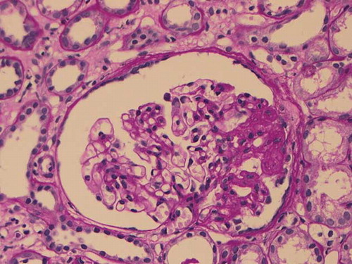
Figure 2. Electron microscopy findings. Myeloid bodies are seen in the glomerular epithelial cells. A few mesangial inclusions are also present (original magnification ×2150).
The presence of myeloid bodies in the glomeruli suggested a diagnosis of Fabry’s disease. We performed an assay of α-galactosidase A activity in leukocytes and plasma, using the method described by Mayes et al.,Citation6 to confirm the diagnosis. The leukocyte α-galactosidase A activities of this patient and his brother were 60.3 and 45.7 nmol/mg protein/h, respectively (normal range: 49.8–116.4 nmol/mg protein/h). The plasma α-galactosidase A activities of this patient and his brother were 12.53 and 8.57 nmol/mL/h, respectively, while that of normal controls was 10.10 nmol/mL/h and that of pooled plasma of Fabry’s disease was 1.48 nmol/mL/h. Based on these results, Fabry’s disease was excluded as a diagnosis. We eventually diagnosed focal segmental glomerulosclerosis associated with myeloid bodies.
In spite of receiving medication, including steroid therapy, his renal function gradually decreased to end-stage renal failure and hemodialysis was initiated in April 2000. Currently he is receiving hemodialysis three times a week in a neighboring hospital and does not exhibit any specific symptoms.
DISCUSSION
We initially suspected a diagnosis of Fabry’s disease with focal segmental glomerulosclerosis, because myeloid bodies in renal biopsy specimens were detected, and the patient had a family history of renal disease. Several reports have demonstrated that the diagnosis of Fabry’s disease can only be made after the detection of myeloid bodies in renal biopsy specimens.Citation7,8 Recently, it is said that males with atypical Fabry’s disease may be more common than previously suspected. Their α-galactosidase A activity is low, but greater than 1%.Citation9 However, the leukocyte and plasma α-galactosidase A activity in this patient were within normal limits, so Fabry’s disease was excluded as a diagnosis.
There were several other factors that were not supportive of the diagnosis of Fabry’s disease, including the absence of extrarenal organ involvement, angiokeratomas, corneal opacity, pain in the extremities, anidrosis, and heart and cerebrovascular dysfunction in this patient.Citation1,2 Second, in our patient, foam cells in the glomeruli were not revealed by light microscopy and only few myeloid bodies were detected by electron microscopy. In patients with Fabry’s disease, myeloid bodies are widespread in distribution and are generally numerous in the affected cells, especially in the glomerular epithelium.Citation1,2 Third, the leukocyte α-galactosidase A activity in our patient was within the normal range, while that of the brother was slightly low. This does not support the diagnosis of Fabry’s disease, in which male hemizygous patients exhibit extremely low α-galactosidase A activity.Citation2,4 Thus, the family history of our patient may be related to a renal disorder other than Fabry’s disease, such as familiar focal segmental glomerulosclerosis.Citation10
As in our patient, normal α-galactosidase A activity with similar inclusions was revealed in the glomerular epithelium of a patient with silicon nephropathy.Citation11 But our patient had no history of silicon exposure. McNamara presented three patients with myeloid bodies without the clinical manifestations of Fabry’s disease.Citation12 is a summary of the characteristics of the patients with glomerular myeloid bodies without Fabry’s disease. Furthermore, similar but not identical ultrastructural features may be observed in the renal glomerular cells of patients treated with chloroquine and in the tubular cells of patients treated with aminoglycoside antibiotics.Citation13 In our patients, hypercholesterolemia might be one of the causes of myeloid body accumulation, as demonstrated by Grone et al.Citation14
Table 1. The characteristics of patients with glomerular myeloid bodies without Fabry’s disease.
Some of the lysosomal storage disorders also show pathological changes similar to Fabry’s disease. Niemann–Pick disease, caused by the deficiency of sphingomyelinase, shows myelin-like lamellae in electron microscopy. But Niemann–Pick disease presents with mental retardation in infancy or early childhood and in most cases, stored material is present in liver and lung.Citation2 Our patient showed no mental retardation and no abnormality in lung and liver.
In conclusion, these findings suggest that occasional myeloid bodies in renal biopsy specimens should be interpreted with caution.
Declaration of interest
The authors report no conflicts of interest. The authors alone are responsible for the content and writing of the paper.
REFERENCES
- Gregory MC, Atkin CL. Alports syndrome, Fabrys disease, and Nail Patella syndrome. In: Schrier RW, Gottschalk CW, eds. Disease of Kidney. Boston: Little Brown; 1997:561–590.
- Finn LS, Bernstein. J. Renal disease caused by familial metabolic and hematologic disease. In: Charles Jannette J, Olson JL, Schwartz MM, Silva FG, eds. Hepitinstall’s Pathology of the Kidney. 6th ed. Philadelphia: Lippincott Williams & Wilkins; 2007:1199–1256.
- Desnick RJ, Astrin KH, Bishop DF. Fabry disease: Molecular genetics of the inherited nephropathy. Adv Nephrol. 1989;18:113–127.
- McGovern MM, Desnick RJ. Lysosomal storage disease. In: Bennett JC, Plum F, eds. Cecil Textbook of Medicine. Philadelphia: Saunders; 1998:1095–1097.
- Gubler MC, Lenoir G, Grunfeld JP, Ulmann A, Droz D, Habib R. Early renal changes in hemizygous and heterozygous patients with Fabry disease. Kidney Int. 1978;13:223–235.
- Mayes JS, Scheerer JB, Sifers RN, Donaldson ML. Differential assay for lysosomal alpha-galactosidases in human tissues and its application to Fabry’s diseases. Clin Chim Acta. 1981;112:247–251.
- Cohen AH. Renal pathology forum. Am J Nephrol. 1985;5: 305–311.
- Kawamura O, Sakuraba H, Itoh K, . Subclinical Fabry’s disease occurring in the context of IgA nephropathy. Clin Nephrol. 1997;47:71–75.
- Metha A, Hughes DA. Fabry disease. In: Pagon RA, Bird TC, Dolan CR, Stephens K, eds. Gene Reviews. Seattle: University of Washington; 2008.
- Colon PJ, Lynn K, Winn MP, . Spectrum of disease in familial focal and segmental glomerulosclerosis. Kidney Int. 1999;56:1863–1871.
- Banks DE, Milutinovic J, Desnick RJ, Grabowski GA, Lapp NL, Boehlecke BA. Silicon nephropathy mimicking Fabrys disease. Am J Nephrol. 1983;3:279–284.
- McNamara TE, Goodloe S, Butkus DE. Myeloid bodies in patients without clinical Fabrys disease. Arch Pathol Lab Med. 1980;104:14–16.
- Cohen AH, Adler SG. Fabrys disease (angiokeratoma corporis diffusum universale). In: Tisher CC, Brenner BM, eds. Renal Pathology with Clinical and Functional Correlation. Philadelphia: Lippincott; 1989:1197–1204.
- Grone HJ, Walli A, Grone E, . Induction of glomerulosclerosis by dietary lipids. A functional and morphologic study in the rat. Lab Invest. 1989;60:433–446.