Abstract
This study was designed to evaluate the preventive role of melatonin (Mel) and 1,25-dihydroxyvitamin D3 (VD3) in biochemical and apoptotic events leading to tissue injury and renal dysfunction after ischemia–reperfusion (I/R). Thirty male Wistar rats were divided into five groups: sham-operated, I/R, Mel + I/R, VD3 + I/R, and Mel + VD3 + I/R. The rats were intraperitoneally administered with Mel (10 mg/kg), VD3 (0.5 μg/kg), or Mel (10 mg/kg) plus VD3 (0.5 μg/kg) each day at 1 week prior to ischemia. Right nephrectomy was initially performed and left renal I/R injury was induced by 45 min of bilateral renal ischemia followed by 45 min of reperfusion. After reperfusion, kidneys and blood were obtained for histopathologic and biochemical evaluation. Mel and VD3 had an ameliorative effect on biochemical parameters such as serum creatinine, blood urea nitrogen, alanine aminotransferase, aspartate aminotransferase, and apoptosis (caspase-3 and terminal deoxynucleotidyl transferase-mediated dUTP–biotin nick end labeling staining) in the kidneys against renal I/R injury in rats. Additionally, VD3 combined with Mel significantly reduced apoptotic and histological alterations when compared with Mel or VD3 alone. This preventive effect on renal tubular apoptosis was remarkable when Mel was combined with VD3.
INTRODUCTION
Acute renal failure induced by ischemia–reperfusion (I/R) injury is a major cause of morbidity and mortality among patients during cardiopulmonary bypass surgery, accidental trauma, sepsis, hydronephrosis, and elective urological operations, especially kidney transplantation.Citation1,2 It is reported that the proximal tubule (namely the S3 segment) is vulnerable to ischemic insult.Citation3
Molecular biological evidence suggests that apoptosis is the primary mechanism of cell death during renal I/R.Citation4 Apoptosis is a complex process including a variety of different signaling pathways and results in multiple changes in the dying cell.Citation5,6 Many of the events occurring during apoptotic process are mediated by a family of cysteine proteases called caspases.Citation5,6 Among identified caspases, caspase-3 is known as a key mediator of apoptotic death.Citation6,7 Caspases take part in two distinct signaling pathways: activation of proinflammatory cytokines and promotion of apoptotic cell death.Citation6 One of the treatments to limit apoptotic cell death and caspase activation in response to I/R injury seems to be antioxidant therapy. Indeed in many studies, some antioxidants such as superoxide dismutase, vitamin E, glutathione, catalase, and melatonin (Mel) were reported to exert protective properties against ischemia-induced tissue damage.Citation6,8 A strong direct free radical scavenger and indirect antioxidant Mel is the major hormone secreted by the pineal gland.Citation9–11 The exogenous Mel was shown to preserve renal function by reducing lipid peroxidation, increasing glutathione levels, and preventing the increase in nitrite levels induced by renal I/R.Citation12 In addition, Mel treatment has been shown to have a protective effect against I/R-induced histopathological changes.Citation13,14
1,25-Dihydroxyvitamin D3 (VD3) is well known for its role in calcium homeostasis, and various additional effects of VD3 have been recently reported.Citation15 VD3 treatment was shown to decrease the progression of renal injuryCitation16 and attenuate glomerulonephritis.Citation17 The renoprotective effects of VD3 in the ischemic kidney have been evaluated only in one study.Citation18 The authors concluded that VD3 exerts a protective effect against I/R injury.Citation18
Furthermore, until this date there has not been any study that compared the biochemical and/or apoptotic effect of both Mel and VD3 in renal I/R injury. Vitamin D3 regulates not only calcium homeostasis but also cellular differentiation and proliferation, and reproduction and immune function processes.Citation19 Our study investigated whether VD3 and Mel or Mel combined with VD3 pretreatment have a significant effect on renal apoptosis caused by I/R injury.
MATERIALS AND METHODS
Animals and Surgery
Thirty adult male Wistar rats, weighing approximately 200–250 g, were housed under a 12-h light/12-h dark cycle. Food and water were available ad libitum. Rats were randomly divided into five groups: control (n = 6), renal I/R (n = 6), Mel + renal I/R (n = 6), VD3 + renal I/R (n = 6), and Mel + VD3 + renal I/R (n = 6). VD3 (Deva, Kocaeli, Turkey) was diluted with sterile saline and adjusted to a final concentration of 0.5 μg/kg.Citation18 A week prior to I/R, VD3 was injected intraperitoneally (i.p.) for 7 days into the VD3+ I/R group. Mel (10 mg/kg)Citation8,12,20 (M-5250 Sigma Chemical Company, St. Louis, MO, USA) was injected i.p. for 7 days into the Mel + I/R group, and Mel (10 mg/kg) in combination with VD3 (0.5 μg/kg) was injected i.p. for 7 days into the Mel + VD3 + I/R group. Following a 12-h fasting period, rats were anesthetized with intramuscular ketamine (50 mg/kg) and xylazine (10 mg/kg). All rats had right nephrectomy through a dorsal flank incision. In the I/R groups, the left renal pedicle was clamped with a noncrushing microvascular clamp (Bulldog Artery Clamp; Harvard Apparatus, Holliston, MA, USA) for 45 min, and the presence of ischemia was visually confirmed by observing blanching of the kidney. The ischemia period was followed by 45 min of reperfusion period, and then the rats were exsanguinated and killed. At the time of death, blood was collected by heart puncture for measurement of biochemical analysis. Serum creatinine (SCr), blood urea nitrogen (BUN), alanine aminotransferase (ALT), and aspartate aminotransferase (AST) levels were measured, and the kidneys were removed for histological and biochemical evaluation.
The experimental protocols were approved by the Animal Ethics Committee of Maltepe University, Faculty of Medicine.
Biochemical and Histological Analysis
SCr, BUN, ALT, and AST levels were determined by spectrophotometric methods using the autoanalyzer (RxL-Max, Siemens, Munich, Germany).
Renal samples were embedded in paraffin, cut into 4-μm sections, and stained with hematoxylin–eosin and periodic acid-Schiff (PAS). The evaluation was performed with light microscopy without knowledge of the study groups. The assessment was carried out by expert observers.
Apoptosis Detection: In Situ DNA End Labeling Method (TUNEL)
Terminal deoxynucleotidyl transferase-mediated dUTP–biotin nick end labeling (TUNEL) assay was performed to detect apoptosis in situ. Briefly, the slides were digested with proteinase K, incubated with terminal deoxyribonucleotidyl transferase enzyme, and subsequently incubated with anti-digoxigenin peroxidase by using the in situ apoptosis detection kit (ApopTag® Peroxidase In Situ Apoptosis Detection Kit, Millipore, MA, USA) according to the protocol. Apoptotic cells had a brown-stained nucleus. Morphometric analysis of the positive cells in tissues stained using TUNEL method was performed under high-power magnification (×400) in a blinded fashion. On each slide, 10 fields were randomly selected. To quantify the extent of apoptosis, we recorded the numbers of apoptotic cells (TUNEL-positive cells) in sections from the four groups. We totaled all TUNEL-positive and intact cells in those fields, and then calculated the apoptotic index by means of an average count per slide. The apoptotic index was calculated according to the formula, as stated by Tunçdemir et al.Citation21: AI = (AC/AC + IC) × 100, where AI is the apoptotic index, AC is the apoptotic cell number, and IC is the intact cell number.
Immunohistochemistry
The renal tissue sections were placed onto slides coated with poly-l-lysine (0.1% w/v in water, Sigma Chemical Company) and were left overnight to dry at 37°C. They were then deparaffinized and rehydrated. Immunoperoxidase staining was performed using Histostain-Plus Bulk kits (Zymed LAB-SA Detection System). Immunostaining procedures were carried out following the guidelines of the manufacturer. After washing with phosphate-buffered saline (PBS), the sections were incubated for 10 min with 3% H2O2 in PBS to block endogenous peroxidase activity. After the sections were washed with PBS, incubation with normal blocking serum was performed. Sections were incubated overnight with rabbit polyclonal caspase-3 (Santa Cruz Biotech Inc., Santa Cruz, CA, USA; Sc-7148, 1:50 dilution) antibody at +4°C, followed by PBS washing, incubation with biotinylated secondary antibody, and a final PBS washing. Then, sections were incubated with a substrate–chromogen solution (AEC; Zymed, San Francisco CA, USA) for 5–6 min before incubation with horseradish peroxidase–streptavidin complex. Sections were counterstained with hematoxylin. To determine the specificity of immunostaining, sections were incubated following the above procedure, except for incubation with primary antibody. Instead of primary antibody, control serum was also used as control.
Statistical Analysis
All statistical analyses were carried out using UNISTAT 5.0 for Windows (Istanbul University) package program. Differences in measured parameters among groups were analyzed using Kruskal–Wallis test. Dual comparisons between groups that present significant values were evaluated using Mann–Whitney U test. The values were expressed as mean ± SEM. A value of p < 0.05 was considered to be statistically significant.
RESULTS
Biochemical Results
There was a significant increase in the SCr, BUN, and ALT levels in the I/R group compared with the control group (p < 0.005). Renal I/R produced increased serum AST level, but the difference was not statistically significant. Administration of Mel, VD3, and Mel + VD3 significantly decreased the SCr, BUN, and ALT levels compared with the I/R group. Serum concentration of AST was significantly decreased in Mel + I/R and Mel + VD3 + I/R groups compared with the I/R group (p < 0.005) ().
Table 1. The serum values of SCr, BUN, ALT, and AST.
Table 2. Immunohistochemical distribution of caspase-3 in the kidney tissue sections in all groups.
Histopathologic Results (PAS)
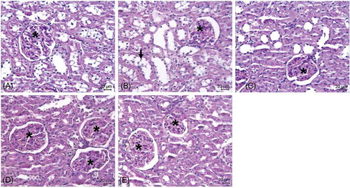
Renal morphology was normal in the control group (A), whereas the kidneys in the I/R group displayed extensive necrosis plus dilation, flattening of epithelium, and loss of brush border in proximal tubuli (arrow, B). In Mel + VD3 + I/R group (E), the histology looked less affected compared with Mel + I/R (C), VD3 + I/R (D), and I/R groups. Finally, all three pretreatment groups showed better histopathologic findings compared with the I/R group.
Figure 1. Histomorphology of kidneys. Light photomicrographs of PAS-stained sections of kidney from control rats and ischemic kidney from rats treated with Mel and VD3. (A) Control group, (B) focal loss of epithelial brush border lining of tubular cells (arrow) in the I/R group, (C) Mel + I/R group, (D) VD3 + I/R group, and (E) Mel + VD3 + I/R group. Bar = 20 μm. Asterisk denotes glomeruli.
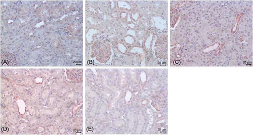
Caspase-3 Immunoreaction
shows the immunohistochemical distribution of caspase-3 in the kidney tissue sections in all groups. Minimal caspase-3 immunoreactivity was observed in the control group (A). Caspase-3 immunoreactivity was intense and moderate in the tubuli and glomeruli in the I/R group, respectively (B). On the other hand, caspase-3 immunoreactivity was weak or moderate in the tubuli and glomeruli in Mel + I/R (C) and VD3 + I/R (D) groups, whereas mild in the Mel + VD3 + I/R group (E).
Figure 2. Immunoreactivity of caspase-3. (A) Control group, (B) I/R group, (C) Mel + I/R group, (D) VD3 + I/R group, and (E) Mel + VD3 + I/R group. Bar = 20 μm.
TUNEL Assay Results
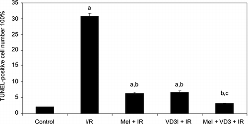
Cell apoptosis was evaluated using TUNEL immunostaining to localize DNA fragmentation. The kidneys from rats in the I/R group showed extensive TUNEL-positive staining (B) compared with the control group ( p < 0.001) (A). The apoptotic cells were observed especially in the proximal and distal tubules where the damage was intense. In contrast, the kidneys from rats in the Mel (C), VD3 (D), and Mel + VD3 treatment groups (E) had very little TUNEL-positive staining compared with the I/R group. Mel + VD3 treatment groups showed significant reduction of the renal injury and cell apoptosis (p < 0.001) (E). Mel + VD3 treatment was superior to Mel or VD3 treatment ().
Figure 3. Effects of Mel and VD3 treatment on apoptosis induced by renal I/R. Apoptosis was evaluated by TUNEL staining (Brown nuclei). (A) Control group, (B) I/R group, (C) Mel + I/R, (D) VD3 + I/R, and (E) Mel + VD + I/R group. Bar = 20 μm.
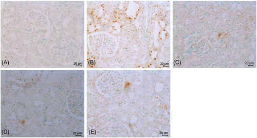
Figure 4. Percentage of TUNEL-positive cells in kidney sections.
Notes: The values are expressed as mean ± SEM. ap < 0.001 versus control group; bp < 0.001 versus I/R group; cp < 0.01 versus Mel + I/R, VD3 + I/R groups.
DISCUSSION
The aim of this study was to determine whether Mel, VD3, and Mel plus VD3 delivered prior to ischemia could protect the kidney from I/R injury in the experimental rat’s model with significant reductions in SCr, BUN, ALT, and AST concentrations and improvement of histological injury with reduced evidence of apoptosis. In order to evaluate biochemical and apoptotic parameters within the reversible first phase of reperfusion injury extending only for a few hours, the reperfusion interval is limited to 45 min in this study. The SCr and BUN levels were used as indicators of impaired glomerular function, and AST was used as an indicator of renal I/R injury.Citation22 Our results clearly showed that prior administration of Mel and VD3 or Mel plus VD3 improved renal function in I/R injury, as evidenced by reduction in the levels of SCr, BUN, and ALT compared with I/R animals without pretreatment. But, AST levels tended to increase in the I/R group compared with the control group; this increase was not statistically significant. However, Mel and Mel combined with VD3 treatments decreased the AST levels compared with the I/R group.
Renal proximal tubules are susceptible to many kinds of insults such as I/R injury and toxic substrates.Citation23 These tubules contain large quantities of mitochondria, which provide energy for reabsorption.Citation23 I/R injury induces fragmentation of the mitochondria, which leads to mitochondrial outer membrane permeabilization, release of apoptogenic factors, and consequent apoptosis.Citation24 Renal tubular cell apoptosis has been observed after renal I/R injury and this process may represent a direct mechanism by which tubule cells are damaged.Citation24 In this study, the numbers of apoptotic cells within the proximal and distal tubules were found to be significantly increased, whereas they were decreased in Mel, VD3, and Mel combined with VD3 compared with the untreated I/R group. Additionally, a combination of Mel and VD3 was shown to reduce apoptosis more efficiently than pretreatment with Mel or VD3 alone.
I/R injury is actually mediated largely by apoptotic pathways and caspase activation.Citation25 Caspase-3, a member of the caspase family, has been shown to be a major execution caspase that acts downstream in the apoptosis pathway.Citation6 In addition, caspases contain an active site or cysteine nucleophile, which is prone to oxidation or thiol alkylation; this suggests that caspase activity is optimal under conditions of reducing environments. It clearly indicates that the redox status of a cell can affect its ability to undergo apoptosis.Citation26 Caspase-3 immunoreactivity was weak or moderate in the tubuli in Mel and VD3-treated groups, whereas weak in the Mel combined with VD3 group compared with the I/R group. At the same time, weak or moderate immunoreactivity in the Mel and VD3 groups and weak immunoreactivity in the Mel plus VD3 group for caspase-3 were seen in glomeruli compared with the I/R group. The pineal hormone Mel is a widespread physiological mediator.Citation20,27 Mel is reported to be a potent free radical scavenger that protects the cells from damage induced by a variety of oxidants, including hydroxyl radical and lipid peroxidation products.Citation20,27 The antioxidant effect of Mel may be partly due to its antiapoptotic effect.Citation26 In various experimental studies, Mel has been shown to decrease apoptosis via inhibiting caspase-3 activity.Citation6,28 In our study, the demonstration that pretreatment of rats with Mel prevents caspase-3-dependent cell apoptosis suggests that the mechanisms of Mel’s action may involve a caspase-signaling cascade. Similar results were reported by Kunduzova et al.Citation6 We may suggest that the antioxidant effect of Mel might be in part of its antiapoptotic action. In contrast to Mel, VD3 in I/R injury has not been frequently studied. The recent discovery of vitamin D receptors in most tissues and cells in the body has provided the most important new insights into the function of this vitamin.Citation19 Many gene-encoding proteins that regulate cellular proliferation, differentiation, and apoptosis are modulated in part by VD3 in the organism.Citation15 However, the exact antiapoptotic mechanism of VD3 in renal I/R remains to be elucidated. VD3 treatment is supposed to accelerate the recovery of renal tubular cells by increasing the proliferation of renal tubular cells in ischemic rat kidneys due to immunomodulatory effect of VD3 on inflammatory cytokines.Citation18 These authors reported that VD3 is potentially useful in the prevention of ischemic renal injury.Citation18 In our study, the demonstration that pretreatment of rats with Mel and VD3 prevents caspase-3-dependent cell apoptosis suggests that the mechanisms of Mel’s and VD3’s actions may involve a caspase-signaling cascade.
In conclusion, although many inhibitors of apoptosis in renal I/R injury have been used in the attempt to reduce organ damage, the mechanisms of preventing apoptosis during I/R are not clearly shown. Taken together, these results suggest that Mel and VD3 treatments prevented renal damage induced by I/R, but combined application of Mel and VD3 revealed more efficient results in preventing renal cell apoptosis and damages. These results represent the initial step toward determining the histopathological parameters by which Mel and/or VD3 prevents renal I/R injury. The demonstration that Mel and VD3 pretreatment prevents caspase-3-dependent cell apoptosis and renal dysfunction after I/R injury may provide new therapeutic implications for the treatment of kidney diseases, in which apoptotic cell death pathway plays the predominant role.
ACKNOWLEDGMENT
Funding. This study was funded by Maltepe University (2011.04).
Declaration of interest: The authors report no conflicts of interest. The authors alone are responsible for the content and writing of the article.
REFERENCES
- Russ AL, Haberstroh KM, Rundell AE. Experimental strategies to improve in vitro models of renal ischemia. Exp Mol Pathol. 2007;83(2):143–159.
- Kadkhodaee M, Golab F, Zahmatkesh M, . Effects of different periods of renal ischemia on liver as a remote organ. World J Gastroenterol. 2009;15(9):1113–1118.
- Witzgall R. The proximal tubule phenotype and its disruption in acute renal failure and polycystic kidney disease. Exp Nephrol. 1999;7(1):15–19.
- Schumer M, Colombel MC, Sawczuk IS, . Morphologic, biochemical, and molecular evidence of apoptosis during the reperfusion phase after brief periods of renal ischemia. Am J Pathol. 1992;140(4):831–838.
- Thornberry NA, Lazebnik Y. Caspases: Enemies within. Science. 1998;281(5381):1312–1316.
- Kunduzova OR, Escourrou G, Seguelas MH, . Prevention of apoptotic and necrotic cell death, caspase-3 activation, and renal dysfunction by melatonin after ischemia/reperfusion. FASEB J. 2003;17(8):872–874.
- Tunçdemir M, Oztürk M. The effects of angiotensin-II receptor blockers on podocyte damage and glomerular apoptosis in a rat model of experimental streptozotocin-induced diabetic nephropathy. Acta Histochem. 2011;113(8):826–832.
- Aktoz T, Aydogdu N, Alagol B, Yalcin O, Huseyinova G, Atakan IH. The protective effects of melatonin and vitamin E against renal ischemia-reperfusion injury in rats. Ren Fail. 2007;29(5):535–542.
- Reiter RJ, Reyes-Gonzales M, Fuentes-Broto L, Tan DX. Melatonin reduces oxidative catastrophe in neurons and glia. Act Nerv Super Rediviva. 2010;52(1):93–103.
- Rodriguez C, Mayo JC, Sainz RM, . Regulation of antioxidants enzymes: A significant role for melatonin. J Pineal Res. 2004;36(1):1–9.
- Ozturk G, Coşkun S, Erbaş D, Hasanoglu E. The effect of melatonin on liver superoxide dismutase activity, serum nitrate and thyroid hormone levels. Jpn J Physiol. 2000;50(1):149–153.
- Rodriguez-Reynoso S, Leal C, Portilla-de Buen E, Castillo JC, Ramos-Solano F. Melatonin ameliorates renal ischemia/reperfusion injury. J Surg Res. 2004;116(2):242–247.
- Yurtcu M, Abasiyanik A, Avunduk MC, Muhtaroglu S. Effects of melatonin on spermatogenesis and testicular ischemia-reperfusion injury after unilateral testicular torsion-detorsion. J Pediatr Surg. 2008;43(10):1873–1878.
- Yurtcu M, Abasiyanik A, Bicer S, Avunduk MC. Efficacy of antioxidant treatment in the prevention of testicular atrophy in experimental testicular torsion. J Pediatr Surg. 2009;44(9): 1754–1758.
- Chu MP, Alagiakrishnan K, Sadowski C. The cure of ageing: Vitamin D-magic or myth? Postgrad Med J. 2010;86(1020):608–616.
- Kuhlmann A, Haas CS, Gross ML, . 1,25-Dihydroxyvitamin D3 decreases podocyte loss and podocyte hypertrophy in the subtotally nephrectomized rat. Am J Physiol Renal Physiol. 2004;286(3):526–533.
- Panichi V, Migliori M, Taccola D, . Effects of 1,25(OH)2D3 in experimental mesangial proliferative nephritis in rats. Kidney Int. 2001;60(1):87–95.
- Kim YO, Li C, Sun BK, . Preconditioning with 1,25-dihydroxyvitamin D3 protects against subsequent ischemia-reperfusion injury in the rat kidney. Nephron Exp Nephrol. 2005;100(2):85–94.
- Khan KA, Akram J, Fazal M. Hormonal actions of vitamin D and its role beyond just being a vitamin: A review article. Int J Med Med Sci. 2011;3(3):65–72.
- Reiter RJ, Tan DX, Poeggeler B, Menendez-Pelaez A, Chen LD, Saarela S. Melatonin as a free radical scavenger: Implications for aging and age-related diseases. Ann N Y Acad Sci. 1994;719:1–12.
- Tunçdemir M, Demirkesen O, Öztürk M, Atukeren P, Gümüştaş MK, Turan T. Antiapoptotic effect of angiotensin-II type-1 receptor blockade in renal tubular cells of hyperoxalouric rats. Urol Res. 2010;38(2):71–80.
- Chatterjee PK, Patel NS, Kvale EO, . Inhibition of inducible nitric oxide synthase reduces renal ischemia/reperfusion injury. Kidney Int. 2002;61(3):862–871.
- Isaka Y, Kimura T, Takabatake Y. The protective role of autophagy against aging and acute ischemic injury in kidney proximal tubular cells. Autophagy. 2011;7(9):1085–1087.
- Brooks C, Wei Q, Cho SG, Dong Z. Regulation of mitochondrial dynamics in acute kidney injury in cell culture and rodent models. J Clin Invest. 2009;119(5):1275–1285.
- Zhang X, Zheng X, Sun H, . Prevention of renal ischemic injury by silencing the expression of renal caspase 3 and caspase 8. Transplantation. 2006;82(12):1728–1732.
- Sainz RM, Mayo JC, Reiter RJ, Tan DX, Rodriguez C. Apoptosis in primary lymphoid organs with aging. Microsc Res Tech. 2003;62(6):524–539.
- Poeggeler B, Reiter RJ, Tan DX, Chen LD, Manchester LC. Melatonin, hydroxyl radical-mediated oxidative damage, and aging: A hypothesis. J Pineal Res. 1993;14(4):151–168.
- Kim SH, Lee SM. Cytoprotective effects of melatonin against necrosis and apoptosis induced by ischemia/reperfusion injury in rat liver. J Pineal Res. 2008;44(2):165–171.