Abstract
Spontaneous nontraumatic rupture of the kidney (Wunderlich syndrome) is an extremely uncommon condition on hemodialysis. We report a case of 44-year-old hemodialysis patient presented with hemorrhagic shock and a right quadrant abdominal pain to the emergency department. There was no history of trauma. A kidney rupture was revealed by abdominal computed tomography, and active bleeding was successfully managed with arterial embolization. This case illustrates the safe and successful application of interventional radiology in the management of nontraumatic renal hemorrhage in the specific group of hemodialyzed patients even in the emergency setting.
INTRODUCTION
Spontaneous nontraumatic perinephric hemorrhage, or Wunderlich syndrome, is a rare but potentially life-threatening condition. Renal tumors and cysts followed by vascular diseases were the most common causes, and nephrectomy was usually performed.Citation1
Recent advances in computed tomography (CT) technology and interventional radiology make conservative treatment a valuable alternative. Angiography and more recently superselective embolization have emerged as effective modalities for diagnosing and treating acute renal injury.Citation2
We report on a case of perirenal hematoma due to spontaneous renal rupture in a patient with chronic renal disease treated with hemodialysis and the successful treatment with selective embolization.
A CASE REPORT
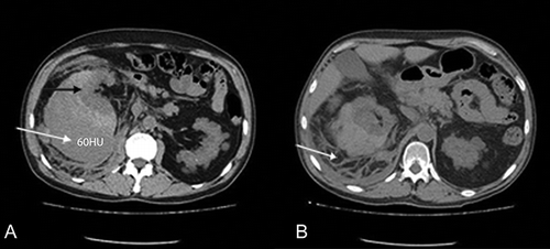
A 44-year-old man was admitted to the emergency department (ED) complaining of dyspnea and right quadrant abdominal pain. On admission, vital signs were blood pressure 85/50 mm Hg, heart rate 125/min, and body temperature 36.5°C. Past medical history included end-stage renal disease (ESRD), 5 years on hemodialysis, arterial hypertension, and hepatitis C. There was no acute or a history of a previous trauma. Physical examination revealed distended abdomen. Routine laboratory tests showed hematocrit of 20%, hemoglobin of 6.6 mg/dL, elevated serum lactate dehydrogenase (LDH) and creatine kinase (CK) levels, and findings consistent with chronic renal disease (urea = 116 mg/dL, creatinine = 7.9). ED ultrasound showed fluid around the right kidney and to the Morrison pouch. After clinical stabilization, the patient was further evaluated with multidetector CT scan without contrast media, which revealed perirenal and giant subcapsular hematoma displacing the right kidney upward (A and B). Furthermore, CT scan showed a shrunken right kidney with multiple small cysts, typical for an acquired cystic kidney disease (secondary cystic degeneration). Immediately afterward, an angiography was performed.
Figure 1. Computed tomography scan on admission: (A) giant subcapsular hematoma, increased density of 60 Hounsfield Units (white arrow) depressing the right kidney upward (black arrow) and (B) perirenal fluid (arrow).

Through the right common femoral artery, using the Seldinger technique, a hydrophilic catheter Cobra 1 type (Terumo Europe, Leuven, Belgium) was used to catheterize the right renal artery. Angiogram showed displacement of the kidney upward. No extravasation of contrast agent was noted at this point. Then, selective catheterization of the right segmental renal arteries using a microcatheter (PROGREAT 2, 7 F) was performed. Further control angiogram revealed small extravasation of contrast agent from a small polar branch of right renal artery (A). Thereafter, the right segmental renal arteries were embolized using microcoils (Balt Extrusion, Montmorency, France). Attention was given to preserve the right inferior adrenal artery (B). Finally, the aorta angiogram confirmed the occlusion of the embolized arteries (C).
Figure 2. (A) Control angiogram showing small extravasation of contrast agent from a small polar branch of the right renal artery (white arrow). (B) During coiling, embolization of right segmental renal arteries with preservation of the right inferior adrenal artery (white arrow). The black arrow indicates the microcoils. (C) After embolization, a control aorta angiogram confirmed the occlusion of the embolized arteries. No accessory right renal artery was identified. (D) CT scan in follow-up verified the complete occlusion of segmental renal arteries (black arrow). A patent inferior adrenal artery was also observed (white arrow).

The operative time was 15 min. No immediate postembolization complications were observed. The postoperative period was uneventful, and the patient was discharged on postoperative day 3. Follow-up with CT scan after 10 days verified the complete occlusion of segmental renal arteries (D).
DISCUSSION
A recent review and meta-analysis by Zhang et al.Citation1 on the etiology of spontaneous perirenal hematoma showed that tumors are involved in the majority of cases (74.9%), followed by vascular disease (17%), idiopathic (6.7%), and infection (2.4%). In addition, among idiopathic cases, especially in hemodialysis patients, anticoagulant therapy (i.e., heparinization during hemodialysis) and uremic thrombocytopathy predispose to hemorrhage.Citation3
Traditional treatment embraces conservative management with blood transfusions and close follow-up. These patients need to be followed in a high dependence unit, because the resuscitation process can easily place them into heart and respiratory failure. On the other hand, surgical intervention in these particular high-risk patients carries high morbidity and mortality rates.Citation4
Arterial embolization is a minimal invasive approach, which has already been used in traumatic rupture of kidney with high technical and clinical success.Citation5,6 However, this intervention, especially for the hemodynamically unstable patients, requires experience on the selective and hyperselective embolism with the availability of the proper facilities in the emergency setting.Citation7
Major complications of the technique are incomplete embolization, coil migration leading to injury of adjacent organs, and postinfraction syndrome including fever, nausea, and vomiting and flank pain. Minor complications include dissection of vessel and groin hematoma, renal intimal dissection, and renal abscess.Citation8 Nowadays, the use of new microvascular catheters and microcoils has refined the technique reducing iatrogenic morbidity.Citation9
In the presented case, a hydrophilic catheter Cobra 2.5 F followed by a microcatheter Progreat 2.7 F for the selective embolization along with microil agent was successfully used. The only risk factor was ESRD due to chronic glomerulonephritis. The patient had no history of trauma and he had received anticoagulants 1 day before the incident. It could be assumed that the secondary cystic degeneration along with derangements of platelets predisposed to the genesis of hematoma. The hemodynamic instability along with the above pathology required an immediate and fast intervention. To our knowledge, this is the first case of nontraumatic rupture of kidney in ESRD treated successfully with selective embolization.
Although further studies are needed to elucidate this approach in this specific group of patients, it seems that it may be the treatment of choice even for hemodynamically unstable patient provided adequate experience.
CONCLUSION
Spontaneous kidney rupture, although rare, should be considered in a chronic hemodialysis patient even in the absence of previous trauma. Segmental renal artery embolization may constitute an effective treatment option in such patients in the emergency setting.
Declaration of interest: The authors report no conflicts of interest. The authors alone are responsible for the content and writing of the paper.
REFERENCES
- Zhang JQ, Fielding JR, Zoos KH. Etiology of spontaneous perirenal hemorrhage: A meta-analysis. J Urol. 2002;167:1593–1596.
- Schwartz MJ, Smith EB, Trost DW, Vaughan Jr. ED. Renal artery embolization: Clinical indications and experience from over 100 cases. BJU Int. 2007;99:881–886.
- Mabjeesh NJ, Matzkin H. Spontaneous subcapsular renal hematoma secondary to anticoagulant therapy. J Urol. 2001;165:1201.
- Bensalah K, Martinez F, Ourahma S, Bitker MO, Richard F, Barrou B. Spontaneous rupture of non-tumoral kidneys in patients with end stage renal failure: Risks and management. Eur Urol. 2003;44:111–114.
- Hagiwara A, Sakaki S, Goto H, . The role of interventional radiology in the management of blunt renal injury: A practical protocol. J Trauma. 2001;51:526–531.
- Breyer BN, McAninch JW, Elliott SP, Master VA. Minimally invasive endovascular techniques to treat acute renal hemorrhage. J Urol. 2008;179:2248–2252.
- Sofocleous CT, Hinrichs C, Hubbi B, . Angiographic findings and embolotherapy in renal arterial trauma. Cardiovasc Intervent Radiol. 2005;28:39–47.
- Dinkel HP, Danuser H, Triller J. Blunt renal trauma: Minimally invasive management with microcatheter embolization experience in nine patients. Radiology. 2002;223:723–730.
- Beaujeux R, Saussine C, Al-Fakir A, . Superselective endo-vascular treatment of renal vascular lesions. J Urol. 1995;153:14–17.
