Abstract
Objectives: This study aimed to investigate the effect of natrium hyaluronate (NH) on fibrous tissue formation and wound healing in experimental pelviureteral anastomosis (PUA). Materials and methods: Eighteen rabbits were divided equally into three groups: surgical (S), sham (Sh), and NH. A 1-cm length of the ureteropelvic segment was resected through a laparotomy incision and then anastomosis was performed. The rabbits were injected with saline (Sh group) and NH (NH group) into anastomoses lines after the surgical procedure. The S group did not receive any medication during their procedure. Intravenous pyelography was carried out on postoperative day 21. The rabbits were sacrificed and dissected under a dissecting microscope and examined for acute inflammation (AI), chronic inflammation (CI), granulation tissue amount (GTA), granulation tissue fibroblast maturation (GTFM), collagen deposition (CD), neovascularization (N), re-epithelialization (R), and peripheral tissue reaction (PTR) in the anastomosis lines 3 weeks later. Main findings: There were no significant differences in the GTFM scores in the S group compared with those in the NH group. In the NH group, N scores were higher than they were in the S group. Re-epithelialization in the NH group was higher than it was in the S group. Principle conclusions: NH did not decrease fibrosis, but increased important parameters in wound healing such as neovascularization and re-epithelialization in an experimental model of PUA in rabbits.
INTRODUCTION
Pelviureteral repair is generally performed with interrupted absorbable suture material in a single layer. The four principles that Foley reported should be applied in open pyeloplasties: (a) the pelviureteral junction should be in funnel form after anastomosis, (b) the ureter should be anastomosed to the lowest point of the pelvis, (c) the anastomosis should be waterproof, and (d) there should be no tension in the anastomosis line. However, renal failure and hypertension, the morbidity and mortality of which are very high, can occur due to pelviureteral junction obstruction (PUJO) (9–20%), which is one of the postoperative complications. Differential function of the kidney that underwent a surgical procedure cannot be corrected in 40% of cases because of contralateral stabilized hypertrophy in spite of successful pyeloplasty carried out in adults and children.Citation1
Ureteral catheter and double-J are used during the primary anastomosis, balloon dilatation, and endopyelotomy in secondary obstructions to prevent these complications.Citation1 Endourological procedures are not appropriate in newborns and infants because there are some problems, such as instrumentation difficulties, frequent radiation, and several anesthesia applications for stent settlement and extraction.Citation1 However, postoperative PUJOs remain despite all these surgical procedures. Natrium hyaluronate (NH) was used to increase the fibrosis quality in several areas,Citation2 but it has not been used simultaneously with primary anastomosis prophylactically to prevent PUJOs.
Although many treatment modalities have been suggested to prevent fibrosis, including Anderson–Hynes dismembered pyeloplasty, Foley Y-V pyeloplasty, and Culp–DeWeerd flap pyeloplasty,Citation1 the optimal management protocol to treat the fibrosis after PUA remains controversial. Because none of these clinical approaches has gained popularity, research regarding pyeloplasty was conducted to reduce the risk of stricture formation.Citation1 When wound healing is considered, NH is a rather useful healing agent in several areas, such as healing of acute tympanic membrane perforation,Citation2–5 after cholesteatoma surgery for reconstruction of the middle ear and cornea epithelial wounds.Citation6
The aim of this study was to investigate the effect of NH on fibrous tissue formation in experimental pelviureteral anastomosis (PUA).
MATERIALS AND METHODS
The experimental protocol was approved by the Ethical Committee of Selcuk University Faculty of Medicine (Konya, Turkey). Eighteen 6-month-old male New Zealand rabbits weighing between 2500 and 3000 g were used. The rabbits were housed in cages with one animal per cage on sawdust bedding at a constant temperature of 21°C and humidity of 55% with 12-h periods of light–dark exposure. The animals were allowed access to standard rabbit chow and water ad libitum. A 3-week period of acclimatization was used.
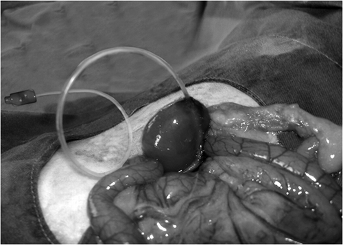
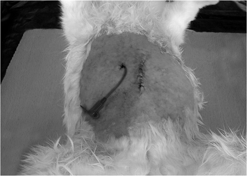
The rabbits were divided into three groups, each containing six rabbits. The animals were fasted for 12 h before the procedures. All surgical procedures were performed under ketamine (50 mg/kg, intramuscularly) and xylazine hydrochloride (5 mg/kg, intramuscularly) anesthesia by sterile technique. First of all, surgery was carried out through an abdominal midline incision to expose the ureter, which was catheterized through the flank approach with a catheter that consisted of a 50-cm length of 2 Fr to allow perioperative identification of the pelviureteral region (). Second, 1 cm of proximal ureteral segments and renal pelvis were isolated from other tissues in all groups.
In the surgical group (S), a 1-cm length of the proximal ureteral segment was resected and then the anastomosis was performed using absorbable suture material [(6–0 DEXON); DAVIS + GECK, Inc., American Cyanamid Company, Manati, PR 00701, USA]. The laparotomy incision was then closed in standard fashion. The animals were not allowed to feed for the next 48 h. The catheter was kept in place and the animals were fed parenterally for the first 2 days and later enterally.
After the same procedures were performed, saline was injected into the anastomose lines in the Sh group, and NH was injected in the NH group. After pelviureteral anastomose, NH group rats were administered 1 mL NH [Orthovisc, 2 mL (30 mg), Anika Therapeutics, Inc., Woburn, MA, USA] + 5 mL saline twice a day at 12-h intervals with a 50-cm length of 2-Fr catheter extending from abdominal wall to anastomose line for 7 days. After pelviureteral anastomose, Sh group rats were administered 5 mL saline twice a day at 12-h intervals with a 50-cm length of 2-Fr catheter extending from abdominal wall to anastomose line for 7 days (). All medical applications were performed under ketamine (50 mg/kg, intramuscularly) anesthesia by sterile technique.
Figure 2. The appearance of the catheter extending from abdominal wall to pelviureteral anastomose line.
Intravenous pyelography was carried out on postoperative day 21 to check whether there was pelviureteral leakage in the groups. The rabbits were fed orally on day 3 on the condition that there was no vomiting. There was no leakage in any of the study groups.
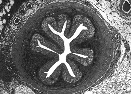
The rabbits were sacrificed after 3 weeks. A 1-cm segment of the ureter, which included both sides of the anastomosis in all groups, was taken (removed) to determine histopathologic findings.
The histopathologic evaluation was performed by a pathologist in a blind manner. Tissue specimens were fixed in 10% neutral buffered formaldehyde, processed routinely by an automatic tissue processor, and embedded in paraffin blocks. Sections of 4-μm thickness were stained with hematoxylin and eosin (H&E) for light microscopic morphologic evaluation. The extent of granulation tissue, granulation tissue fibroblast maturation (GTFM), and collagen deposition (CD) were evaluated both immunohistochemically and light microscopically. FGFR1 (Abcam, Rabbit polyclonal FGFR1 (ab10646) and collagen type III (Abcam, Rabbit polyclonal to Col III. Ab7778) antibodies were used for immunohistochemistry.Citation7–12 The specimens were scored for the differentiation of the healing and the repairing of the PUA. Histopathological parameters such as acute inflammation (AI) (presence of polymorphonuclear leukocytes at the anastomosis site), chronic inflammation (CI) (presence of lymphocytes and plasma cells), extent of granulation tissue (amount of granulation tissue), granulation tissue fibroblast maturation (maturation of granulation tissue, namely young, plump fibroblasts within a basophilic background or spindle fibroblasts compressed within a hyalinized background), collagen deposition (collagen matrix deposition between fibroblast bundles), re-epithelialization (urothelial re-epithelializationover the anastomosis site), neovascularization (amount of vascular proliferation during healing phase), and peripheral tissue reaction (PTR) (amount of foreign body reaction and reactive changes in the surrounding tissues of the anastomosis site) were evaluated according to a four-tiered system (score 0–3). We used a modified histologic scoring system developed specifically for this study based on the scoring system suggested in previous studies.Citation7,9 According to the scoring system, the extent of polymorphonuclear leukocyte infiltration at the anastomosis site was graded as “0” if none, and graded as “3” if extensive. All other morphologic parameters were evaluated according to the same principle. Histopathologic images were photographed by Zeiss Axio Imager D1 Microscope (Carl Zeiss Micro imaging GmbH 37081, Göttingen, Germany) with a computerized digital camera system attached to it.
Data concerning chronic inflammation, collagen deposition, neovascularization, re-epithelialization, and PTR were evaluated by Kruskal–Wallis variance analysis. Comparisons for AI, granulation tissue amount (GTA), and GTFM were analyzed by Kruskal–Wallis variance analysis, followed by Bonferroni-adjusted Mann–Whitney U-test. p-Values less than 0.05 were considered statistically significant for all tests (p-values less than 0.01 were considered statistically significant for Bonferroni-adjusted Mann–Whitney U-test). In all computations, SPSS version 17.0 (SPSS Inc., Chicago, IL, USA) was used.
RESULTS
AI scores in the Sh (1.333 ± 0.516) group were significantly higher than those in the S (0.000 ± 0.000) and NH (0.333 ± 0.5160) groups (p < 0.002) (). GTA scores in the Sh (2.166 ± 0.408) group were significantly higher than those in the S (1.166 ± 0.408) group (p < 0.014) ().
Table 1. The results of AI, GTA, and GTFM scores in fresh specimens.
There were no significant differences in the CI scores in the Sh (1.666 ± 0.516) group compared with those in the S (1.500 ± 0.547) and NH (2.333 ± 0.516) groups (p = 0.052) ().
Table 2. The results of CI, CD, N, R, and PTR scores in fresh specimens.
GTFM scores in the S (2.666 ± 0.516) group were significantly higher than those in the Sh (1.333 ± 0.516) group (p < 0.009) (). There were no significant differences in the GTFM scores in the S (2.666 ± 0.516) group compared with those in the NH (2.166 ± 0.752) group (p = 0.310) (). There were no significant differences in the GTFM scores in the Sh (1.333 ± 0.516) group compared with those in the NH (2.166 ± 0.752) group (p = 0.093) ().
There were no significant differences in the CD scores in the Sh (1.166 ± 0.408) group compared with those in the S (1.166 ± 0.408) and NH (1.166 ± 0.408) groups (p = 1.000) ().
There were no significant differences in the PTR scores in the Sh (2.500 ± 0.547) group compared with those in the S (2.166 ± 0.752) and NH (2.333 ± 0.516) groups (p = 0.686) ().
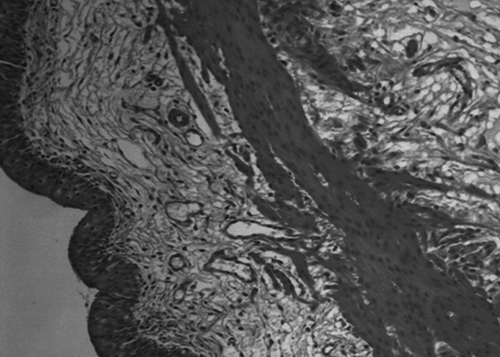
Neovascularization (N) scores in the NH (2.000 ± 0.632) group were higher than those in the S (1.166 ± 0.408) and Sh (1.500 ± 0.547) groups (p = 0.063) ().
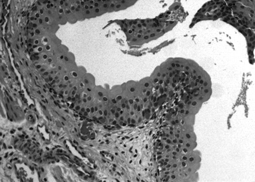
Re-epithelialization (R) scores in the NH (2.500 ± 0.836) group were higher than those in the S (2.328 ± 1.029) and Sh (2.333 ± 1.032) groups (p = 0.298) ().
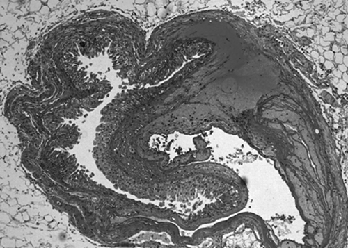
NH did not decrease fibrosis, but increased important parameters in wound healing such as neovascularization (p = 0.063) () ( and ) and re-epithelialization (p = 0.298) () ( and ) in an experimental model of PUA in rabbits.
DISCUSSION
In this pyeloplasty model, we used NH as a wound healing agent, which has not been tried before in such a model. We demonstrated that NH administration did not decrease fibrosis, but increased important parameters in wound healing such as re-epithelialization and neovascularization in an experimental model of PUA in rabbits. It is of interest that on postoperative day 21, PUA sections from NH-treated rabbits appear more normal than those of untreated rabbits and this confirmed the protective effect of NH.
Early postoperative complications such as leakage and stricture seen after PUA are a major clinical problem with a high degree of morbidity. Most clinicians still use surgical procedures such as Anderson–Hynes dismembered pyeloplasty, Foley Y-V pyeloplasty, and Culp–DeWeerd flap pyeloplasty, facing the problem of recurrent ureteropelvic stricture after the surgery. However, these various surgical procedures have been performed to prevent postoperative PUJOs, but it remains an important problem.
Hyaluronic acid or hyaluronan is a high molecular weight linear polysaccharide composed of repeating disaccharide units of N-acetyl glucosamine and glucuronic acid. It is synthesized by fibroblast cell membranes and then extruded into the extracellular space.Citation13 Hyaluronan is a ubiquitous compound, nonimmunogenic, and nontoxic.Citation14 In addition, hyaluronate can stimulate TLR4, which, except antigen-presenting cells, is also expressed in both epithelial cells and fibroblasts.Citation15 TLR4 stimulation in fibroblasts does not affect fibrosis-related factors but it enhances MCP-1 and IL-8 production, which leads to attraction of monocytes and neutrophils, respectively, in the area. Then TLR4 stimulant induces monocytes to produce TGF-b, which in turn promotes fibroblasts transformation to myofibroblasts and collagen formation.Citation16 A previous study has shown the ability of hyaluronan to accelerate the rate of healing and diminish scar formation in tympanic membrane perforations. The mechanism is not well understood, but it is thought to enhance cell migration and facilitate alignment of collagen fibrils to allow more rapid epithelialization and diminish connective tissue proliferation.Citation17 However, NH has not been used simultaneously with primary anastomosis prophylactically to prevent postoperative PUJOs.
The healing rate and degree of scar tissue formation were dependent on the concentration and number of applications of hyaluronan. Serial applications of concentrated hyaluronan accelerated healing and decreased scar tissue formation.Citation18 An agent such as NH, which has been shown to reduce connective tissue overgrowth in healing wounds, might be expected to alter the pyeloplasty healing process in the rabbit model and thereby provide additional insight into the pathogenesis of PUJOs. AI, CI, GTA, GTFM, CD, re-epithelialization (R), neovascularization (N), and PTR in anastomosis lines could be considered reliable and objective anastomoses markers in scar tissue formation.Citation7,9
Our results showed that the local application of NH significantly increased AI scores in the NH group when compared with the S group and significantly increased GTA in the NH group when compared with the S group. NH did not decrease fibrosis, but increased important parameters in wound healing such as re-epithelialization and neovascularization in an experimental model of PUA in rabbits.
In conclusion, our results indicate that NH facilitates better PUA lines. NH can be used simultaneously with pyeloplasty prophylactically to prevent postoperative PUJOs in children when re-epithelialization and neovascularization are considered. We think that increased re-epithelialization and neovascularization in the NH group are positive in terms of fibrous tissue formation. Further experimental and clinical studies are needed for detailed evaluation of NH in PUA.
ACKNOWLEDGMENTS
We thank Dr. T. Kemal Şahin for his help with the statistical analysis. This study was supported by Selcuk University Scientific Research Projects (grant 2011.09401092).
Declaration of interest: The authors report no conflicts of interest. The authors alone are responsible for the content and writing of this article.
REFERENCES
- Joyner BD, Mitchell ME. Ureteropelvic junction obstruction. In: Grosfeld JL, O’Neill JA, Fonkalsrud EW, Coran AG, eds. Pediatric Surgery. Philadelphia, PA: Mosby Elsevier; 2006:1723–1740.
- Spandow O, Hellstrom S. Healing of tympanic membrane perforation—a complex process influenced by a variety of factors. Acta Otolaryngol Suppl. 1992;492:90–93.
- Laurent C, Hellstrom S, Fellenius E. Hyaluronan improves the healing of experimental tympanic membrane perforations. Arch Otolaryngol Head Neck Surg. 1988;114(12):1435–1441.
- Hellstrom S, Laurent C. Hyaluronan and healing of tympanic membrane perforations. An experimental study. Acta Otolaryngol Suppl. 1987;442:54–61.
- Guneri EA, Tekin S, Yilmaz O, . The effects of hyaluronic acid, epidermal growth factor, and mitomycin in an experimental model of acute traumatic tympanic membrane perforation. Otol Neurotol. 2003;24(3):371–376.
- Nakamura M, Hikida M, Nakano T. Concentration and molecular weight dependency of rabbit corneal epithelial wound healing on hyaluronan. Curr Eye Res. 1992;11(10):981–986.
- Abramov Y, Golden B, Sullivan M, . Histologic characterization of vaginal vs. abdominal surgical wound healing in a rabbit model. Wound Rep Reg. 2007;15:80–86.
- Greenhalgh DG, Sprugel KH, Murray MJ, . PDGF and FGF stimulate wound healing in the genetically diabetic mouse. Am J Pathol. 1990;136:1235–1246.
- Fedakar-Senyucel M, Bingol-Kologlu M, Vargun R, . The effects of local and sustained release of fibroblast growth factor on wound healing in esophageal anastomoses. J Pediatr Surg. 2008;43:290–295.
- Zhang H, Dessimoz J, Beyer TA, . Fibroblast growth factor receptor 1-IIIb is dispensable for skin morphogenesis and wound healing. Eur J Cell Biol. 2004;83(1):3–11.
- Takenaka H, Yasuno H, Kishimoto S. Immunolocalization of fibroblast growth factor receptors in normal and wounded human skin. Arch Dermatol Res. 2002;294(7):331–338.
- Volk SW, Wang Y, Mauldin EA, Liechty KW, Adams SL. Diminished type III collagen promotes myofibroblast differentiation and increases scar deposition in cutaneous wound healing. Cells Tissues Organs. 2011;194(1):25–37.
- Laurent TC, Fraser JR. Hyaluronan. FASEB. 1992;6:2397–2404.
- Bagger-Sjoback D. Sodium hyaluronate application to the open inner ear: an ultrastructural investigation. Am J Otol. 1991;12:35–39.
- Eleftheriadis T, Pissas G, Liakopoulos V, Stefanidis I, Lawson BR. Toll-like receptors and their role in renal pathologies. Inflamm Allergy Drug Targets. 2012; November 8 [Epub ahead of print].
- Eleftheriadis T, Liakopoulos V, Lawson B, Antoniadi G, Stefanidis I, Galaktidou G. Lipopolysaccharide and hypoxia significantly alters interleukin-8 and macrophage chemoattractant protein-1 production by human fibroblasts but not fibrosis related factors. Hippokratia. 2011;15(3):238–243.
- Lacarte PR, Casasin T, Pumarola F, Alonso A. An alternative treatment for the reduction of tympanic membrane perforations: sodium hyaluronate: a double blind study. Acta Otolaryngol (Stockh). 1990;110:110–114.
- White SJ, Wright CG, Robinson KS, Meyerhoff WL. Effect of topical hyaluronic acid on experimental cholesteatoma. Am J Otolaryngol. 1995;16(5):312–318.

