Abstract
Nephrotoxicity is a major complication of gentamicin (GEN), which is widely used in the treatment of severe Gram-negative infections. Reactive oxygen species are important mediators of GEN-induced nephrotoxicity. Because of the strong antioxidant properties of pomegranate extract (PE), we evaluated the protective effect of PE against GEN-induced nephrotoxicity. Thirty-two adult male rats were randomly divided into four equal groups: (1) controls; (2) treated with GEN for 14 consecutive days (100 mg/kg/day); (3) treated with GEN plus distilled water; and (4) treated with GEN plus PE (100 μL). After 15 days, the rats were killed and their kidneys were taken, and blood analysis was performed. Tubular necrosis and interstitial fibrosis scores were determined histopathologically; and biochemically, nitric oxide (NO), malondialdehyde (MDA), and reduced glutathione (GSH) levels in kidneys were determined. Urea, creatinine, Na+, and K+ levels were investigated in the blood analysis. Statistical analyses were made by the chi-square test and analysis of variance. Serum urea and creatinine levels were significantly higher in rats treated with GEN alone than rats in the control and the GEN + PE-treated groups. The GSH level in renal tissue of only GEN-treated rats was significantly lower than those in the control group, and administration of PE to GEN-treated rats significantly increased the level of GSH. The group that was given GEN and PE had significantly lower MDA levels in kidney cortex tissue than those given GEN alone. There was no significant difference of NO levels between the groups. In rats treated with GEN + PE, despite the presence of mild tubular degeneration and tubular necrosis is less severe, and glomeruli maintained a better morphology when compared with the GEN-treated group. We think that PE prevents kidney damage by decreasing oxidative stress in kidney.
INTRODUCTION
Gentamicin (GEN) is an antibiotic of aminoglycoside group and is widely used in the treatment of Gram-negative infections. A major complication of this drug is nephrotoxicity, and it has been estimated that approximately 10–20% of cases are treated with aminoglycoside therapy.Citation1 Despite the introduction of less nephrotoxic antibiotics against Gram-negative microorganism, it is still used because of its low cost and efficacy against resistant beta-lactam positive microorganisms.Citation2 GEN is not metabolized in the body but is essentially eliminated by glomerular filtration and partially reabsorbed by proximal tubular cells.Citation3 No conclusive evidence exists of tubular secretion of GEN, and consequently, most aminoglycosides are excreted in the urine correspond to filtrates.Citation4 The specificity of GEN for renal toxicity is apparently related to its preferential accumulation in the renal proximal convoluted tubules, reaching a concentration of 5–50 times higher than plasma in the tubular renal cell.Citation5
It has been demonstrated that nephrotoxicity induced by GEN is characterized by direct tubular necrosis, which is localized mainly in the proximal tubules. The exact mechanisms of GEN-induced nephrotoxicity still remain unclear. However, several studies demonstrated that reactive oxygen species (ROS) may be important in GEN-induced nephrotoxicity.Citation6 Moreover, GEN has also been shown to enhance the generation of ROS. Lipid peroxidation (LPO) mediated by ROS has been suggested as a causative agent of cell death in different pathological states including various models of renal diseases.Citation7,8 Besides their direct damaging effects on tissues, ROS seem to trigger the accumulation of leukocytes in the tissue involved, and thus cause tissue injury indirectly through activated neutrophils. It has been shown that activated neutrophils secrete enzymes such as myeloperoxidase, elastase, and proteases and liberate oxygen radicals.Citation9 On the other hand, in vivo and in vitro studies have shown that the scavengers of reactive oxygen metabolites are protective in GEN-induced renal failure.Citation10,11
Pomegranate extract (PE) and its derivatives have been used for centuries to confer health benefits in a number of inflammatory diseases. Edible parts of pomegranate fruit represent 52% of total fruit weight, comprising 78% juice and 22% seeds.Citation12 Fresh juice is rich in vitamin C, and polyphenolic compounds such as anthocyanins, punicalagin, and ellagic and gallic acids.Citation13,14 PE is a rich source of potent polyphenolic, flavonoid antioxidants (anthocyanins). The soluble polyphenol content in PE varies within the limits of 0.2%–1.0% depending on the variety and includes mainly anthocyanins that have been shown to possess antiatherogenic properties. Anthocyanins were shown to be effective inhibitors of LPO, production of nitric oxide (NO), and inducible nitric oxide synthase (iNOS) activity in different model systems.Citation15,16
Pomegranate has become more popular because of the attribution of important physiological properties, such as anticancer,Citation17,18 cholesterol-lowering, cardioprotective,Citation19 and so on. Many investigators have reported that pomegranate and its derivatives have free radical scavenger and potent antioxidant activity.Citation20–22 It has also been shown that pomegranate can suppress nuclear factor kappa B (NF-κB) activation through a novel mechanism in vascular endothelial cells.Citation23
As a result of this information, in this study we investigated the possible inhibitory effects of pomegranate against the GEN-induced oxidative stress and renal injury in rat models. The nephroprotective effect of PE is previously established in many studies, however, to our knowledge this is the first study in literature concerning the protective role of pomegranate against GEN nephrotoxicity.
MATERIALS AND METDODS
Drugs
GEN was purchased from Bilim Pharmaceuticals (Istanbul, Turkey), and PE (100% pure, pasteurized pomegranate juice, 250 mL; Elite Natural Beverage Co., Ankara, Turkey) was purchased from a local store. GEN was dissolved in saline and injected intraperitoneally. PE was dissolved in distilled water and administered via nasogastric gavage. The average of 2.5 mL diluted PE contains 100-μL PE, which is equivalent to 2.8 μmol of total polyphenol per day.
Animals
Adult male Wistar albino rats (200–250 g) were housed in clean plastic cages in a temperature and humidity-controlled facility with a constant 12 h light/dark cycle with free access to food and water. The use of animals and the experimental protocol were approved by the Institutional Animal Care and Use Committee, and animals were treated in accordance with the Guide for the Care and Use of Laboratory Animals of Research Council.
Treatment and Experimental Design
After a quarantine period of 7 days, 32 rats were randomly divided into four equal groups, each consisting of eight animals as follows: (1) controls; (2) intraperitoneally injected with GEN for 14 consecutive days (100 mg/kg/day); (3) treated with GEN plus distilled water via nasogastric gavage for 14 days; (4) treated with GEN plus PE (100 μ/L) for 14 days. Rats were treated for 14 days. After 15 days, rats were killed and their kidneys were taken, and blood analysis was performed. Tubular necrosis and interstitial fibrosis scores were determined histopathologically in a part of kidneys; NO, malondialdehyde (MDA), and reduced glutathione (GSH) levels were determined in the other part of kidneys. Urea, creatinine, Na+, and K+ levels were investigated in a blood analysis.
Sample Collection and Biochemical Assays
Twenty-four hours after the administration of last doses of GEN and PG, on the 15th day, the rats were anesthetized by intraperitoneal injection of ketamine and were sacrificed. Twenty-four-hour urine collections were obtained in standard metabolic cages a day before the rats were killed. Renal cortical tissues were separated into two parts for biochemical analysis and light microscopic examination. Blood samples were also obtained by cardiac puncture to assess the serum levels of urea, creatinine, Na+, and K+ concentrations. The tissues were shock-frozen in liquid nitrogen and were kept in −80°C. In the frozen tissues, MDA, end product of LPO, reduced GSH, nonenzymatic antioxidant, and total nitrite (NOx), a stable product of NO, were evaluated biochemically as a means of oxidative stress.
Renal impairment was assessed by serum urea and creatinine levels, as well as by the kidney histology. Serum urea and creatinine levels were determined using an autoanalyzer (SYNCHRON LX20, Beckman Coulter Inc., Ireland) by using commercial Becman Coulter diagnostic kits. Kidney tissue (300 mg) was homogenized in ice-cold tamponade containing 150 mM KCl for determination of MDA. MDA levels were assayed for products of LPO. MDA referred to as thiobarbituric acid reactive substance was measured with thiobarbituric acid at 532 nm using a spectrofluorometer, as described previously.Citation24 GSH was determined by the spectrophotometric method, which was based on the use of Ellman’s reagent.Citation25
NOx was quantified by the Griess reactionCitation26 after incubating the supernatant with nitrate reductase from Escherichia coli to convert nitrate (NO3) to nitrite (NO2). Griess reagent (1 mL—1% sulfanilamide, 0.1% naphthyl-ethylenediamine hydrochloride, and 2.5% phosphoric acid; Sigma Chemical Co., St. Louis, MO, USA) was then added to 1 mL of supernatant. The absorbance was read at 545 nm after 30-min incubation. The absorbance was compared with the standard graph of NaNO2, obtained from the reduction of NaNO3 (1–100 lmol/L). The accuracy of the assay was checked in two ways; the interassay and intraassay coefficients of variation were 7.52% and 4.61%, respectively. To check conversion of nitrate to nitrite (recovery rate), known amounts of nitrate were added to control plasma samples; these samples were deproteinized and reduced as above.
Histopathological Examinations
Histopathological evaluation of the kidney tissues was done. Paraffin-embedded specimens were cut into 6-μm thickness and stained with Hematoxylin–Eosin stain for light microscopic examination using a conventional protocolCitation27 (Olympus BH-2, Olympus, Tokyo, Japan). A semiquantitative evaluation of renal tissues was accomplished by scoring the degree of severity according to previously published criteria.Citation28 All sections of the kidney samples were examined for parietal cell hyperplasia, tubular vacuolization, and tubular necrosis. Briefly, minimum of 50 proximal tubules associated with 50 glomeruli were examined for each slide, and an average score was obtained. Severity of lesion was graded from 0 to 3 according to the percentage of tubular involvement. Slides were examined and assigned for severity of changes using scores on scale of none (0), mild (1), moderate (2), and severe (3) damages, in which (0) denotes no change; grade (1) changes affecting <25% tubular damage (mild); grade (2) changes affecting 25%–50% of tubules (moderate); grade (3) changes affecting >50% of tubules (severe).
To evaluate kidney fibrosis, the specimens obtained from kidney were embedded in paraffin, sectioned at 6-μm sections, and stained with Masson’s trichrome. Specimens were scored after painting with, in brief, (−) no fibrosis, (+) fibrosis in <25% of total kidney tissue (mild), (++) fibrosis in 25%–50% of total kidney tissue (moderate), (+++) fibrosis in >50% of total kidney tissue (serious).Citation29
Statistical Analyses
Results of all groups were shown as mean values ± standard deviation (SD). Statistical analyses of the histopathological evaluation of the groups were carried out by the chi-square test, and biochemical data were analyzed by the one-way analysis of variance (ANOVA). The significance between two groups was determined by the Dunnett’s multiple comparison test, and p < 0.05 was accepted as statistically significant value.
RESULTS
No deaths or remarkable signs of external toxicity were observed in the groups of rats given GEN either alone or combination with PE. The biochemical and histopathological results were similar for the control and PE groups, and we decided to consider them without distinction and report only the control group.
Urinary Volume
The 24-h urine volume in the GEN-treated group was significantly higher than in the group control (p < 0.01), indicating the presence of GEN-induced polyuria, whereas in the group treated with GEN + PE, it was not different from that of the group control, pointing out the protective role of PE against acute tubular necrosis ().
Table 1. Effects of GEN alone and its combination with PE on plasma urea, creatinine, Na+, K+, and 24-h urine volume levels in rats.
Biochemical Variables in Plasma and Tissue
Na+ and K+ concentrations among four groups were similar. Serum urea and creatinine levels were significantly higher in rats treated with GEN alone than rats in the control and GEN + PE-treated groups (p < 0.01). Administration of PE to the GEN-treated rats caused decrease in serum urea and creatinine levels ().
The GSH level in renal tissue of only GEN-treated rats was significantly lower than those in the control group (p < 0.05), and administration of PE to GEN-treated rats significantly increased the level of GSH (p < 0.05; ).
Table 2. Effects of PE on NO, MDA, and GSH levels of rat kidney.
The group that was given GEN and PE had significantly lower MDA levels in kidney cortex tissue than those given GEN alone. There was no significant difference for any levels between the groups ().
Results of Histopathological Examinations
The histopathological examination of kidney showed no pathologic findings in the control group (A). In rats treated with GEN and GEN + vehicle, there were mild and severe tubular necrosis, tubular degeneration, and epithelial vacuolization in the proximal tubules, and parietal cell hyperplasia compared with the control group (B and C). In rats treated with GEN + PE, despite the presence of mild tubular degeneration and epithelial vacuolization in the proximal tubules, parietal cell hyperplasia and tubular necrosis are less severe, and glomeruli maintained a better morphology when compared with the GEN group (D). These changes are summarized in .
Figure 1. (A) Normal tubules and glomeruli in kidney cortex, staining with H&E ×100 (control group). (B) Severe tubular necrosis, tubular degeneration, and epithelial vacuolization in the proximal tubules, staining with H&E ×100 (GEN-treated group). (C) Moderate tubular necrosis, tubular degeneration, and epithelial vacuolization in the proximal tubules, staining with H&E ×100 (GEN + vehicle-treated group). (D) Mild epithelial vacuolization in the proximal tubules and normal glomeruli, staining with H&E ×100 (GEN + PE-treated group).
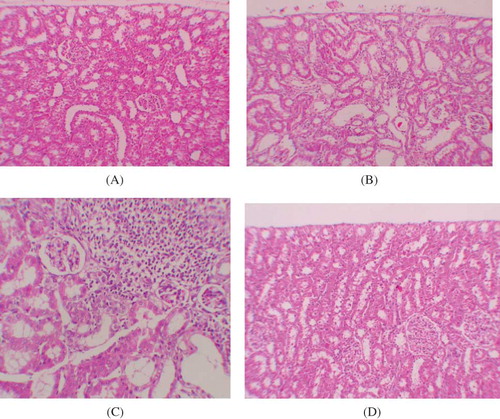
Figure 2. (A) No fibrosis in the control group, staining with Masson’s trichrome ×100. (B) Mild fibrosis in interstitium, staining with Masson’s trichrome ×400 (GEN-treated group). (C) Mild fibrosis in interstitium, staining with Masson’s trichrome ×200 (GEN + vehicle-treated group). (D) No fibrosis in GEN + PE-treated group, staining with Masson’s trichrome ×100.
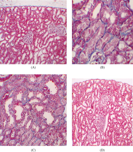
Table 3. Semiquantitative analysis of tubular necrosis, tubular vacuolization, parietal cell hyperplasia in the control, GEN-, GEN + vehicle-, and GEN + PE-treated rats.
Table 4. Analysis of kidney fibrosis in the control, GEN-, GEN + vehicle-, and GEN + PE-treated rats.
After staining with Masson’s trichrome, no statistical difference was found between the groups in kidney fibrosis scores. However, in GEN-treated rats, there was more mild and moderate fibrosis in kidney specimen (; A and D).
DISCUSSION
The kidneys are easily susceptible to damage from drugs because of larger perfusion and accumulation of excreted compounds that occur in renal tubular cells during absorption and secretion. Aminoglycoside is an antibiotic whose clinical use is limited by its nephrotoxicity. GEN is a widely used aminoglycoside antibiotic and has been shown to cause marked histological damage in particular to renal proximal convoluted tubules,Citation30,31 resulting in swelling, vacuolization, and necrosis of epithelial cells and accumulation of myelin-like bodies.Citation32
Furthermore, the results of many studies have shown that altered concentrations of various biochemical indicators of oxidative stress in kidney tissue are due to GEN.Citation33 Because of the obvious responsibility of ROS in GEN-induced renal damage, several antioxidant agents have been used to prevent GEN nephrotoxicity.Citation34 PE is rich in antioxidants of the polyphenolic class, which includes tannins and anthocyanins. These antioxidants are more potent, on a molar basis, than many other antioxidants, including vitamins C and E, coenzyme Q-10, and lipoic acid. The antioxidant levels in PE were found to be higher than that in other natural juices, such as blueberry, cranberry, and orange, as well as in red wine.Citation15
Pomegranate has become more popular because of the attribution of important physiological properties, such as anticancer, cholesterol-lowering, and cardioprotective. This study demonstrated ameliorative effects of PE, a phenolic antioxidant, on GEN-induced nephrotoxicity, in line with the consideration that oxygen-free radicals are important mediators of GEN-induced acute renal failure. The understanding of aminoglycoside nephrotoxicity is clinically important; such nephrotoxicity is typically associated with anoliguric acute renal failure, that is, azotemia in the presence of 1–2 L/day urine output. In this study, the 24-h urine volume in the GEN group was significantly higher than in the control group, indicating the presence of GEN-induced polyuria, whereas in the GEN + PE-treated group, it was not different from that of the control group, suggesting the protective role of PE against acute tubular necrosis. In addition, increased serum urea and creatinine levels in GEN-treated rats reflect the renal damage. In contrast to the previous studies, serum K+ levels were similar between the GEN-induced nephrotoxicity and the control groups. The final studies show that K+ levels were not increased in rats with GEN-induced nephrotoxicity.Citation35–37 Administration of PE protects the kidney function from GEN as indicated by preventing an increase in serum urea and creatinine levels. PE restores the renal function by preserving the structural integrity of renal cells against GEN challenge, evidenced by significantly preventing an increase in the levels of serum creatinine and urea.
Since brush border membrane (BBM) and other intracellular organelles such as mitochondria and lysosomes are known GEN target,Citation38,39 the structural/functional integrity was assessed by the status of their respective biomarker enzymes. So, here, we measured the MDA, GSH, and NO, as a means of oxidative stress. Our findings corroborate those of the earlier studies demonstrating that an enhanced endogenous oxidative stress has a major role in the severity of GEN-induced acute renal failure.Citation40,41 MDA, a stable lipid hydroperoxide, provides an index of the LPO in biological tissues.Citation42 In this study, we found increased MDA levels in the GEN-treated group, and as a protective effect of PE, lower MDA levels were found in the group determined by GEN + PE. The GSH antioxidant system is considered as the most notable cellular protective mechanism. GSH has a very important role in protecting against oxygen-free radical damage by providing reducing equivalents for several mechanism, as well as scavenging hydroxyl radicals and singlet oxygen. Its depletion is a common consequence of increased formation of ROSCitation5 like GEN-induced nephrotoxicity. In the group treated with GEN + PE, we found increased GSH levels. However, our study has shown that PE does not affect NO levels protectively in contrast to some previous studies with different antioxidant agents.Citation3,43 So, it may enhance LPO by a different way. These findings strongly indicate that PE is important in protecting the kidney from GEN-induced injury through improvement in oxidant status.
GEN treatment provokes acute tubular necrosis and acute renal failure in about 30% of high-risk patients.Citation6 Animal models of aminoglycoside nephrotoxicity also present residual areas of interstitial fibrosis in the renal cortex and progressive tubular injury.Citation44,45 After staining with Masson’s trichrome, no statistical difference was found between the groups in kidney fibrosis scores. However, in GEN-treated rats, there was more mild and moderate fibrosis in kidney specimen as previous studies. In this study, the histopathological examination of kidneys showed severe and extensive damage in GEN-treated rats which have tubular necrosis and edema. This could be due to the formation of highly reactive radicals as a consequence of oxidative stress caused by GEN. The kidneys of the control group showed normal histological features, but the GEN-treated group revealed more extensive and marked tubular necrosis. On the other hand, the tubules from rats of the GEN + PE-treated group were nearly normal in histological appearance except for a slight desquamation and atrophy of the tubular epithelial cells. Similar changes were also reported by some studies, which demonstrated structural changes in renal tissue of GEN-treated animals and its reversal by various agents.Citation3,46,47
In conclusion, the data indicated that GEN-induced nephrotoxicity might be related to oxidative damage. The results obtained in this study suggested that PE has an overall protective effect against GEN-induced nephrotoxicity in rat model. The observed protective effects can be attributed to the antioxidant properties of PE. So, PE is a highly free-radical scavenger agent and offers protection against GEN-induced acute renal failure.
Declaration of interest: The authors report no conflicts of interest. The authors alone are responsible for the content and writing of this article.
REFERENCES
- Humes HD, Weinberg JM. Toxic nephropathies. In: Brenner BM, Rector FC, eds. The Kidney. Philadelphia: WB Saunders Co.; 1986:1491–1532.
- Edson RS, Terrell CL. The aminoglycosides. Mayo Clin Proc. 1999;74:519–528.
- Karadeniz A, Yildirim A, Simsek N, Kalkan Y, Celebi F. Spirulina platensis protects against gentamicin-induced nephrotoxicity in rats. Phytother Res. 2008;22:1506–1510.
- De la Cruz Rodriguez LC, Araujo CR, Posleman SE, Rey MR. Attenuation of gentamicin-induced nephrotoxicity: trimetazidine versus N-acetyl cysteine. J Appl Toxicol. 2010;30:343–353.
- Abdel-Raheem IT, Abdel-Ghany AA, Mohamed GA . Protective effect of quercetin against gentamicin-induced nephrotoxicity in rats. Biol Pharm Bull. 2009;32:61–67.
- Cuzzocrea S, Mazzon E, Dugo L, . A role for superoxide in gentamicin-mediated nephropathy in rats. Eur J Pharmacol. 2002;450:67–76.
- Grune T, Sommerburg O, Petras T, . Postanoxic formation of aldehydic lipid peroxidation products in human renal tubular cells. Free Radic Biol Med. 1995;18:21–27.
- Baliga R, Ueda N, Walker PD, . Oxidant mechanisms in toxic acute renal failure. Drug Metab Rev. 1999;31:971–997.
- Winterbourn CC, Pichorner H, Kettle AJ. Myeloperoxidase-dependent generation of a tyrosine peroxide by neutrophils. Arch Biochem Biophys. 1999;338:15–21.
- Fryer MJ. Vitamin E may slow kidney failure owing to oxidative stress. Redox Rep. 1997;3:259–261.
- Pedraza-Chaverrı´ J, Maldonado PD, Medina-Campos ON, . Garlic ameliorates gentamicin nephrotoxicity: relation to antioxidant enzymes. Free Radic Biol Med. 2000;29:602–611.
- Tsuda T, Horio F, Cyanidin OT. 3-O-beta-d-glucoside suppresses nitric oxide production during a zymosan treatment in rats. J Nutr Sci Vitaminol (Tokyo). 2002;48:305–310.
- Ellman GL. Tissue sulfhydryl groups. Arch Biochem Biophys. 1959;82:70–77.
- de Nigris F, Williams-Ignarro S, Lerman LO, . Beneficial effects of pomegranate juice on oxidation-sensitive genes and endothelial nitric oxide synthase activity at sites of perturbed shear stress. Proc Natl Acad Sci USA. 2005;102:4896–4901.
- Aviram M, Dornfeld L, Kaplan M, . Pomegranate juice flavonoids inhibit low-density lipoprotein oxidation and cardiovascular diseases: studies in atherosclerotic mice and in humans. Drugs Exp Clin Res. 2002;28:49–62.
- Tsuda T, Horio F, Osawa T. Cyanidin 3-O-beta-d-glucoside suppresses nitric oxide production during a zymosan treatment in rats. J Nutr Sci Vitaminol (Tokyo). 2002;48:305–310.
- Kaplan M, Hayek T, Raz A, . Pomegranate juice supplementation to atherosclerotic mice reduces macrophage lipid peroxidation, cellular cholesterol accumulation and development of atherosclerosis. J Nutr. 2001;131:2082–2089.
- Houghton DC, Plamp CE III, DeFehr JM, Bennett WM, Porter G, Gilbert D. Gentamicin and tobramicin nephrotoxicity. A morphologic and functional comparison in the rat. Am J Pathol. 1978;93:137–152.
- Buege JA, Aust SD. Microsomal lipid peroxidation. Methods Enzymol 1978;52:302–310.
- Chen WC, Chen HY, Wu HC, Wu MC, Hsu CD, Tsai FJ. Vascular endothelial growth factor gene polymorphism is associated with calcium oxalate stone disease. Urol Res. 2003;31:218–222.
- Esen T, Akinci M, Aytekin Y, Altug T, Hekim N. The effects of citrate, polyphosphates and pridoxin on intratubular crystallization in hyperoxaluric rats. Turk J Urol.1990;16:365–370.
- Selvam R. Calcium oxalate stone disease: role of lipid peroxidation and antioxidants. Urol Res. 2002;30:35–47.
- Itoh Y, Yasui T, Okada A, Tozawa K, Hayashi Y, Kohri K. Preventive effects of green tea on renal stone formation and the role of oxidative stress in nephrolithiasis. J Urol. 2005;173(1):271–275.
- Villegas I, Mart´n AR, Toma W, . Rosiglitazone, an agonist of peroxisome proliferator-activated receptor c, protects against gastric ischemia–reperfusion damage in rats: role of oxygen free radicals generation. Eur J Pharmacol. 2004;505:195–203.
- Wasowicz W, Neve J, Peretz A. Optimized steps in fluorometric determination thiobarbituric acid-reactive substances in serum: importance of extraction pH and influence sample preservation and storage. Clin Chem. 1993;39:2522–2528.
- Beutler E. Glutathione in Red Blood Cell Metabolism. A Manual of Biochemical Methods. New York: Grune and Stratton; 1984:112–114.
- Sun Y, Oberley LW, Li Y. A simple method for clinical assay of superoxide dismutase. Clin Chem. 1988;34:497–500.
- Allen CT. Laboratory methods in histochemistry. In: Prophet EB, Mills B, Arrington JB, Sobin LH, eds. American Registry of Pathology. 1st ed. Washington DC: Armed Forces Institute of Pathology; 1992:53.
- Ayyıldız A, Nuhoğlu B, Gülerkaya B, . Effect of intraurethral mitomycin C on healing and fibrosis in rats with experimentally induced urethral stricture. Int J Urol. 2004;11:1122–1126.
- Humes HD, Connor RPO. Aminoglycoside nephrotoxicity. In: Schrier RW, Gottschalk CW, eds. Diseases of the Kidney. Vol. 2, no. 4. Boston, MA: Little Brown; 1988:1229–1273.
- Abdel-Gayoum AA, Ali BH, Abdel-Razig AA, Bashir AA, Ghywarsha K. Effect of gentamicin-induced nephrotoxicity on some carbohydrate metabolic pathways in the rat renal cortex. Arch Toxicol. 1994;68:643–647.
- Ali BH, Bashir AA. Effect of fish oil treatment on gentamicin nephrotoxicity in rats. Anal Nutr Metab. 1994;38:336–339.
- Humes HD. Aminoglycoside nephrotoxicity. Kidney Int. 1988;33:900–911.
- Simmons CF Jr, Bogusky RT, Humes HD. Inhibitory effects of gentamicin on renal mitochondrial oxidative phosphorylation. J Pharmacol Exp Ther. 1980;214:709–715.
- Silan C, Uzun O, Comunoglu NU, Gokcen S, Bedirhan S, Cengiz M. Gentamicin-induced nephrotoxicity in rats ameliorated and healing effects of resveratrol. Biol Pharm Bull. 2007;30:79–83.
- Cronin RE, Thompson JR . Role of potassium in the pathogenesis of acute renal failure. Miner Electrolyte Metab. 1991;17:100–105.
- Said MM. The protective effect of eugenol against gentamicin-induced nephrotoxicity and oxidative damage in rat kidney. Fundam Clin Pharmacol. 2011; 25:708–716.
- Farooq N, Priyamvada S, Khan F, Yusufi ANK. Time dependent effect of gentamicin on enzymes of carbohydrate metabolism and terminal digestion in rat intestine. Hum Exp Toxicol. 2007;26:587–593.
- Banday AA, Farooq N, Priyamvada S, Yusufi ANK, Khan F. Time dependent effects of gentamicin on the enzymes of carbohydrate metabolism, brush border membrane and oxidative stress in rat kidney tissues. Life Sci. 2008;82(9):450–459.
- Martinez-Salgado C, Lopez-Hernandez FJ, Lopez-Novoa JM. Glomerular nephrotoxicity of aminoglycosides. Toxicol Appl Pharmacol. 2007;223:86–98.
- Walker PD, Shah SV. Gentamicin enhanced production of hydrogen peroxide by renal cortical mitochondria. Am J Physiol. 1987;253:C495–C499.
- Kuhad A, Tirkey N, Pilkhwal S, . Effect of Spirulina, a blue green algae, on gentamicin-induced oxidative stress and renal dysfunction in rats. Fundam Clin Pharmacol. 2006;202:121–128.
- Ozbek E, Cekmen M, Ilbey YÖ, . Atorvastatin prevents gentamicin-induced renal damage in rats through the inhibition of p38-MAPK and NF-kB pathways. Renal Fail. 2009;31:382–392.
- Houghton DC, English J, Bennett WM. Chronic tubulointerstitial nephritis and renal insufficiency associated with longterm “subtherapeutic” gentamicin. J Lab Clin Med. 1988;112:694–703.
- Fuhr J, Kaczmarczyk J, Kruttgen CD. A simple colorimetric method of inulin determination in renal clearance studies on metabolically normal subjects and diabetics. Klin Wochenschr. 1955;33:729–730.
- Kumar KV, Shifow AA, Naidu MU, . Carvedilol: a beta blocker with antioxidant property protects against gentamicin-induced nephrotoxicity in rats. Life Sci. 2000;66:2603–2611.
- Nakakuki M, Yamasaki F, Shinkawa T, . Protective effect of human ulinastatin against gentamicin-induced acute renal failure in rats. Can J Phys Pharmacol. 1996;74:104–111.