Abstract
Ultrasound-guided right internal jugular vein catheterization (RIJV) should be the first choice to decrease the catheter-related complications in high-risk hemodialysis patients. For this procedure, clinicians should identify the optimum positions of the RIJV, including its lower overlap with the carotid artery (CA) and high cross-sectional area of the vein. The aim of this prospective randomized study to evaluate the effects of mild ipsilateral head rotation combined with Trendelenburg position on RIJV cross-sectional area and its relation to the CA in adult patients. Forty ASA I–II patients who were undergoing elective surgery were enrolled for this study. The subjects were asked to remain supine in the 15–20° Trendelenburg position. Two-dimensional ultrasound was then used to measure the degree of overlap between the RIJV and CA, the cross-sectional area of the RIJV. These measurements were compared between head rotation to the >30° left, <30° left, neutral, and <30° right positions. When the head was in the >30° left position, overlap was seen in 38 of 40 patients (95%). As the head was rotated from >30° left to <30° right, the CA-RIJV overlap (from 95% to 57.5%), and the cross-sectional area (from 14.2 mm to 8.7 mm) significantly decreased. In conclusion, when the head was turned to <30° right, the CA-RIJV overlap significantly decreased, and the cross-sectional area also decreased. When clinicians determine the optimal head position before RIJV cannulation, it is important to consider the advantages and disadvantages of the different head positions from >30° left to <30° right.
INTRODUCTION
Internal jugular vein (IJV) catheters are commonly employed for temporary vascular access for hemodialysis.Citation1 However, hemodialysis patients are generally are at an increased risk of catheter-related complications.Citation2 Owing to the high procedural success rate of right IJV (RIJV), the shorter and more direct route to the superior vena cava, larger vessel diameter, lower risk of carotid artery (CA) puncture, low incidence of anatomic variation, and the absence of the thoracic duct in right side, it is preferred over left IJV.Citation3–5 Inadvertent CA puncture is the most frequent complication of IJV catheter insertion; which can result in serious complications, even death.Citation1–3 Clinicians should find the optimum positions of IJV, including larger veins and lower overlap with the CA in order to perform a safe and successful cannulation. Therefore, researchers should aim to develop new maneuvers or techniques to engorge IJV and decrease CA puncture risk during central venous catheterization.
The Trendelenburg position, Valsalva maneuver, humming,Citation6 positive end-expiratory pressure (PEEP),Citation7 skin-traction method,Citation8 and lifting the probe straight upwardCitation9 combine the advantages of the short- and long-axis approaches while US-guided IJV cannulationCitation10,Citation11 primarily focuses on dilating the IJV.
Head rotations not only facilitate the visibility of anatomical landmarks for RIJV cannulation by clearing the chin,Citation12 but they also change the position of the RIJV with respect to the CA.Citation13–17 As the head is rotated from the midline, the IJV becomes more directly anterior to the CA, resulting in a greater overlap of the IJV and CA, which has been postulated to increase the rate of CA puncture as compared with a neutral head position.Citation13–17 However, no studies have specifically evaluated how the ipsilateral head rotation affects the IJV-CA overlap. The authors hypothesized that head rotation to a mild (<30°) ipsilateral position can decrease the IJV-CA overlap.
The primary aim of this prospective randomized study was to evaluate the effects of mild ipsilateral head rotation combined with the Trendelenburg position on the cross-sectional area of the RIJV and its relation to the CA in adult patients.
MATERIAL-METHODS
The study protocol was approved by the Institutional Review Board of the Medical Faculty Hospital, Selcuk University, before the start of the study. Between July 2012 and December 2012, a total of 40 ASA I–II patients between 18 and 65 years of age who were scheduled for elective surgery (general and orthopedic surgery) were eligible for this study. Patients were excluded from the study if thrombosis of IJV, anatomical neck abnormalities, neck masses, history of previous neck surgery, huge goiters, and lymphadenopathies were observed. Written informed consents were obtained from all the participants prior to the start of the study.
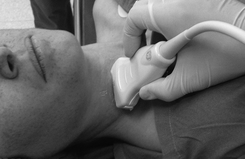
None of the patients received premedication and intravenous fluid replacement before the operation. Routine hemodynamic monitoring was carried out during the operation. The patient was positioned in the 15–20° Trendelenburg position with neck extension without a pillow. Before the administration of anesthesia, the RIJV and CA were ultrasonographically measured using a LOGIQ Book XP (GE Healthcare, Wauwatosa, WI) with a linear (7.5 MHz) probe. Ultrasound images of the RIJV were obtained in a transverse orientation at the cricoid level. The RIJV was depicted in the middle of the ultrasound image. Probe pressure was kept as low as possible to avoid compression of the RIJV.
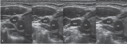
Each patient’s head was positioned in a stepwise manner in the following positions: >30° left, <30° left, neutral, and <30° right (). After the images were obtained for each position, the probe was moved in the same transverse plane until the center of the RIJV was at the center of the ultrasonography image and then fixed in this position. The images for each head position were frozen and stored. After freezing the real-time image, the circumference of the RIJV was delineated using an electronic marker, and the cross-sectional area of the RIJV was calculated using a program pre-loaded into the ultrasound unit.
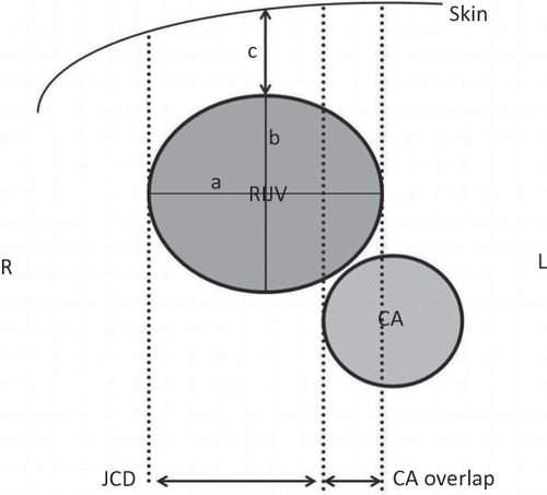
No patient was hypotensive or hemodynamically unstable at the time of data collection. The following measurements were carried out at each head position: (1) The degree of overlap between the RIJV and CA, (2) the cross-sectional area of the RIJV, (3) the transverse and anteroposterior diameters of the RIJV (4) the margin of safety, and (5) the depth of the RIJV from the skin ().
Figure 2. Anatomical illustration of study measurements. RIJV, right internal jugular vein; CA, carotid artery; JCD, jugular to carotid distance (safety margin); a, transverse diameter; b, anteroposterior diameter; c, depth from the skin; R, right; L, left.

Figure 3. Ultrasound images of cross sectional area of RIJV and relative positions of the RIJV and CA. (A) >30° left head rotation, (B) <30° left head rotation, (C) Neutral position, (D) <30° right head rotation. RIJV, right internal jugular vein; CA, carotid artery.

The overlap between the RIJV and CA was categorized on the basis of the percentage of overlap: (1) 0% (no overlap) (2) the RIJV overlapped <25% of the diameter of the CA; (3) the RIJV overlapped 25–50% of the diameter of the CA; or (4) the RIJV overlapped >50% of the diameter of the CA.
The transverse and anteroposterior diameters of the RIJV were measured by drawing a line between the furthest two points of the vein wall in the transverse and anteroposterior planes, respectively. The margin of safety was defined as the distance from the lateral-most border of the IJV and the lateral-most border of the CA at which the IJV could be punctured without touching the CA The depth of the RIJV from the skin was measured by drawing a line between the skin and the closest margin of the vein to the skin’s surface ().
Statistical analysis was performed using SPSS version 16.0 (SPSS Inc., Chicago, IL). The data were tested for normality using the Kolmogorov–Smirnov test. The results are expressed as mean ± SD, number of patients, and percentage. Categorical data were analyzed using the χ2 test. Statistical analysis included one-way analysis of variance with repeated measures in each head position. Post-hoc Tukey’s test was used when significant differences were found between the groups. p-values of <0.05 were considered statistically significant.
RESULTS
Data were collected for all the 40 patients enrolled in the study. Patient characteristics are shown in . There were more men than women in the study (26 of 40). The body mass index values were similar between both male and female patients.
Table 1. Demographic characteristics of the study population. Results are shown as mean ± SD.
The study measurements are summarized in . As the head was rotated from >30° left to <30° right, the overlap (from 95 to 57.5%), and the cross-sectional area (from 14.2 to 8.7 mm), and anteroposterior diameter of the RIJV (from 10.9 to 7.9 mm) was significantly decreased. The depth of the RIJV from the skin was 9.9 mm in the >30° left position, and it increased to 11.9 mm when the head was turned to the <30° right position (p = 0.007) ().
Table 2. Percentage overlap of the carotid artery and the right internal jugular vein at four different head positions. Values indicate the number of patients (%).
Table 3. Results of the ultrasonographic measurements. Results are shown as mean ± SD.
DISCUSSION
Our results demonstrated that when the head was turned from >30° left to <30° right, not only the CA-RIJV overlap tended to decrease (from 95 to 57.5%), but the cross-sectional area also decreased from 14.2 to 8.7 mm in adult patients. The reduced cross-sectional area may offset the advantage offered by the decreased overlap of the CA, thereby decreasing the probability of successful IJV cannulation.
It is important to avoid CA puncture during the IJV cannulation in dialysis patients,Citation18 which can occur even when performed under ultrasound guidance. Head rotation can increase the visibility of anatomical landmarks, thus facilitating skin puncture and cannulation.Citation3,Citation12,Citation18 However, Lamperti et al.Citation12 reported that the perception of difficulty in patients in the neutral and head turned positions during IJV cannulation did not significantly differ among the anesthesiologists (9% in the neutral position vs. 11% in the head turned position) when performed under ultrasound guidance.
When the CA is overlapped by the RIJV, double wall puncture predisposes an increased risk of CA puncture.Citation19 Troianos et al.Citation16 recorded images of 1100 patients in far lateral head turn, and found that the incidence of overlap increased to 90%. In the present study, we found overlap in 38 of 40 patients (95%) when the head was in the >30° left position. Previous ultrasonographic studies have reported that the neutral position was associated with the smallest amount of overlap, when compared with when the head was rotated to the left.Citation13–17 Our data complement and extend the findings of previous studiesCitation13–17 and showed decreased incidence of overlap from 95 to 77.5% and 63.5% from the >30° left, neutral, and <30° right head positions, respectively.
The rate of successful first-pass catheterization of IJV is directly correlated with the size of the vein.Citation19 Several methods that result in the largest possible cross-sectional area of the IJV can reasonably be assumed to give optimum conditions.Citation17,Citation20 Suarez et al.Citation17 demonstrated no differences in the cross-sectional area of the RIJV during 0°, 20°, and 58° head rotations combined with the Trendelenburg position in 24 adults. In our study, the cross-sectional area and diameter of the RIJV reached the lowest values when the head was in the < 30° right position, although patients were placed in the Trendelenburg position. The significant decrease in the right IJV cross-sectional area with the <30° right position in our study may be attributed to the increased compression of the IJV by the neck muscles.
The margin of safety was defined as the distance from the lateral-most border of the IJV and the lateral-most border of the CA at which the IJV could be punctured without touching the CA; these distances were found to be similar between the different head positions in the present study (9, 10, and 11 mm in the >30° left, neutral, and <30° right position, respectively; ). However, Wank et al.Citation13 defined the margin of safety as the absolute distance between the midpoint of the RIJV and the lateral-most border of the CA, and showed that the margin of safety significantly decreased when the head was turned to 90°.Citation13 Differences between the two studies may be associated with the different definitions of the safety margin.
IJV is generally encountered approximately 1.0–1.5 cm below the skin in adult patients.Citation21 Our data complement this knowledge, as the IJV was found to be 11 mm below the skin in the neutral position. Although there was a statistically significant difference in the depth of the IJV between the >30° left and <30° right positions (9.4 and 11.7 mm, respectively, p = 0.007), we opine that these differences have no clinical importance.
Our study has several limitations. First, since the patients recruited in this study had ASA I–II, the study did not include all scenarios in which IJV cannulation is required for hemodialysis. Further studies should be planned in hemodialysis patients to support our results. Second, we evaluated the effects of head rotation only at the cricoid level. The outcome measurements (cross-sectional area, diameters, and depth) of the RIJV and the degree of CA overlap may differ according to the site of measurement. Third, although meticulous care was taken to reduce venous compression by the ultrasound probe, there may have been minimal errors in measurement due to some venous compression by the probe. Finally, it was impossible to keep the ultrasound examiner completely blinded to the head position and the measurements obtained because of visual contact with the patients and screen, which is necessary to obtain adequate images.
In conclusion, when the head was turned to <30° right, not only the CA-RIJV overlap significantly decreased, but the cross-sectional area also decreased. When clinicians determine the optimal head position before RIJV cannulation, it is important to consider the advantages and disadvantages of the different head positions from >30° left to <30° right.
ACKNOWLEDGMENTS
The authors thank Dr. Kayhan Ozturk and Dr. Burhan Apiliogullari for their valuable comments.
Declaration of interest
The authors report no conflicts of interest.
REFERENCES
- Bansal R, Agarwal SK, Tiwari SC, Dash SC. A prospective randomized study to compare ultrasound-guided with non-ultrasound-guided double lumen internal jugular catheter insertion as a temporary hemodialysis access. Ren Fail. 2005;27:561–564.
- Bahcebasi S, Kocyigit I, Akyol L, et al. Carotid-jugular arteriovenous fistula and cerebrovascular infarct: a case report of an iatrogenic complication following internal jugular vein catheterization. Hemodial Int. 2011;15:284–287.
- Apiliogullari B, Kara I, Apiliogullari S, Arun O, Saltali A, Celik JB. Is a neutral head position a safe alternative to neck rotation during landmarks-guided internal jugular vein cannulation? Results of a randomized controlled clinical trial. J Cardiothorac Vasc Anesth. 2012;26:985–988.
- Ishizuka M, Nagata H, Takagi K, et al. Right internal jugular vein is recommended for central venous catheterization. J Invest Surg. 2010;23:110–114.
- Lim CL, Keshava SN, Lea M. Anatomical variations of the internal jugular veins and their relationship to the carotid arteries: a CT evaluation. Australas Radiol. 2006;50:314–318.
- Lewin MR, Stein J, Wang R, et al. Humming is as effective as Valsalva’s maneuver and Trendelenburg’s position for ultrasonographic visualization of the jugular venous system and common femoral veins. Ann Emerg Med. 2007;50:73–77.
- Lee SC, Han SS, Shin SY, Lim YJ, Kim JT, Kim YH. Relationship between positive end-expiratory pressure and internal jugular vein cross-sectional area. Acta Anaesthesiol Scand. 2012;56:840–845.
- Sasano H, Morita M, Azami T, et al. Skin-traction method prevents the collapse of the internal jugular vein caused by an ultrasound probe in real-time ultrasound-assisted guidance. J Anesth. 2009;23:41–45.
- Yoshida H, Yaguchi S, Kudo T, Ono T, Hirota K. A new method for longitudinally increasing the internal jugular vein diameter during ultrasound-guided cannulation. Anesth Analg. 2012;115:738–739.
- Dilisio R, Mittnacht AJ. The “medial-oblique” approach to ultrasound-guided central venous cannulation—maximize the view, minimize the risk. J Cardiothorac Vasc Anesth. 2012;26:982–984.
- Ho AM, Ricci CJ, Ng CS, et al. The medial-transverse approach for internal jugular vein cannulation: an example of lateral thinking. J Emerg Med. 2012;42:174–177.
- Lamperti M, Subert M, Cortellazzi P, et al. Is a neutral head position safer than 45-degree neck rotation during ultrasound-guided internal jugular vein cannulation? Results of a randomized controlled clinical trial. Anesth Analg. 2012;114:777– 784.
- Lieberman JA, Williams KA, Rosenberg AL. Optimal head rotation for internal jugular vein cannulation when relying on external landmarks. Anesth Analg. 2004;99:982–988.
- Wang R, Snoey ER, Clements RC, Hern HG, Price D. Effect of head rotation on vascular anatomy of the neck: an ultrasound study. J Emerg Med. 2006;31:283–286.
- Saitoh T, Satoh H, Kumazawa A, et al. Ultrasound analysis of the relationship between right internal jugular vein and common carotid artery in the lefthead-rotation and head-flexion position. Heart Vessels. 2012. September 12 E pub.
- Troianos CA, Kuwik RJ, Pasqual JR, Lim AJ, Odasso DP. Internal jugular vein and carotid artery anatomic relation as determined by ultrasonography. Anesthesiology. 1996;85:43–48.
- Suarez T, Baerwald JP, Kraus C. Central venous access: the effects of approach, position, and head rotation on internal jugular vein cross-sectional area. Anesth Analg. 2002;95:1519–1524.
- Ozbek S, Apiliogullari S, Erol C, et al. Optimal angle of needle entry for internal jugular vein catheterization with a neutral head position: a CT study. Ren Fail. DOI:10.3109/0886022X.2013.774672.
- Gordon AC, Saliken JC, Johns D, Owen R, Gray RR. US-guided puncture of the internal jugular vein: complications and anatomic considerations. J Vasc Interv Radiol. 1998;9:333–338.
- Blaivas M, Adhikari S. An unseen danger: frequency of posterior vessel wall penetration by needles during attempts to place internal jugular vein central catheters using ultrasound guidance. Crit Care Med. 2009;37:2345–2349.
- American Association of Clinical Anatomists, Educational Affairs Committee. The clinical anatomy of several invasive procedures. Clin Anat. 1999;12:43–54.

