
Abstract
The present study was conducted to elucidate the protective role of p-coumaric acid, a common dietary polyphenol against cadmium induced nephrotoxicity in rats. For the purpose of comparison, a standard reference drug silymarin (50 mg/kg b. wt) was used. In this experiment, the animals were divided into four groups, with each consisting of six animals. The animals in Group I animals received saline and served as a control group and those in Group II received cadmium chloride (3 mg/kg b. wt) subcutaneously once daily for 3 weeks, but Group III and IV animals received cadmium chloride followed by p-coumaric acid (100 mg/kg b. wt, oral) and silymarin (50 mg/kg b. wt, oral), respectively, daily for 3 weeks. At the end of the treatment, the animals were sacrificed, and the blood and kidney samples were collected. The results obtained in this study revealed the fact that the levels of lipid peroxidation, lysosomal enzymes, glycoprotein, cadmium and metallothionein were increased in the cadmium chloride alone treated rats and antioxidant status was found to be decreased, when compared to the control group. The levels of kidney functional markers (urea, uric acid and creatinine) were also found to be abnormal in serum and urine of cadmium chloride alone treated rats. On the other hand, the administration of p-coumaric acid along with cadmium chloride significantly protected the biochemical alterations as observed in the cadmium chloride alone treated rats as evidenced by histopathology. Thus, the oral administration of p-coumaric acid significantly protected the cadmium-induced nephrotoxicity in rats.
Introduction
Cadmium (Cd) is one of the non-essential hazardous heavy metals, which is known for its non-corrosive nature. It is mainly used in paints, dyes, cement and phosphate fertilizers.Citation1 Industrial usage of cadmium began in 1941 and each year’s cadmium production has reached 15,000 tons. It is estimated that the anthropogenic sources of cadmium add 3–10 folds more cadmium to the atmosphere than natural sources.Citation2 Cadmium contamination of soil and water is of increased concern due to its high bioaccumulation ability and long half-life of 10–30 years.Citation3 Hence, cadmium-polluted environment leads to severe damage to the human organs such as liver, kidney, lungs, bone and testes.Citation4 Humans are exposed to cadmium by occupational exposure such as working with cadmium containing pigments, plastics, metal alloys, glasses, nickel–cadmium batteries and non-occupational exposure such as food, water and cigarette smoke. The cadmium exposure from the environment occurs into the body and it is mostly through by pulmonary and enteral pathways.Citation5 Several studies in experimental animals and on humans have identified kidney as one of the important target organs for cadmium-induced toxicity.
Metallothioneins are thiol-containing, cysteine-rich heavy metal-binding proteins that sequester metals, thus preventing accumulation of potentially toxic-free metal ions within the cell.Citation6 Cadmium toxicity is believed to be results from non metallothionein bound cadmium or free cadmium and metallothionein bounded cadmium is considered biologically inactive.Citation7 Directly after the absorption, cadmium is bound to the apoprotein thionein and forms holoprotein metallothionein and filtered by the glomerulus into the urinary space where it is endocytosed by the proximal tubular cells and degraded by the lysosomes that result in the release of cadmium. The intracellular release of cadmium is responsible for the generation of reactive oxygen species, depletion of glutathione, lipid peroxidation, protein cross-linking, DNA damage and culminating ultimately in oxidant-induced cell death and renal toxicity.Citation8
p-Coumaric acid (trans-4-hydroxycinnamic acid) is a dietary polyphenol, mainly present in plants and forms a part of human diet. It has its wide existence in fruits and vegetables such as apples, pears, potatoes and tomatoes, beverages such as tea, coffee and beer.Citation9 Recent studies from our laboratory and others have reported that p-coumaric acid possesses anti-oxidant,Citation10 cardioprotective,Citation11 antimelanogenic,Citation12 antimutagenic,Citation13 antiplatelet,Citation14 anti-inflammatory and immunomodulatory properties.Citation15 Furthermore, it has been reported that it reduces the growth of stomach, breast and colon cancer cells Citation16,Citation17 and down regulates the mRNA expression levels of the key angiogenic factors.Citation18 Considering the potential beneficial properties of p-coumaric acid, the present study was undertaken to investigate the nephroprotective potential of p-coumaric acid against cadmium chloride-induced toxicity in rats. For purpose of comparison, a standard reference drug silymarin (50 mg/kg b. wt) was used.
Materials and methods
Animals
Either sex of Wistar albino rats, 170–200 g were obtained from the Animal House, VIT University, Vellore, India. They were acclimatized in a light and temperature-controlled room with a 12 h light–dark cycle and fed with commercial pelleted feed from Hindustan Lever Ltd. (Mumbai, India), and water was freely available. The animals used in this study were treated and cared for in accordance with the guidelines recommended by the Committee for the Purpose of Control and Supervision of Experiments on Animals (CPCSEA), VIT University, Vellore, India.
Drugs and chemicals
Cadmium chloride was obtained from MERCK, Bangalore, India. p-coumaric acid (≥98% HPLC) and silymarin, a standard hepatoprotective drug were obtained from the Sigma Aldrich, St. Louis, MO and Micro Labs Ltd, Goa, India, respectively. All other chemicals and reagents used were of analytical grade and purchased locally.
Experimental design
Rats were divided into four groups with each group consisting of six animals.
Group I: Control rats administered with saline subcutaneously daily for 21 days.
Group II: Rats subcutaneously administered with cadmium chloride (3 mg/kg b.wt/day) dissolved in saline for 21 days.
Group III: Rats subcutaneously administered with cadmium chloride (3 mg/kg b.wt/day) followed by an oral administration of p-Coumaric acid (100 mg/kg b.wt/day) for 21 days
Group IV: Rats subcutaneously received with cadmium chloride (3 mg/kg b. wt/day) followed by an oral administration of silymarin (50 mg/kg b. wt/day) for 21 days.
On the 22nd day, the end of the experimental period, the urine samples were collected over a period of 24 h, using metabolic cages. The animals were cervically decapitated. The trunk blood was collected and centrifuged for serum and plasma separation. The kidney tissues were dissected out and washed, using ice-cold Phosphate buffered saline (pH 7.4). A 10% homogenate of the washed tissues was prepared in PBS, pH 7.4 for the biochemical assays.
Assay of kidney functional markers
Urea, uric acid and creatinine in the serum and 24-h urine samples were estimated spectrophotometrically using commercially available kits purchased from Span Diagnostics, Bangalore, India.
Assay of lipid peroxidation and antioxidant status
Lipid peroxidation in kidney was determined by the procedure of Ohkawa et al.Citation19 Malondialdehyde, formed as an end product of the peroxidation of lipids, served as an index of oxidative stress. Superoxide dismutase (SOD) activity in kidney was assayed according to the method of Marklund and Marklund.Citation20 The enzyme activity is defined as the enzyme required to give 50 % inhibition of pyrogallol auto-oxidation. Catalase (CAT) was estimated by measuring the chromic acetate formed in the presence of H2O2 according to Sinha.Citation21 Glutathione peroxidase (GPx) was determined by the method of Rotruk et al.Citation22 Glutathione reductase (GR) was assayed by the method of Bellomo et al.Citation23 Glutathione-S-transferase (GST) and total reduced glutathione (GSH) were estimated by the method of Habig et al.Citation24 and Moron et al.,Citation25 respectively. The protein level was determined by the method of Lowry et al.Citation26 using bovine serum albumin as a standard.
Assay of lysosomal enzymes and glycoproteins
Acid phosphatase was assayed by the method of KingCitation27 using disodium phenyl phosphate as the substrate. The enzyme activity was expressed as μmol phenol liberated/min/mg protein. N-acetyl glucosaminidase activity was assessed by the method of MaruhnCitation28 using 4-nitrophenyl-N-acetyl glucosaminide as the substrate and its activity was expressed as μmol p-nitrophenol formed/h/mg protein. The activity of β-galactosidase was assessed by the method of Rosenblit et al.Citation29 using 4-nitrophenyl-N-acetyl glucopyranoside as the substrate and its activity was expressed as μmol p-nitrophenol liberated/h/mg protein. Cathepsin D activity was calculated by the method of Biber et al.Citation30 using hemoglobin as the substrate and the activity expressed as μmol of tyrosine liberated/h/mg protein.
The kidney tissue samples were defatted before the estimation of glycoproteins. A weighed amount of defatted tissue was suspended in 3 ml of 2 M HCl and heated at 90 °C for 4 h. The sample was cooled and neutralized with 3 ml of 2 M NaOH. Aliquots from this were used for the estimation of sialic acid and hexosamine. Sialic acid was determined by the method of AminoffCitation31 with modifications by Niebes.Citation32 An aliquot of tissue homogenate was mixed with 0.25 M periodate (in 0.1 N H2SO4), and the reaction was inhibited after 30 min by arsenite solution. Then thiobarbituric acid was added and the contents were heated. The pink color that developed on cooling was measured at 540 nm. Hexosamine was estimated by the method of Wagner.Citation33 The acetyl acetone reagent consisting of trisodium phosphate and potassium tetraborate with acetyl acetone was added to tissue homogenate and boiled. After cooling, ehrlich’s reagent was added and the pink color developed was measured at 540 nm.
Estimation of metallothionein and cadmium content
Metallothionein content present in kidney tissues was estimated according to the method of Viarengo et al.Citation34 with modification by Petrovic et al.Citation35 MT concentration was estimated utilizing GSH as standard and expressed as nmol of GSH/mg protein. Cadmium content in kidney tissues was estimated by atomic absorption spectrophotometry. The kidney samples were prepared with 65% nitric acid for total dissolution. An aliquot of individual samples was used to evaluate cadmium content. The cadmium level was determined at 228 nm by flame atomic absorption spectrometer.
Histopathological analysis
Immediately after sacrifice, a portion of the kidney was fixed in 10% formalin, then washed, dehydrated in descending grades of isopropanol and finally rinsed with xylene. The tissues were then embedded in molten paraffin wax. Sections were cut at 5 μm thickness, stained with hematoxylin and eosin, and observed under microscope for histopathological changes.
Statistical analysis
Results were expressed as mean ± SD, and statistical analysis was performed using ANOVA, to determine the significant differences between the groups, followed by Student’s Newman–Keul’s test; p < 0.05 implied significance.
Results
Effect of p-coumaric acid on kidney functional markers
and represent the effect of p-coumaric acid on the level of kidney functional markers in serum and urine of control and experimental rats. The results showed that urea, uric acid and creatinine levels were found to be significantly elevated in serum and decreased in urine samples of cadmium chloride alone treated rats compared to the control and drug treated rats. However, p-coumaric acid treatment along with cadmium chloride administration restored the levels of kidney functional markers when compared to cadmium chloride alone treated rats.
Table 1. Effect of p-coumaric acid on serum kidney markers in cadmium chloride-intoxicated rats.
Table 2. Effect of p-coumaric acid on urinary kidney markers in cadmium chloride-intoxicated rats.
Effect of p-coumaric acid on lipid peroxidation and antioxidant status
depicts the effect of p-coumaric acid on lipid peroxidation and antioxidant status in the kidney tissues of control and experimental rats. The lipid peroxidation levels were found to be increased and the activity of antioxidants such as superoxide dismutase, catalase, glutathione-s-transferase, glutathione reductase, glutathione peroxidase and reduced glutathione levels were found to be decreased in the kidney of cadmium chloride alone treated rats compared to the control rats. In rats treated with p-coumaric acid in addition to cadmium chloride administration, the lipid peroxidation levels were found to be decreased and the antioxidant levels were found to be significantly increased similar to that of silymarin treatment in the kidney tissue.
Table 3. Effect of p-coumaric acid on kidney lipid peroxidation levels and antioxidant status in cadmium chloride-intoxicated rats.
Effect of p-coumaric acid on lysosomal enzymes
shows the protective effect of p-coumaric acid on the activities of lysosomal enzymes in control and experimental rats. The activity of the lysosomal enzymes such as acid phosphatase, N-acetyl glucosaminidase, β-galactosidase and cathepsin D were found to be significantly elevated in the kidney of cadmium chloride alone treated groups compared to the control rats, whereas in rats treated with p-coumaric acid in addition to cadmium chloride administration, the enzyme levels were found to be significantly decreased similar to that of silymarin treatment.
Table 4. Effect of p-coumaric acid on lysosomal enzymes in kidney of cadmium chloride-intoxicated rats.
Effect of p-coumaric acid on glycoproteins
represents the protective effect of p-coumaric acid on the level of glycoproteins in control and experimental rats. Glycoproteins levels (hexosamine and sialic acid) were found to be significantly higher in cadmium chloride alone treated rats than the control rats. On the other hand, the rats treated with p-coumaric acid in addition to cadmium chloride, the glycoprotein levels (hexosamine and sialic acid) were found to be significantly restored to near normal level similar to that of silymarin treatment.
Table 5. Effect of p-coumaric acid on glycoproteins in kidney of cadmium chloride-intoxicated rats.
Effect of p-coumaric acid on metallothionein and cadmium content
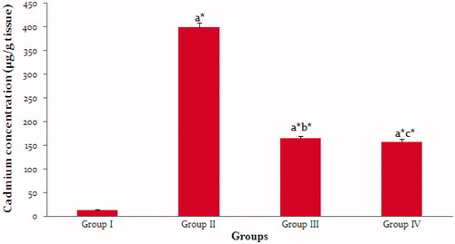
and illustrate the effect of p-coumaric acid on the level of metallothionein and cadmium in the kidney tissues of control and experimental rats. The cadmium and metallothionein content in the kidney tissues were increased in cadmium chloride alone treated rats compared to the control rats. Nevertheless, the administration of p-coumaric acid along with cadmium chloride in rats decreased the cadmium content in kidney tissues. On the other hand, the metallothionein level was found to be increased further significantly in p-coumaric acid treated cadmium chloride exposed rats than the cadmium chloride alone treated rats.
Figure 1. Effect of p-coumaric acid on metallothionein concentration in kidney tissues of rats exposed to cadmium chloride. Group I – Control rats, Group II – Cadmium chloride-intoxicated rats, Group III – Cadmium chloride-intoxicated rats treated with p-coumaric acid (100 mg/kg b.wt) and Group IV – Cadmium chloride-intoxicated rats treated with Silymarin (50 mg/kg b.wt). Each value represents the mean ± SD of six rats. Comparisons were made as follows: aGroup I versus Group II, III and IV, bGroup II versus Group III and cGroup II versus Group IV. The symbols represent statistical significance at: *p < 0.05. Statistical analysis was calculated by one-way ANOVA followed by Student’s Newman–Keul’s test.
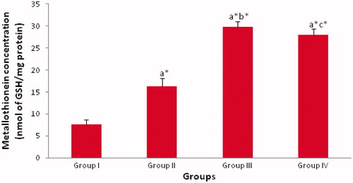
Figure 2. Effect of p-coumaric acid on cadmium concentration in kidney tissues of rats exposed to cadmium chloride. Group I – Control rats, Group II – Cadmium chloride-intoxicated rats, Group III – Cadmium chloride-intoxicated rats treated with p-coumaric acid (100 mg/kg b.wt) and Group IV – Cadmium chloride-intoxicated rats treated with Silymarin (50 mg/kg b.wt). Each value represents the mean ± SD of six rats. Comparisons were made as follows: aGroup I versus Group II, III and IV, bGroup II versus Group III and cGroup II versus Group IV. The symbols represent statistical significance at: *p < 0.05. Statistical analysis was calculated by one-way ANOVA followed by Student’s Newman–Keul’s test.
Histopathological analysis
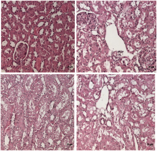
represents the effect of p-coumaric acid on histopathological alterations in kidney tissues of control and experimental rats. The kidney sections of control rats () showed normal renal glomerular and tubular structure, whereas cadmium chloride alone treated rats showed severe tubular epithelial cell degeneration (). On the other hand, p-coumaric acid and silymarin treatment to the rats along with cadmium chloride reduced these histopathological changes remarkably in the kidney tissue ().
Figure 3. Histopathological analysis of the nephroprotective potential of p-coumaric acid in cadmium chloride-intoxicated rats (H&E 20X). (a) Control rats showing normal rat kidey – normal glomeruli and tubular structure. (b) Cadmium chloride-intoxicated rats showing severe kidney tubular epithelial cell degeneration. (c,d) Cadmium chloride followed by p-coumaric acid (100 mg/kg b.wt) and silymarin (50 mg/kg b.wt) treated rats, respectively, showing improvement over cadmium intoxicated group with mild tubular epithelial cell degeneration.
Discussion
The present study was carried out to investigate whether p-coumaric acid, a common dietary polyphenol, could render protection against cadmium chloride-induced nephrotoxicity in rats by examining the different biochemical parameters in serum, urine and kidney of rats.
The serum levels of urea, uric acid and creatinine can be used as an indicator of renal dysfunction and damage. Our present study has shown that renal functional markers such as urea, uric acid and creatinine levels in the serum were increased in cadmium chloride alone treated rats, whereas, these marker levels were found to be decreased in the urine. In renal diseases, the rate of serum urea production exceeds more than the rate of clearance which results in uremia, Moreover, increased blood urea is known to be correlated with an elevated protein catabolism in mammals or the conversion of ammonia to urea as a result of increased synthesis of arginase enzyme involved in urea production as well as cadmium inhibits the incorporation of amino acids into protein causing an increase in urea level.Citation36 In our study, the finding of elevated serum uric acid concentrations may reflect the bodily response to an increased production of endogenous oxygen species, because uric acid is a potent scavenger of peroxynitrite.Citation37 Creatinine is mostly derived from endogenous sources by tissue creatine breakdown. The plasma creatinine concentrations in normal individuals are usually affected by a number of factors such as the muscle mass, high protein diet, and catabolic state.Citation38 Hence, our results confirm that administration of cadmium generates a progressive tubular damage as evidenced by the kidney histopathology (), since the proximal tubules are specifically sensitive to cadmium exposure due to their high reabsorptive activity and results in the abnormalities.Citation39 However, the administration of p-coumaric acid along with cadmium chloride inhibits the kidney dysfunction and restores the urea, uric acid and creatinine levels to near normal in serum and urine. In addition, histological studies on the kidney tissue of the p-coumaric acid treated cadmium chloride exposure rats showed mild tubular epithelial cell degeneration similar to that of silymarin treatment. This shows the nephroprotective effect of p-coumaric acid.
During cadmium exposure, the interactions of cadmium with intracellular sites like mitochondria, peroxisomes and microsomes results in the generation of free radicals, which ultimately leads to lipid peroxidation.Citation40 Antioxidants are the important substances that neutralize free radicals or their actions. These include superoxide dismutase that catalyzes the dismutation of superoxide to H2O2 and catalase that breaks down H2O2 to water.Citation41 Glutathione peroxidase catalyzes the oxidation of reduced glutathione to oxidized glutathione by hydroperoxides.Citation42 The glutathione-s-transferase are the enzymes which bind glutathione to the electrophilic molecules and have a main role in xenobiotics, drugs and carcinogens and thus protects the cells against oxidative stress.Citation43 Glutathione reductase, one of the markers of oxidative stress, is involved in the maintenance of the optimum level of oxidized and reduced glutathione.Citation44 During cadmium accumulation, excessive lipid peroxidation can cause increased glutathione consumptionCitation45 and the other antioxidant enzymes become inactive by the direct binding of cadmium to the sulfhydryl (–SH) groups in the enzyme active sites as well as displacement of metal cofactors from the active sites of the enzymes.Citation46 Administration of p-coumaric acid significantly increased the activities of these antioxidants and decreased the lipid peroxidation in kidney tissues which could be due to the free radical scavenging role of p-coumaric acid and also by inducing the gene expression of endogenous antioxidants.Citation47
Lysosomes are sub-cellular organelles, which catabolize intracellular materials to generate smaller biomolecules for biological processes.Citation48 In our study, the activities of lysosomal enzymes are found to be elevated in the extracellular fluid occur as a result of decreased lysosomal membrane stability and kidney damage. The elevated levels of lysosomal enzymes during cadmium exposure eventually affect the metabolism of different connective tissue constituents, viz glycosaminoglycans, glycoproteins, collagen and results in irreversible tissue damage. The elevation in the levels of glycoprotein components of kidney tissue is due to the secretion of cell membrane glycoconjugates into the cytosol of kidney tissue respectively. On the other hand, administration p-coumaric acid significantly decreased the lysosomal enzyme release and glycoprotein levels in cadmium chloride alone treated rats. This result shows the membrane stabilizing effect of p-coumaric acid.
In our study, the metallothionein levels were found to be increased in p-coumaric acid treated cadmium chloride exposed rats compared to the cadmium chloride alone treated rats. It has been reported that metallothionein transgenic mice showed decreased susceptibility to cadmium toxicity and metallothionein-null mice showed increased susceptibility to cadmium toxicity.Citation49 In concordance with these reports, the increased metallothionein concentration found in the kidney tissue of p-coumaric acid treated cadmium chloride exposed rats reduces the cadmium toxicity. The possible protective mechanism of metallothionein against oxidative damage could be due to the fact that the cysteine residues of the metallothionein are the primary target for the reaction of oxygen free radicals.Citation50
In the present study, cadmium content in the renal tissue was found to be increased in the cadmium chloride alone treated rats compared to the control group, which indicates the greater affinity of cadmium to kidneys. However on p-coumaric acid treatment to the cadmium chloride induced rats, the cadmium levels were found to be decreased which might be due to its metal chelating activity as well as increase in the levels of metallothionein.
In conclusion, from the results obtained, the present study suggests that p-coumaric acid, a common dietary polyphenol possesses significant protective role against cadmium chloride-induced nephrotoxicity in rats.
Declaration of interest
The authors declare no conflicts of interest. The authors alone are responsible for the content and writing of this article.
References
- Jarup L. Hazards of heavy metal contamination. Br Med Bull. 2003;68:167–182
- Waalkes MP. Cadmium carcinogenesis. Mutat Res. 2003;533(1–2):107–120
- Satarug S, Baker JR, Urbenjapol S, et al. A global perspective on cadmium pollution and toxicity in non-occupationally exposed population. Toxicol Lett. 2003;137(1–2):65–83
- Domingo JL. Metal-induced developmental toxicity in mammal: a review. J Toxicol Environ Health. 1994;42(2):123–141
- Waisberg M, Joseph P, Hale B, Beyersmann D. Molecular and cellular mechanisms of cadmium carcinogenesis. Toxicol. 2003;192(2–3):95–117
- Ybarra GR, Webb R. Differential responses of GROEL and metallothionein genes to divalent metal cations and the oxyanions of arsenic in the cyanobacterium Synecoccus sp. strain PCC 7942. Proceedings of the 1998 Conference on Hazardous Waste Research, Snowbird, Utah, May 18–21, 1998:76–82
- Kagi JHR, Vallee BL. Metallothionein: a cadmium and zinc-containing protein from equine renal cortex. J Biol Chem. 1960;235:3460–3465
- Thevenod F. Nephrotoxicity and the proximal tubule. Insights from cadmium. Nephron Physiol. 2003;93(4):87–93
- King A, Young G. Characteristics and occurrence of phenolic phytochemicals. J Am Diet Assoc. 1999;99(2):213–218
- Castelluccio C, Paganga G, Melikian N, et al. Antioxidant potential of intermediates in phenylpropanoid metabolism in higher plants. FEBS Lett. 1995;368(1):188–192
- Prasanna N, Krishnan DN, Rasool M. Sodium arsenite-induced cardiotoxicity in rats: protective role of p-coumaric acid, a common dietary polyphenol. Toxicol Mech Methods. 2013;23(4):255–262
- An SM, Koh JS, Boo YC. p-coumaric acid not only inhibits human tyrosinase activity in vitro but also melanogenesis in cells exposed to UVB. Phytother Res. 2010;24(8):1175–1180
- Ferguson LR, Lim IF, Pearson AE, Ralph J, Harris PJ. Bacterial antimutagenesis by hydroxycinnamic acids from plant cell walls. Mutat Res. 2003;542(1–2):49–58
- Luceri C, Giannini L, Lodovici M, et al. p-Coumaric acid, a common dietary phenol, inhibits platelet activity in vitro and in vivo. Br J Nutr. 2007;97(3):458–463
- Pragasam SJ, Venkatesan V, Rasool M. Immunomodulatory and anti-inflammatory effect of p-coumaric acid, a common dietary polyphenol on experimental inflammation in rats. Inflammation. 2013;36(1):169–176
- Ferguson LR, Shuo-tun Z, Harris PJ. Antioxidant and antigenotoxic effects of plant cell wall hydroxycinnamic acids in cultured HT-29. Mol Nutr Food Res. 2005;49(6):585–593
- Hudson EA, Dinh PA, Kokubun T, Simmonds MS, Gescher A. Characterization of potentially chemopreventive phenols in extracts of brown rice that inhibits the growth of human breast and colon cancer cells. Cancer Epidemiol Biomarkers Prev. 2000;9(11):1163–1170
- Kong CS, Jeong CH, Choi JS, Kim KJ, Jeong JW. Antiangiogenic effects of p-coumaric acid in human endothelial cells. Phyther Res. 2013;27(3):317–323
- Ohkawa H, Ohishi N, Yagi K. Assay for lipid peroxides in animal tissues by thiobarbituric acid reaction. Anal Biochem. 1979;95(2):351–358
- Marklund SL, Marklund G. Involvement of superoxide anion radical in the autoxidation of pyrogallol and a convenient assay for superoxide dismutase. Eur J Biochem. 1974;47(3):469–474
- Sinha AK. Colorimetric assay of Catalase. Anal Biochem. 1972;47(2):389–394
- Rotruk JT, Pope AL, Ganther HE, Swanson AB, Hafeman DG, Hoekstra WG. Selenium, biochemical role as a component of glutathione peroxidase purification and assay. Science. 1973;179(4073):588–590
- Bellomo G, Mirabelli F, DiMonte D, et al. Formation and reduction of glutathione-mixed disulfides during oxidative stress. Biochem Pharmacol. 1987;36(8):1313–1320
- Habig WH, Pabst MJ, Jakoby WB. Glutathione-S-transferases. The first enzymatic step in mercapturic acid formation. J Biol Chem. 1974;249(22):7130–7139
- Moron MS, Depierre JW, Mannervik B. Levels of glutathione, glutathione reductase and glutathione-S-transferase activities in rat lung and liver. Biochem Biophys Acta. 1979;582(1):67–78
- Lowry OH, Rosebrough NJ, Farr AL, Randall RJ. Protein measurement with the folin phenol reagent. J Biol Chem. 1951;193(1):265–275
- King J. The hydrolases-acid and alkaline phosphatases. In: Van D, ed. Practical Clinical Enzymology. London: Nostrand Company Limited; 1965:191–208
- Maruhn D. Rapid colorimetric assay of beta-galactosidase and N-acetyl-beta-glucosaminidase in human urine. Clin Chim Acta. 1976;73(3):453–461
- Rosenblit PD, Metzyer RP, Wick AN. Effect of Streptozotocin diabetes on acid phosphatase and selected glycosidase activities of serum and various rat organs. Proc Soc Exp Biol Med. 1974;145(1):244–248
- Biber J, Stieger B, Haase W, Murer H. A high yield preparation for rat kidney brush-border membranes, different behaviors of lysosomal markers. Biochim Biophys Acta. 1981;647(2):169–176
- Aminoff D. Methods for the quantitative estimation of N-acetyl neuraminic acid and their application to hydrolysates of sialomucoids. Biochem J. 1961;81(2):384–392
- Niebes P. Determination of enzymes and degradation products of glycosaminoglycan metabolism in the serum of healthy and varicose subjects. Clin Chim Acta. 1972;42(2):399–408
- Wagner WD. A more sensitive assay discriminating galactosamine and glucosamine in mixtures. Anal Biochem. 1979;94(2):394–396
- Viarengo A, Ponzano E, Donder F, Fabbri R. A simple spectrophotometric method of metallothionein evaluation in marine organisms: an application to Mediterranean and Antarctic mollusks. Mar Env Res. 1997;44(1):69–84
- Petrovic S, Ozretic B, Krajnovic-Ozretic M., Bobinac D. Lysosomal membrane stability and metallothioneins in digestive gland of mussels (Mytilus galloprovincialis Lam.) as biomarkers in a field study. Mar Pollut Bull. 2001;42(12):1373–1378
- Tormanen CD. Inhibition of rat liver and kidney arginase by cadmium ion. J Enzyme Inhib Med Chem. 2006;21(1):119–123
- Hooper DC, Spitsin S, Kean RB, et al. Uric acid, a natural scavenger of peroxynitrite, in experimental allergic encephalomyelitis and multiple sclerosis. Proc Natl Acad Sci. 1998;95(2):675–680
- Mayne PD. The kidneys and renal calculi. In: Zilva JF, Pannall PR, eds. Clinical Chemistry in Diagnosis and Treatment. 6th ed. London: Edward Arnold Publications; 1994:2–24
- Thiruchelvi R, Arul D, Meenakshi S, Subramanian K. Protective effects of Terminalia chebula fruit extract against cadmium-induced nephrotoxicity in rats. Int J Environ Biol. 2012; 2(3):108–112
- Stohs SJ, Bagchi D, Hassoun E, Bagchi M. Oxidative mechanisms in the toxicity of chromium and cadmium ions. J Environ Pathol Toxicol Oncol. 2000;19(3):201–213
- Sies H. Antioxidants in Disease, Mechanisms and Therapy. New York: Academic Press; 1996
- Halliwell B, Gutteridge JMC. Free Radicals in Biology and Medicine. Oxford: Oxford University Press; 1999
- Mates JM. Effects of antioxidant enzymes in the molecular control of reactive oxygen species toxicology. Toxicol. 2000;153(1–3):83–104
- Winston GW, Di Giulio RT. Prooxidant and antioxidant mechanisms in aquatic organisms. Aquat Toxicol. 1991;19(2):137–161
- Onyema OO, Farombi EO, Emerole GO Ukoha AI, Onyeze GO. Effect of Vitamin E on monosodium glutamate induced hepatotoxicity and oxidative stress in rats. Ind J Biochem Biophys. 2006;43(1):20–24
- Renuga devi J, Prabu SM. Naringenin protects against cadmium-induced oxidative renal dysfunction in rats. Toxicol. 2009;256(1–2):128–134
- Yeh CT, Ching LC, Yen GC. Inducing gene expression of cardiac antioxidant enzymes by dietary phenolic acids in rats. J Nutr Biochem. 2009;20(3):163–171
- Versteeg DJ, Giesy JP. (1985). Lysosomal enzyme release in the bluegill sunfish (Lepomis macrochirus Rafinesque) exposed to cadmium. Arch Environ Contam Toxicol. 1985;14(5):631–640
- Liu Y, Liu J, Iszard MB, Andrews GK, Palmiter RD, Klaassen CD. Transgenic mice that overexpress metallothionein-1 are protected from cadmium lethality and hepatotoxicity. Toxicol Appl Pharmacol. 1995;135(2):222–228
- Thornally PJ, Vasak M. Possible role of metallothionein in the protection against radiation-induced oxidative stress. Kinetic and mechanisms of its reaction with superoxide and hydroxyl radicals. Biochem Biophys Acta. 1985;827(1):36–44