
Abstract
Renal ischemia reperfusion injury (IRI) is a major problem, currently without treatments in clinical use. This reflects the failure of animal models to mimic the severity of IRI observed in clinical practice. Most described models lack both the ability to inflict a permanent reduction in renal function and the sensitivity to demonstrate the protective efficacy of different therapies in vivo. To test novel cell-based therapies, we have developed a model of renal IRI in Fisher 344 rats. Animals were subjected to 120 min of unilateral warm ischemia, during which they underwent an intra-renal artery infusion of therapeutic agents or vehicle. At either 2 or 6 weeks post-surgery, animals underwent terminal glomerular filtration rate (GFR) studies by inulin clearance to most accurately quantify renal function. Harvested kidneys underwent histological analysis. Compared to sham operations, saline treated animals suffered a long-term reduction in GFR of ≈50%. Histology revealed short- and long-term disruption of renal architecture. Despite the injury severity, post-operative animal losses are <5%. This model produces a severe, consistent renal injury that closely replicates the pathological processes encountered in clinical medicine. Renal artery infusion mimics the route likely employed in clinical transplantation, where the renal artery is accessible. Inulin clearance characterizes GFR, allowing full assessment of therapeutic intervention. This model is useful for screening therapeutic agents prior to testing in a transplant model. This reduces animal numbers needed to test drugs for clinical transplantation and allows for refinement of dosing schedules.
Introduction
Ischemia reperfusion injury (IRI) is a leading cause of acute kidney injury and represents a major clinical problem with high associated levels of morbidity and mortality.Citation1–5 IRI to native kidneys results from pathological processes that cause organ hypo-perfusion such as trauma, dehydration and sepsis. Renal IRI also commonly occurs in patients undergoing aortic or cardiac surgery.Citation6 Furthermore during kidney transplantation, IRI that results from allograft implantation has been shown to contribute to early organ dysfunction and poor long-term outcomes.Citation7 Cell-based therapy has emerged as a potential treatment for patients at risk of renal IRI. It is postulated that such treatments may reduce renal damage by down regulating inflammatory pathways, promote tissue regeneration and consequently contribute to improved outcomes in the long-term.Citation5,Citation8–10 Unfortunately no such treatments are currently available for clinical use, with supportive therapy being the mainstay of treatment.Citation11,Citation12 Given the prevalence and morbidity associated with renal IRI, the development of such therapies should be considered a priority.Citation13
While the mechanisms by which IRI occurs are relatively well understood, further insight into the efficacy of possible therapies might be provided by developing experimental models that mimic the severe IRI that is often seen in the clinical setting. In vitro models of renal IRI typically use renal epithelial cell lines or isolated perfused kidneys. Such models provide valuable information, but lack the physiological complexity of in vivo models and are thus limited in the data they provide.Citation14 In vivo rodent models of renal IRI may broadly be divided into two types: bilateral or unilateral renal injury. Unilateral injury models may be further divided into models with or without a contralateral nephrectomy. Models using bilateral injury or unilateral injury with contralateral nephrectomy are the most commonly studied, probably because blood analysis may demonstrate trends in renal function throughout the experimental time course.
In both of the commonly used models, warm ischemic times of between 30 and 60 min have been shown to cause acute renal dysfunction,Citation15–18 but this is reported to resolve within a week of surgery.Citation17–19 Longer periods of warm ischemia are associated with unacceptable numbers of post-procedure animal deaths.Citation20,Citation21 Using conventional rodent models, it therefore appears difficult to inflict a longstanding renal injury, as rodent kidneys appear to regenerate to function normally after non-fatal periods of renal ischemia. The benefit of testing the long-term effects of novel therapies in conventional rodent models is therefore questionable.
Unilateral injury models without contralateral nephrectomy leave animals with a healthy functioning kidney that serve as ‘protection’ against acute renal failure. Consequently, such models allow the study of prolonged ischemic times in the injured kidney, without excessive animal loses. Unfortunately unilateral injury models without nephrectomy do not allow functional elements of the ischemic injury to be studied using serial blood analysis, as renal function markers in such models are affected by the filtration of uninjured kidney.
Herein we described a novel rat model of renal IRI designed to address the problems described above. We employ a unilateral injury without contralateral nephrectomy model. To produce a high local dose of treatment, and to mimic the therapeutic route most likely employed in clinical transplantation, we inject therapy directly into the renal artery. Animals subsequently undergo inulin clearance studies as a secondary procedure to most accurately determine glomerular filtration rate (GFR) responses to injury ± intervention. Kidneys are later analyzed using histological methods to provide further insight into structural renal damage.
Methods
IRI procedure
Animals
We developed a model of renal IRI in Fisher 344 male rats, aged between 12 and 14 weeks, weighing 225–250 g.
Anesthesia
After placing the animal in an anesthetic chamber, induction was achieved using 5% inhaled isoflurane. Anesthesia was maintained with 5% isoflurane, given via facemask, mixed with 1 L of oxygen/min.
Position and temperature control
The rat was placed supine on a far infrared heating pad with the midline shaved and cleaned with chlorhexidine. The core temperature was measured regularly using an infrared thermometer and the heat mat was adjusted accordingly to maintain this between 36.5 and 37 °C.
Surgical procedure
A high midline incision was made. Bowel was wrapped in damp swabs and retractors were placed to enable access to the left renal pedicle. Division of the lateral peritoneal attachments mobilized the left colon, thereby exposing the renal vasculature. The renal artery was separated from the renal vein using blunt dissection with cotton tips and/or forceps. Full exposure of the renal artery from its origin on the aorta to the renal hilum necessitated tying of the suprarenal vein. Once exposed, an atraumatic vascular clamp was placed on the artery’s aortic origin, signifying the start of the ischemic period. A color change in the kidney confirmed correct placement of the clamp.
After 90 min had elapsed, an arteriotomy was made in the renal artery. Via this, the kidney was flushed with a 0.8 mL intra-arterial injection given via a 30G Ryecroft cannula. A thin rubber sloop was placed around the cannula and artery, preventing back-leak while an assistant gently injected the contents of the syringe. A color change in the cortex of the kidney confirmed infiltration of the kidney parenchyma with the desired agent or saline vehicle.
The artery was fully transected, then re-anastomosed in a standardized triangulated fashion, using 12 interrupted 10/0 nylon sutures.Citation22 A frame clamp was used to secure the arterial ends during anastomosis and background material was placed to prevent damage to posterior structures. The arterial clamps were removed after exactly 120 min of ischemia and reperfusion of the kidney was confirmed visually.
Wound closure
The incisions were closed in two layers, using a continuous 4/0 polyglactin suture (Vicryl®).
Post-surgical care
The animal was injected with 2.5 mL of subcutaneous 0.9% saline post-operatively. Buprenorphine analgesia was injected subcutaneously, at 0.0045 mg per 100 g of body weight. This was repeated as necessary every 12 h for a maximum duration of 48 h post-surgery. The animal was placed in a warming box at 38 °C for 60 min. It was then returned to its cage and allowed free access to rodent chow and water. The animal was weighted daily.
Quantifying renal injury
Determination of GFR with inulin clearance studies
We have adapted a CIC modelCitation23 that allows the GFR of each kidney to be calculated individually. Animals may undergo inulin studies at any point after the IRI procedure – we have typically chosen time points of 2 or 6 weeks.
Preparation of inulin solution
Fluorescein Isothiocyanate Inulin (FITC-Inulin) (Sigma-Aldrich) solution (0.2%) was made by dissolving 40 mg of FITC Inulin in 20 mL of warmed 0.9% saline solution. One percent FITC Inulin was made by dissolving 10 mg FITC Inulin per mL of 0.9% physiological saline solution. The solutions were dialyzed with physiological saline overnight through a 1 kDa semipermeable membrane, removing any unbound fluorescein.
Anesthesia
Induction was with isoflurane as described previously. Anesthesia was maintained with an initial intraperitoneal injection of thiobutabarbitol sodium (Inactin®) using 12 mg per 100 g of body weight. Further maintenance doses of 2.4 mg per 100 g of body weight were given as required. This provided stable cardiovascular parameters.Citation24
Surgical procedure
Positioning and temperature control are as described above. The femoral artery and vein were fully dissected. After clamping the femoral vein proximally and ligating distally, microsurgical polyethylene tubing (0.96 mm external diameter (ED)) was used to cannulate the vein. The tubing was secured using 5/0 ligatures. A 0.5 mL bolus of 1% inulin solution was given before starting a constant infusion of 0.2% FITC inulin using a syringe pump at a rate of 3 mL/h.
A microsurgical catheter (0.8 mm ED) was then placed in the femoral artery. This was used to take blood samples during the experiment and provided continuous blood pressure monitoring to ensure hemodynamics stability. Between sampling, the line was locked with heparin-saline solution.
Ureteric cannulation
The abdomen was then re-opened along the line of the previous midline incision. The bowel was wrapped in damp swabs, the ureters were dissected and cannulated using a 7 cm length of microsurgical tubing (0.61 mm ED). Ligatures were applied to secure the catheters’ position.
Via these cannulas, urine was collected into separate tubes during each 30-min interval. In the middle of each 30 min collection period, an 80 μL blood sample was taken via the arterial line. Urinary volume for each 30-min period was measured using a Gilson’s pipette.
Serum and urinary fluorescence was calculated spectrofluorometrically using a fluorescence microplate reader (BIO-TEK flx-800) measuring emission and absorbance wavelengths of 495 nm and 430 nm, respectively. Forty microliters of 500 mM HEPES buffer was mixed with each 10 μL sample of serum or urine, since fluorescence of biofluids is known to vary across a pH range.Citation25
Steady state was reached in the plasma when the amount of inulin infused was equal to the amount filtrated by the kidneys. This is defined as a <5% change in inulin plasma concentration over a 30-min period.Citation26 Our data indicated that steady state is typically reached 60–90 min after the inulin infusion had commenced.
The experiment was terminated after six timed urinary/blood samples had been collected. The animal was killed and both kidneys, liver, spleen, heart and lungs were taken and stored in 10% formation, liquid nitrogen and RNA later.
All serum samples with the animal at steady state, along with the corresponding urine collections, were then used to calculate GFR using the following equation:
where V = urine volume/min, U = urinary fluorescence and P = plasma fluorescence.
Tips for achieving reliable GFR results
Avoiding unnecessary blood loss is a key to ensure accurate GFR results. To achieve this, it is advisable to ligate all branches of the femoral vessels, and to ensure proximal and distal control of vessels before making an incision for cannulation. Bleeding points on the re-opened abdominal wall should be cauterized or over-sewn. We also advise cauterization of small retroperitoneal vessels encountered during dissection of the ureter, as these may bleed profusely over the time course of the study if left uncontrolled.
Tips for successful microsurgical cannulation
Cannulation is easier if a precise hole is made in the front wall of the structure to be cannulated. A horizontal cut with a pair of pointed microsurgical scissors is most effective. This maneuver allows the structure to remain under tension, while the created defect gapes open, allowing easy access for the passage of the cannula (. Fully dividing a structure causes proximal retraction, whereby cannulation becomes extremely problematic.
Figure 1. (a) Femoral vein prepared for cannulation. Note ligatures loosely applied, ready to be tightened immediately post-cannula insertion. (b) Venotomy made distally and dilated for insertion of cannula. (c) Inulin infusion into cannulated femoral vein, with catheter secured by ligatures and vascular clamp. (d) Microtubing cut using a scalpel blade (top) and scissors (bottom) viewed at high magnification with the operating microscope. Note the ragged edge of the scissor cut tube.
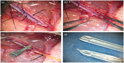
It is useful to have ligatures to secure a catheter loosely applied prior to cannula insertion. This allows simple tightening of the ligature as soon as the cannula is inserted, reducing the chance of the cannula inadvertently slipping out.
When the microsurgical tubing is cut to size, the use of a scalpel blade allows a smooth, angled but non-pointed end of the tube to be fashioned. The use of scissors leads to a ragged pointed tube that is difficult to insert, and will tear holes in vessel walls when advanced.
Histological methods of assessing renal injury
Hematoxylin and eosin staining and histopathology scoring
Formalin fixed specimens are paraffin embedded, sectioned at 5 μm and stained with hematoxylin and eosin for light microscopy analysis. We use an IRI scoring system that examines for tubular epithelial damage, tubular dilation and hyaline cast formation described by a previous study.Citation27 Typically 10 non-overlapping cortical high power fields are examined and the mean scores are compared between treated and untreated kidneys.
Results
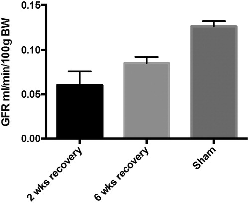
In more than 50 rats subjected to 120 min of unilateral warm renal ischemia, we have only experienced one post-procedure animal death. Furthermore, we have only failed to successfully anastomose the renal artery on one occasion. One hundred and twenty minutes of renal ischemia produces a permanent, severe and reproducible injury that results in long-term GFR deficit (see ).
Figure 2. Left-sided renal function after 120 min IRI at 2 and 6 weeks recovery. We observe a severe reduction in GFR (∼50%) after 2 weeks compared to sham operated animals (p = 0.0025). GFR is typically ∼65% of sham values after 6 weeks recovery and is subjected to less variability (p = 0.0012). (Error bars represent SEM.).
Histological examination of kidney tissues as described above reveals significant disruption in renal architecture in those animals subjected to 120 min of warm ischemia ().
Figure 3. (a) Renal histology from sham operated rat at 2 weeks post-surgery. A glomerulus is marked with an arrow. (b) Histology from rat subjected to 120 min IRI, 2 weeks post-surgery. Note marked tubular dilation (green asterisk) with epithelial thinning and breaks (arrowheads). Also, present are hyaline casts (black dots). A glomeruli with an increased Bowman’s space is marked with an arrow. (c) Histology from rat subjected to 120 min IRI 6 weeks post-surgery. Hyaline casts are noted, but tubular dilation and epithelial thinning are less pronounced. (d) Renal histology from rat subjected to 120 min IRI, 6 weeks post-surgery. The animals were treated with intra-arterial stem-cell based therapy. Histological damage is less marked than with saline treated animals.
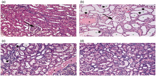
Figure 4. Renal histology shows that after 2 weeks recovery there are significantly more epithelial breaks in rats subjected to 120 min IRI when compared to sham controls (p < 0.0001). Similarly more epithelial cell breaks are observed in the 6 week group than in sham operated animals. (p < 0.0001; Error bars represent SEM.).
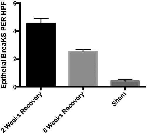
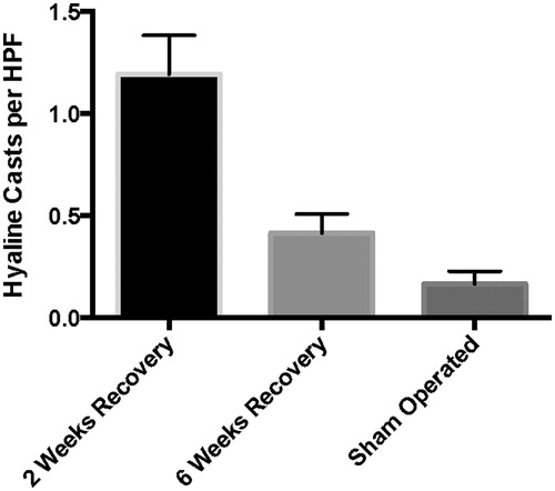
Figure 5. More hyaline casts are observed in both the 2 week recovery group \r\n (p < 0.0001) and the 6 week group (p = 0.0285) when compared to sham operated animals. (Error bars represent SEM.).
Discussion
Cell-based therapy has emerged as a potential treatment for acute renal injury affecting either native or transplanted kidneys. Unfortunately no such treatments are in widespread clinical use. This is a particular problem in the field of transplantation where changing trends in organ donation have led clinicians to meet demand by utilizing organs from older and less fit deceased donors.Citation28,Citation29 Such kidneys may be subjected to longer periods of warm ischemia during retrieval and consequently are more susceptible to IRI during implantation.Citation30–33 Developing strategies, including pharmacological interventions to improve the outcomes of these ‘extended criteria’ allografts is of the upmost importance.
The absence of interventions for the treatment of renal IRI reflects the lack of relevant animal models in which novel therapies may be easily studied. Many common rodent models used to induce renal dysfunction do not rely on warm ischemia as a mechanism of injury. These include models that utilize reduction of renal mass (5/6 nephrectomy), administration of nephrotoxins, or renal injury caused by ureteric obstruction. However, warm renal ischemia and the injury that follows reperfusion are the most common causes of renal dysfunction encountered in clinical practice.Citation34,Citation35 Establishing authentic animal models that mimic the pathological process of renal IRI are of vital importance in evaluating the feasibility and efficacy of novel therapeutic interventions. Unfortunately, commonly used rodent models of renal IRI lack the capacity to inflict a severe, long-standing renal injury without excessive post-operative animal deaths. To address this, Wang et al. investigated the use of prolonged warm ischemic times in order to create a severe and sustained renal injury model.Citation36 They observed warm ischemic times of up to 90 min which were associated with long-term disruption of renal architecture, increased levels of apoptosis and renal fibrosis. However, they reported prolonged ischemia resulted in almost 80% animal deaths at 4 weeks, although renal function as determined by serum creatinine, had normalized by this time.
Rodent kidneys subjected to prolonged periods of warm ischemia have been reported to display abnormal histology after several days’ recovery, while the serum creatinine/blood urea nitrogen (BUN) was found to be normal.Citation18,Citation19,Citation36–39 This paradox may be explained by the relative insensitivity creatinine and BUN at quantifying renal function in rodents.Citation40–43 While convenient, these surrogate markers of glomerular filtration do not become significantly elevated until 50–75% of kidney function is lost.Citation40 In addition, creatinine secretion varies in acute renal failure. Creatinine clearance studies are reported to be more accurate than serum analysis alone, but necessitate the timed collection of urine in conjunction with serum analysis. This makes creatinine clearance an impractical tool for estimating renal function in post-operative rodents.Citation26 Furthermore, such studies have been shown to overestimate GFR by as much as 10%.Citation41,Citation44
With blood analysis as it was unable to accurately determine both short and long-term renal function in IRI experiments, any conclusion regarding the efficacy of therapies based on such markers is questionable. In order to accurately quantify the effect of novel treatments, other methods of demonstrating renal function should be employed.
Glomerular filtration rate (GFR) is accepted as the best overall measure of kidney function.Citation45,Citation46 Many different experimental approaches have been used to calculate GFR, with constant infusion clearance (CIC) or bolus clearance techniques, widely accepted methods. Both approaches are based on the clearance of exogenous renal markers, with inulin clearance regarded as the ‘gold standard.’Citation47,Citation48 Inulin is an ideal GFR reporter since it is freely filtered at the glomerulus, but not secreted nor reabsorbed by tubular epithelial cells. Using a CIC approach, a continuous inulin infusion achieves a constant plasma inulin concentration, and timed urinary and blood collection are used to determine GFR. However, a number of different CIC protocols are described in rodent experimentation, without a single recognized optimized procedure enjoying widespread use.Citation46
The model we describe goes some way to addressing the problems encountered by Wang et al.,Citation36 and additionally has several advantages over commonly used in vivo models. We are able to inflict a reproducible and severe renal IRI without excessive post-operative mortality, and by cannulating both ureters in a terminal procedure, are able to accurately calculate the GFR of each kidney by inulin clearance. This is the most sensitive method of characterizing the injury that results from a given ischemic time and also of assessing the reno-protective effect of any therapeutic intervention.
Given the severity of the ischemic insult inflicted, a major advantage of our model is the low number of post-operative animal deaths we observed. This probably reflects the relative lack of metabolic disturbance that results from the filtration of the uninjured right kidney. It may be argued that this produces an artificial setting, leaving an injured kidney to recover in a non-uremic environment. However, a non-uremic state after renal IRI has been reported to increase fibrosis in injured kidneysCitation49, hindering long-term renal recovery by arresting tubular epithelial cells in G2/M phase of the cell cycle.Citation50 We are not aware of any studies that show such a model to be unreliable.
Furthermore, we believe this is the first description of therapy given directly into the renal artery in an in vivo animal model. Intra-renal artery infusion is a more technically difficult procedure than either intravenous injection or aortic injection via carotid cannulation, but with practice it is possible to achieve arterial anastomotic patency rates approaching 100%. By using this route, we are able to minimize systemic drug distribution and provide a high local concentration of therapy to the damaged kidney. Importantly, this method mimics the therapeutic route likely to be employed in clinical transplantation, where the renal artery is easily accessible for drug administration prior to implantation. This route could easily be utilized in a rodent transplant model, however results obtained using our model are less variable as there is no need for venous and ureteric anastomosis. Hence, we are able to screen potential therapies’ efficacy prior to their use in a more technically complex transplant model. This reduces animal numbers needed to test potential drugs for clinical transplantation and allows refinement of dosing schedules.
| Abbreviations | ||
| BUN | = | Blood Urea Nitrogen |
| CRH | = | Compensatory renal hypertrophy |
| CIC | = | Constant infusion clearance |
| ED | = | External diameter |
| GFR | = | Glomerular filtration rate |
| IRI | = | Ischemia reperfusion injury |
| SEM | = | Standard Error of the Mean. |
Disclosure statement
The authors report no conflicts of interest.
References
- Ali T, Khan I, Simpson W, et al. Incidence and outcomes in acute kidney injury: A comprehensive population-based study. J Am Soc Nephrol. 2007;18:1292–1298.
- Chertow GM, Burdick E, Honour M, Bonventre JV, Bates DW. Acute kidney injury, mortality, length of stay, and costs in hospitalized patients. J Am Soc Nephrol. 2005;16:3365–3370.
- Coca SG, Yusuf B, Shlipak MG, Garg AX, Parikh CR. Long-term risk of mortality and other adverse outcomes after acute kidney injury: A systematic review and meta-analysis. Am J Kidney Dis. 2009;53:961–973.
- Hsu CY, McCulloch CE, Fan D, Ordonez JD, Chertow GM, Go AS. Community-based incidence of acute renal failure. Kidney Int. 2007;72:208–212.
- Semedo P, Palasio CG, Oliveira CD, et al. Early modulation of inflammation by mesenchymal stem cell after acute kidney injury. Int Immunopharmacol. 2009;9:677–682.
- Lameire NH, Vanholder R. Pathophysiology of ischaemic acute renal failure. Best Pract Res Clin Anaesthesiol. 2004;18:21–36.
- Land W. Impact of the reperfusion injury on acute and chronic rejection events following clinical cadaveric renal transplantation. Clin Investig. 1994;72:719.
- Humphreys BD, Bonventre JV. Mesenchymal stem cells in acute kidney injury. Annu Rev Med. 2008;59:311–325.
- Wise AF, Ricardo SD. Mesenchymal stem cells in kidney inflammation and repair. Nephrology. 2012;17:1–10.
- Bajwa A, Kinsey GR, Okusa MD. Immune mechanisms and novel pharmacological therapies of acute kidney injury. Curr Drug Targets. 2009;10:1196–1204.
- Chatterjee PK. Novel pharmacological approaches to the treatment of renal ischemia-reperfusion injury: A comprehensive review. Naunyn-Schmiedeberg's Arch Pharmacol. 2007;376:1–43.
- Chatterjee PK, Thiemermann C. Emerging drugs for renal failure. Expert Opin Emerg Drugs. 2003;8:389–435.
- Furuichi K, Shintani H, Sakai Y, et al. Effects of adipose-derived mesenchymal cells on ischemia-reperfusion injury in kidney. Clin Exp Nephrol. 2012;16:679–689.
- Lieberthal W, Nigam SK. Acute renal failure. II. Experimental models of acute renal failure: Imperfect but indispensable. Am J Physiol Renal Physiol. 2000;278:F1–F12.
- Jo SK, Yun SY, Chang KH, et al. alpha-MSH decreases apoptosis in ischemic acute renal failure in rats: Possible mechanism of this beneficial effect. Nephrol Dialysis Transplant. 2001;16:1583–1591.
- Nemoto T, Burne MJ, Daniels F, et al. Small molecule selectin ligand inhibition improves outcome in ischemic acute renal failure. Kidney Int. 2001;60:2205–2214.
- Ysebaert DK, De Greef KE, Vercauteren SR, et al. Identification and kinetics of leukocytes after severe ischemia/reperfusion renal injury. Nephrol Dialysis Transplant. 2000;15:1562–1574.
- Jablonski P, Howden BO, Rae DA, Birrell CS, Marshall VC, Tange J. An experimental model for assessment of renal recovery from warm ischemia. Transplantation. 1983;35:198–204.
- Forbes JM, Hewitson TD, Becker GJ, Jones CL. Ischemic acute renal failure: Long-term histology of cell and matrix changes in the rat. Kidney Int. 2000;57:2375–2385.
- Zager RA. Partial aortic ligation: A hypoperfusion model of ischemic acute renal failure and a comparison with renal artery occlusion. J Lab Clin Med. 1987;110:396–405.
- Zager RA. Adenine nucleotide changes in kidney, liver, and small intestine during different forms of ischemic injury. Circulat Res. 1991;68:185–196.
- Acland RD. Microsurgical Practice Manual. St Louis: C.V Mosby; 1979.
- Barber HE, Bourne GR. Determination of the renal clearance of inulin in rats: Lowered values at low urine flow rates. Br J Pharmacol. 1971;43:874–876.
- Brammer A, West CD, Allen SL. A comparison of propofol with other injectable anaesthetics in a rat model for measuring cardiovascular parameters. Lab Anim. 1993;27:250–257.
- Lorenz JN, Gruenstein E. A simple, nonradioactive method for evaluating single-nephron filtration rate using FITC-inulin. Am J Physiol. 1999;276:F172–F177.
- Sturgeon C, Sam AD II, Law WR. Rapid determination of glomerular filtration rate by single-bolus inulin: A comparison of estimation analyses. J Appl Physiol. 1998;84:2154–2162.
- Melnikov VY, Faubel S, Siegmund B, Lucia MS, Ljubanovic D, Edelstein CL. Neutrophil-independent mechanisms of caspase-1- and IL-18-mediated ischemic acute tubular necrosis in mice. J Clin Investig. 2002;110:1083–1091.
- Schold JD, Kaplan B, Baliga RS, Meier-Kriesche HU. The broad spectrum of quality in deceased donor kidneys. Am J Transplant. 2005;5:757–765.
- Tuttle-Newhall JE, Krishnan SM, Levy MF, McBride V, Orlowski JP, Sung RS. Organ donation and utilization in the United States: 1998–2007. Am J Transplant. 2009;9:879–893.
- Port FK, Bragg-Gresham JL, Metzger RA, et al. Donor characteristics associated with reduced graft survival: An approach to expanding the pool of kidney donors. Transplantation. 2002;74:1281–1286.
- Koffman G, Gambaro G. Renal transplantation from non-heart-beating donors: A review of the European experience. J Nephrol. 2003;16:334–341.
- Metcalfe MS, Butterworth PC, White SA, et al. A case-control comparison of the results of renal transplantation from heart-beating and non-heart-beating donors. Transplantation. 2001;71:1556–1559.
- Metcalfe MS, White SA, Saunders RN, et al. Long-term results of renal transplantation using organs from non-heart-beating donors. Transplant Proc. 2001;33:826.
- Giraud S, Favreau F, Chatauret N, Thuillier R, Maiga S, Hauet T. Contribution of large pig for renal ischemia-reperfusion and transplantation studies: The preclinical model. J Biomed Biotechnol. 2011;2011:532127.
- Jang HR, Ko GJ, Wasowska BA, Rabb H. The interaction between ischemia-reperfusion and immune responses in the kidney. J Mol Med. 2009;87:859–864.
- Wang HJ, Varner A, AbouShwareb T, Atala A, Yoo JJ. Ischemia/reperfusion-induced renal failure in rats as a model for evaluating cell therapies. Renal Failure. 2012;34:1324–1332.
- Basile DP, Donohoe D, Roethe K, Osborn JL. Renal ischemic injury results in permanent damage to peritubular capillaries and influences long-term function. Am J Physiol Renal Physiol. 2001;281:F887–F899.
- Chen YT, Sun CK, Lin YC, et al. Adipose-derived mesenchymal stem cell protects kidneys against ischemia-reperfusion injury through suppressing oxidative stress and inflammatory reaction. J Transl Med. 2011;9:51.
- Marshall VJP, Howden B, Leslie E, Rae D, Tange J. Recovery of renal function in the rat after warm ischemia: Functional and morphological changes. Org Preserv. 1982;1:69–76.
- Finco DR, Duncan JR. Evaluation of blood urea nitrogen and serum creatinine concentrations as indicators of renal dysfunction: A study of 111 cases and a review of related literature. J Am Vet Med Assoc. 1976;168:593–601.
- Harvey AM, Malvin RL. Comparison of creatinine and inulin clearances in male and female rats. Am J Physiol. 1965;209:849–852.
- Katayama R, Yamaguchi N, Yamashita T, et al. Calculation of glomerular filtration rate in conscious rats by the use of a bolus injection of iodixanol and a single blood sample. J Pharmacol Toxicol Methods. 2010;61:59–64.
- Meyer MH, Meyer RA, Jr, Gray RW, Irwin RL. Picric acid methods greatly overestimate serum creatinine in mice: More accurate results with high-performance liquid chromatography. Anal Biochem. 1985;144:285–290.
- Namnum P, Insogna K, Baggish D, Hayslett JP. Evidence for bidirectional net movement of creatinine in the rat kidney. Am J Physiol. 1983;244:F719–F723.
- Meneton P, Ichikawa I, Inagami T, Schnermann J. Renal physiology of the mouse. Am J Physiol Renal Physiol. 2000;278:F339–F351.
- Schock-Kusch D, Shulhevich Y, Xie Q, et al. Online feedback-controlled renal constant infusion clearances in rats. Kidney Int. 2012;82:314–320.
- Hagemann I, Wustenberg PW. Methods for determining the glomerular filtration rate in experiments with small animals: Position report of the Animal Experiment Diagnosis of Kidney Function Study Group of the Society of Nephrology of East Germany. Zeitschrift Urol Nephrol. 1987;80:605–610.
- Onodera T, Furuhama K. Determination of inulin and PAH clearance in different types of nephropathy rats. Dev Toxicol Environ Sci. 1983;11:443–446.
- Jablonski P, Howden B, Rae D, et al. The influence of the contralateral kidney upon recovery from unilateral warm renal ischemia. Pathology. 1985;17:623–627.
- Yang L, Besschetnova TY, Brooks CR, Shah JV, Bonventre JV. Epithelial cell cycle arrest in G2/M mediates kidney fibrosis after injury. Nat Med. 2010;16:535–543, 1p following 143.