
Abstract
Many laboratories are working to develop in vitro models that will replace in vivo tests, but occasionally there remains a regulatory expectation of some in vivo testing. Historically, cigarettes have been tested in vivo for 90 days. Recently, methods to reduce and refine animal use have been explored. This study investigated the potential of reducing animal cigarette smoke (CS) exposure to 3 or 6 weeks, and the feasibility of separate lung lobes for histopathology or the Comet assay. Rats were exposed to sham air or CS (1 or 2 h) for 3 or 6 weeks. Respiratory tissues were processed for histopathological evaluation, and Alveolar type II cells (AEC II) isolated for the Comet assay. Blood was collected for Pig-a and micronucleus quantification. Histopathological analyses demonstrated exposure effects, which were generally dependent on CS dose (1 or 2 h, 5 days/week). Comet analysis identified that DNA damage increased in AEC II following 3 or 6 weeks CS exposure, and the level at 6 weeks was higher than 3 weeks. Pig-a mutation or micronucleus levels were not increased. In conclusion, this study showed that 3 weeks of CS exposure was sufficient to observe respiratory tract pathology and DNA damage in isolated AEC II. Differences between the 3 and 6 week data imply that DNA damage in the lung is cumulative. Reducing exposure time, plus analyzing separate lung lobes for DNA damage or histopathology, supports a strategy to reduce and refine animal use in tobacco product testing and is aligned to the 3Rs (replacement, reduction and refinement).
Introduction
Cigarette smoke (CS) is a complex and dynamic mixture of more than 7000 chemicals (Rodgman & Perfetti, Citation2013) and smoking was linked to a number of pathologies such as lung cancer, emphysema, chronic bronchitis and cardiovascular disease (Aoshiba & Nagai, Citation2003; DeMarini, Citation2004; Institute of Medicine, Citation2001). In the lung, two types of alveolar epithelial cells (AEC) are present; AEC I and AEC II. Lung disease is linked to AEC II injury (Kamp et al., Citation1998) and AEC II have been identified as one of the targets of CS exposure (Aoshiba & Nagai, Citation2003; Kamp et al., Citation1998). Recently, CS induced in vivo DNA damage has been quantified by the Comet assay in isolated rat lung AEC II (Dalrymple et al., Citation2015).
The Organization for Economic Co-operation and Development (OECD) Guideline 413 (OECD, Citation2009), a rodent nose-only sub-chronic inhalation toxicity guideline, can be used with minor modifications to test both cigarettes (Gaworski et al., Citation2009; Schramke et al., Citation2014) and products that heat tobacco (Fujimoto et al., Citation2015). The OECD 413 protocol stipulates that inhalation should be for 90 days, however, CS induced pathology can be identified within the rat respiratory tract prior to 90 days (Fujimoto et al., Citation2015; Kogel et al., Citation2014; Piade et al., Citation2014; Walker et al., Citation1978a,b). Numerous studies have detailed the characteristic histopathological respiratory tract lesions that develop following long-term CS exposure (Baker et al., Citation2004; Gaworski et al., Citation2009; Potts et al., Citation2007; Werley et al., Citation2013). In the nasal cavity, CS induced responses include respiratory epithelial reserve (basal) cell hyperplasia and squamous epithelial metaplasia, the loss of nerve bundles at the olfactory epithelium and the loss of goblet cells plus atrophy in the nasal septum. In the larynx, reserve (basal) cell epithelial hyperplasia, squamous epithelial metaplasia and cornification can be identified. In the trachea, reserve (basal) cell hyperplasia, squamous epithelial metaplasia and goblet cell hyperplasia are observed. Inflammation occurs in the lung, resulting in an increased number of unpigmented and pigmented macrophages. In some tobacco testing studies, pigmented macrophage nests are detected in the alveolar lumen (Ayres et al., Citation2001; Fujimoto et al., Citation2015; Gaworski et al., Citation2009; Walker et al., Citation1978a). Hyperplasia of lung bronchial goblet cells is also a characteristic response to CS exposure.
One of the main endpoints of the OECD 413 protocol is pathology, although a number of pathways are postulated to be activated by CS prior to the onset of pathology. Recent research within the tobacco industry has focused on molecular and protein pathways that may be regulated prior to the onset of disease and pathology (Baxter et al., Citation2015; Kuehn et al., Citation2015; Talikka et al., Citation2014). Understanding the pathways that are activated by CS could assist in the risk assessment of novel tobacco and nicotine products.
As yet, a suitable in vitro alternative is not available to replace a standard OECD 413 90-day inhalation study (OECD, Citation2009). In support of a product claim or risk assessment, regulators may therefore request rat inhalation data to be included in a product dossier. In preparation for future regulatory needs, we have reviewed our in vivo inhalation strategy with the aim to refine the standard rat 90-day in vivo inhalation protocol, maximize the information that can be obtained if a study is initiated and comply, where feasible, with the 3Rs (replacement, reduction or refinement of animal usage). Recent changes to genotoxicity assay regulatory guidelines have also highlighted the refinement of in vivo protocols. The International Conference on Harmonization guidelines (International Conference On Harmonization, Citation2011) and UK Committee for Mutagenicity test guidelines (UK Committee on Mutagenicity, Citation2011) identified that an ex vivo study undertaken at the same time as in vivo work is preferable to a second in vitro study in mammalian cells. The Comet assay developed in isolated AEC II cells is a potential ex vivo assay that can be added to a rat 90-day inhalation study (Dalrymple et al., Citation2015). Other endpoints of interest are the Pig-a gene mutation and in vivo micronucleus assays, which have been used to measure the effect of genotoxic compounds in rodents (Gollapudi et al., Citation2015; Labash et al., Citation2016).
In this study, we investigated the effect of reducing exposure time from the standard 90 days (13 weeks) traditionally used for tobacco product testing to 3 or 6 weeks. To maximize study information, the suitability of the following ex vivo end points were assessed: DNA damage as measured by the Comet assay, Pig-a mutation and in vivo micronucleus levels. Finally, histopathology and the Comet assay were conducted on separate lobes of the same lung to reduce animal numbers. The study demonstrated that exposure to Kentucky reference 3R4F CS resulted in the formation of characteristic histopathological respiratory tract lesions and DNA damage in isolated AEC II following 3 and 6 weeks of exposure. These data suggest that 3 weeks of exposure is sufficient for cigarette testing. Pig-a mutation or micronucleus levels were not increased by 3R4F CS exposure. Separate lung lobes were effectively used for histopathology and the Comet assay, which has the potential to reduce animal numbers significantly. The methods detailed herein support a strategy to reduce and refine animals use in tobacco product testing.
Materials and methods
Chemicals and reagents
All chemicals and reagents were purchased from Sigma-Aldrich (Madrid, Spain) unless otherwise stated. Formamidopyrimidine DNA glycosylase, for the Modified Alkaline Comet assay, was purchased from New England Biolabs (Hitchin, UK).
Reference cigarettes
3R4F reference cigarettes were obtained from the University of Kentucky (Louisville, KY). Prior to use, 3R4F cigarettes were conditioned for a minimum of 48 h and a maximum of 10 days at 22 ± 1 °C and 58 ± 3% relative humidity, according to International Organization for Standardization (ISO) 3402 (International Organization for Standardization, Citation1999).
Animals
Sprague-Dawley rats (60 males and 60 females, aged 8–9 weeks) were supplied by Charles River Laboratories (Barcelona, Spain), and acclimatized for 8 days prior to use in accordance with OECD 413 (OECD, Citation2009). The animal housing and procedures used were in compliance with Spanish Law RD 53/2013 (Boletín Oficial Del Estado Citation2013) and European Directive 2010/63/UE (European Union, Citation2010). Prior to exposure, animals were distributed by the body weight stratification method into six different experimental groups (10 males and 10 females per group). Animals were exposed for 3 weeks (15 days) or 6 weeks (30 days) to filtered conditioned fresh air (sham air exposure group) or 600 μg/L of 3R4F reference cigarette mainstream smoke wet total particulate matter (WTPM) for 1 or 2 h/day. Animals exposed to 3R4F CS for 2 h/day had the following exposure regime: 1 h 3R4F CS, followed by 1 h fresh air within their cages, and then 1 h 3R4F CS. The exposure protocol was five consecutive days followed by two exposure-free days.
Smoke inhalation method
3R4F reference cigarettes were smoked to ISO standards 3308 and 4387 (International Organization for Standardization, Citation2000a,Citationb) on 30-port smoking machines with an active sidestream exhaust (type SM85i) as described recently (Dalrymple et al., Citation2015). 3R4F CS was diluted with filtered, conditioned fresh air (21 °C, 65% relative humidity) to obtain 600 μg/L of WTPM. Exposure chambers type EC-FPC-232 (Borgwaldt Körber Solutions GmBH, Hamburg, Germany) equipped with individual glass exposure tubes were used. Sham exposed rats were exposed to filtered conditioned fresh air (21 °C, 65% relative humidity) in a separate exposure chamber. Body weight was recorded on the day of distribution to groups, before the first exposure, weekly thereafter, and prior to sacrifice. Behavior and clinical observations were recorded daily.
To characterize the test atmosphere and to check the reproducibility of mainstream smoke generation and dilution, the following analytical parameters were determined at defined intervals: WTPM (twice per exposure hour), carbon monoxide (CO; continuously, daily) and nicotine (three times over the 3- or 6-week inhalation period). The temperature, relative humidity and flow through the exposure chamber were measured continuously. Particle size distribution was measured twice throughout the 3- or 6-week inhalation period by using a cascade impactor (Model I-1 L, Pixe International Corporation, Tallahassee, FL). Mass median aerodynamic diameter (MMAD) and geometric standard deviation (GSD) values were determined.
To estimate the smoke concentration and dose inhaled, steady-state blood carboxy-hemoglobin (HbCO) concentration and serum nicotine/cotinine concentration were determined. Before sacrifice (3–7 days), blood was collected following isofluorane (ISOFLO®, Laboratorio ESTEVE VETERINARIA, Barcelona, Spain) anesthetization from five male and five female animals by retro-orbital sinus puncture. Blood was transferred directly to EDTA tubes (Henry Schein España, S.A., Madrid, Spain). Carboxyhemoglobin concentration was determined by means of a CO-oxymeter (ABL80 FLEX analyzer; Radiometer Medical ApS, Brφnshφj, Denmark). An additional sample of whole blood (∼1.0 mL) was also collected into an Eppendorf tube (Sarstedt, S.A., Barcelona, Spain) for serum nicotine/cotinine determination by standard LC–MS/MS methods at Analytisch-biologisches Forschungslabor GmbH (München, Germany; http://www.abf-lab.com). Data were tested for homogeneity of variance by using Bartlett’s test. Data that were homogeneous were compared by means of a one-way analysis of variance (ANOVA) followed by the Dunnett post hoc test.
Anesthetization and blood collection for Pig-a mutation and micronucleus analysis
Immediately following the final exposure, rats were anesthetized by an intramuscular injection of ketamine/medetomidine (100 mg/kg and 0.5 mg/kg, Merial Laboratorios, S.A/Esteve Veterinaria, Barcelona, Spain), followed by exsanguination via the abdominal aorta. At the 6-week timepoint only, blood was collected from each animal and immediately transferred to individual K2 EDTA Vacutainer tubes supplied by Litron Laboratories (Rochester, NY). Samples were shipped to, processed and analyzed at Litron Laboratories within 48 h. For the Pig-a mutation analysis, samples were processed and analyzed according to the method of Dertinger et al. (Citation2012). MN samples were prepared/analyzed according to the In Vivo Rat MicroFlow Kit manual instructions (v130712) as described in Dertinger et al. (Citation2004).
Tissue processing for histopathological analysis
Only respiratory tract organs were collected. Lungs with larynx and trachea attached were removed, left and right lung lobes were separated distal to the tracheal bifurcation, and right lobes were processed for AEC II isolation (detailed below). Following respiratory tract removal, a gross necropsy was performed to examine the cranial, abdominal and thoracic cavities and contents. To fix left lung lobes, a cannula was inserted in the main bronchus and infused at a constant fluid pressure of 25 cm with ethanol glycerol acetic acid formaldehyde solution (EGAFS), and lung lobes subsequently submerged in EGAFS for a minimum of 24 h. The trachea and the larynx were fixed by immersion in EGAFS. Following eyes and the brain removal, nasal cavities were fixed in EGAFS for 24 h, followed by decalcification in formic acid for 7 days. Tissues were processed using an automatic processor (Shandon Excelsior ES®, Thermo Scientific, Madrid, Spain). Following processing, a nose section, taken between the incisive teeth and the incisive papilla, a transverse section of the trachea with attached thyroid glands, the larynx and left lung from each animal were embedded separately in paraffin wax (Casa Álvarez, Madrid, Spain).
Histological sections (4 μm, ) were subsequently prepared from the nasal cavity region that included olfactory epithelium at the dorsal meatus, the maxillo-turbinates with respiratory epithelium, and the transition to squamous epithelium at the ventral meatus aspect, from the trachea with thyroid glands present, from the larynx at base of epiglottis and at arytenoid projections and from the left lung at a section best representing the intrapulmonary main bronchus with secondary bronchi. All sections were stained with hematoxylin and eosin (H&E); additional slides were prepared from the nasal cavity, trachea, and the left lung and stained with Alcian Blue-Period Acid Schiff’s reagent for goblet cell quantification. Only five tissue levels were prepared per animal as detailed in ; this differs from OECD 413 (OECD, Citation2009), where a minimum of 11 tissue levels are prepared from respiratory tract organs plus all other organs collected at necropsy.
Table 1. Comparison of OECD 413 histopathological levels analyzed by the tobacco industry and the current study.
Histopathological evaluation
Histopathological evaluation was performed blind by light microscopy, according to a semi-quantitative five-step severity grading (from minimal to severe). Epithelial thickness of the laryngeal epithelium was measured morphometrically at the ventral floor of the larynx (lateral to the ventral depression) and at the lower medial region of the vocal cords (arytenoid projections).
Statistical analysis of histopathology data
Statistical analysis was applied to histopathology and morphometry data. The mean value, the standard error of the mean (SEM) and the incidence per exposure group were determined. Group comparison was applied in order to investigate exposure effects compared to sham air exposure, differences in effects between the daily 3R4F CS exposure doses (1 versus 2 h), and differences in effects between exposure durations (3 versus 6 weeks). The Cochran–Mantel–Haenzel (CMH) test and the t-test were applied to histopathology scores and morphometry data, respectively. Differences in groups were considered significant at p ≤ 0.05. Data from male and female animals were pooled for statistical analysis and are reported herein as gender-specific differences were not observed for any endpoints investigated.
Alveolar type II cell isolation and identification
AEC II were isolated, purity determined and viability were assessed as described in Dalrymple et al. (Citation2015). In brief, following anesthesia and lung removal (detailed above), the right lobe was artificially ventilated by perfusion of the pulmonary artery with 0.15 M NaCl, and bronchoalveolar lavage was performed with 0.15 M NaCl (3 × 4 °C and 3 × 37 °C). Immediately thereafter, lung lobes were trypsinized, lung parenchyma cut into 1-mm pieces on ice and then filtered on ice through a 150 μm nylon filter (Laborat S.L., Madrid, Spain). To limit DNA repair, the time for AEC II isolation was set at 45 ± 3 min (Dalrymple et al., Citation2015).
Comet assay in vitro positive controls
To ensure that AEC II isolated from control animals were responsive to exogenous agents, AEC II isolated from two sham air-treated animals were incubated with 750 μM methyl methane-sulfonate (MMS) for 1 h at 37 °C and included in each Comet tank. As described in Dalrymple et al. (Citation2015), two slides containing NCI-H292 cells (American Type Culture Collection, Barcelona, Spain, CRL-1848™) were included in each Comet tank to control for the Comet assay procedure and for assay acceptance (20% of historical control data).
Comet assay
The Alkaline and Modified Alkaline Comet assays were performed as described in Dalrymple et al. (Citation2015). At least two slides per treatment/animal containing 20,000 to 100,000 isolated AEC II were prepared and stained with Vectashield Mounting Medium containing 4′,6-diamidino-2-phenyl-indole (DAPI, Vector Laboratories Inc., supplied by Palex Medical, S.A., Barcelona, Spain).
Comet visualization and statistical analysis
Nuclei were visualized using a fluorescence microscope (20× magnification). Where possible, 100 nuclei/slide were scored, and DNA damage determined using Comet Assay IV image analysis software (Perceptive Instruments, Harverhill, UK). Percentage tail intensity (TI) was recorded. Mean TI and standard deviation (SD) values were subsequently calculated using Microsoft Excel. All data were interpreted using a parametric statistical approach published by Bright et al. (Citation2011) and software supplied by SAS Institute Incorporated (Cary, NC, version 9.3). A p value ≤ 0.05 was considered significant. Data from male and female animals were pooled for statistical analysis as no significant gender-specific differences were observed.
Pig-a mutation and micronucleus level statistical analysis
Mutant reticulocyte (RET) per 106 total RET (Pig-a) and % MN-RET (micronucleated reticulocytes, in vivo micronucleus) values were supplied by Litron Laboratories and analyzed by ANOVA with software supplied by SAS Institute Inc. (v.9.3). Data from male and female animals were pooled for statistical analysis as no significant gender-specific differences were observed.
Basal, MMS and 3R4F CS induced DNA damage in left and right lung lobes
Prior to initiation of the inhalation study, additional method development was performed to determine whether the right and left lung lobes had comparable basal, MMS induced and 3R4F CS induced DNA damage. To measure MMS induced DNA damage in vitro, Sprague-Dawley rats (8 females, 9–11 weeks, Charles River Laboratories) were acclimatized for 2 days, and AEC II were isolated from the left and right lobes as detailed above. Isolated AEC II were incubated with PBS or 750 μM MMS for 1 h at 37 °C, and the level of DNA damage was identified by the Alkaline Comet assay (Dalrymple et al., Citation2015). To measure 3R4F CS induced DNA damage in lung left and right lobes, Sprague-Dawley rats (12 females, 9–11 weeks, Charles River Laboratories) were acclimatized for 8 days in accordance with OECD 413 (OECD, Citation2009). Animals (six per group) were subsequently exposed for 5 days to sham air or 800 μg/L of 3R4F CS WTPM for 1 h/day, as described above. AEC II were isolated from right and left lobes, and the level of DNA damage was identified by the Alkaline and Modified Alkaline Comet assays as described in Dalrymple et al. (Citation2015).
Results
Smoke inhalation method
Values obtained for WTPM, carbon monoxide, nicotine, temperature, relative humidity, air-flow through the exposure chamber and puff volume demonstrated the generation of relatively stable and homogeneous diluted 3R4F CS (data not shown). These values were also comparable to 3R4F CS historical data obtained at the testing laboratory. Particle MMAD and GSD values were, respectively, 0.41 ± 0.03 μm and 2.16 ± 1.22 (1 h 3R4F CS) and 0.59 ± 0.29 μm and 3.51 ± 2.42 (2 h 3R4F CS), all within the repairable size range. Blood HbCO concentration was significantly higher in 3R4F CS exposed animals when compared to sham air exposed animals (p < 0.01). HbCO values (mean ± SEM) at the 3-week timepoint were: 1.4 [± 0.1]% (sham air), 30.3% [± 0.9]% (1 h 3R4F CS), 38.3 [± 1.3]% (2 h 3R4F CS) and 1.3 [± 0.1]% (sham air), 28.0 [± 1.3]% (1 h 3R4F CS), 36.3 [± 1.4]% (2 h 3R4F CS) at the 6-week timepoint. Blood nicotine and cotinine concentrations were also significantly higher in 3R4F CS-exposed animals when compared to sham air-exposed animals (p < 0.01). Nicotine values (mean ± SEM) at the 3-week timepoint were (all as ng/ml): 2.2 [± 0.4] (sham air), 309.0 [± 15.5] (1 h 3R4F CS), 533.9 [± 34.1] (2 h 3R4F CS) and 1.0 [± 0.4] (sham air), 231.9 [± 23.0] (1 h 3R4F CS), and 400.0 [± 33.7] (2 h 3R4F CS) at the 6-week timepoint. Cotinine values (mean ± SEM) at the 3-week timepoint were (all ng/ml): 2.2 [± 0.5] (sham air), 131.2 [± 5.5] (1 h 3R4F CS), 376.1 [± 15.6] (2 h 3R4F CS) and 0.7 [± 0.1] (sham air), 128.1 [± 12.6] (1 h 3R4F CS), and 308.4 [± 15.3] (2 h 3R4F CS) at the 6-week timepoint.
Clinical observations
Normal behavior was observed in all animals following exposure to sham air or 3R4F CS. As routinely observed in rat inhalation studies, chromodacryorrhea (red eye secretion or Harderian gland secretion), chromorhinorrea, piloerection and wet fur were observed in all animals, regardless of group. Excessive salivation and the presence of breathing noises or tremors were observed in 3R4F CS exposed groups, all are routinely observed in CS inhalation studies. Neither the length of daily exposure (1 versus 2 h) nor the duration of the exposure period (3 or 6 weeks) affected the severity or duration of the clinical signs.
Two female animals exposed to 3R4F CS for 2 h/day died on day 11 of the study; the cause of death could not be determined at gross necropsy or after histopathological examination. For all other animals, no macroscopic observations were observed at necropsy in the respiratory tract or any other organs.
Body weight
The mean body weight of male and female animals increased continuously throughout the study period. At the 3- and 6-week timepoints, neither female mean body weight nor percentage mean body weight gain was affected by the two 3R4F CS doses (1 or 2 h/day, ). At the 3-week timepoint, male body weight did not differ significantly among the groups. At the 6-week timepoint, 3R4F CS for 1 h/day did not affect mean male animal body weight, however, 3R4F CS exposure for 2 h a day significantly decreased mean body weight, with respect to sham air exposed males, from weeks 1 to 6 (, p < 0.001). From week 2 until week 6, a significantly lower mean body weight was also observed for male rats exposed to 3R4F CS for 2 h/day when compared with male rats exposed to 3R4F CS for 1 h/day (, p = 0.001). Percentage body weight gain (body weight at day 1 of the study versus the day of sacrifice) was also affected in male rats exposed to CS ().
Table 2. Body weight and body weight gain on the day of sacrifice following sham air or 3R4F reference cigarette exposure for 3 or 6 weeks.
Histopathological evaluation of the nasal cavity
In the nasal cavity, histopathological lesions were observed following 3 and also 6 weeks of 3R4F CS exposure (, and ). Following 1 h 3R4F CS exposure/day, at the 3- and 6-week timepoints, the level of atrophy, loss of nerve bundles and squamous epithelial metaplasia in the olfactory epithelium of the dorsal meatus were not significantly different from that in sham air exposed animals (). However, 3R4F CS exposure for 2 h/day for 3 and 6 weeks resulted in statistically significant differences, when compared to sham air exposed animals, for these endpoints and all others analyzed (). Following 1 h of 3R4F CS/day, statistically significant differences, when compared to sham air exposed animals, were observed only for the endpoints of reserve (basal) cell hyperplasia and squamous epithelial metaplasia of respiratory epithelium at the maxillo-turbinates and lateral aspect of the nose, and loss of respiratory epithelial goblet cells at the nasal septum.
Figure 1. The histopathological response of nasal cavity endpoints following sham air or 3- and 6-week 3R4F CS exposure. Values plotted are mean severity score ± SEM of reserve (basal) cell hyperplasia (respiratory epithelium), squamous epithelial metaplasia (respiratory epithelium), loss of goblet cells (septum) and atrophy (olfactory epithelium). Statistical significance when compared to sham air (*), 1-h 3R4F exposure (+) or 3-week exposure time point (#) are detailed.
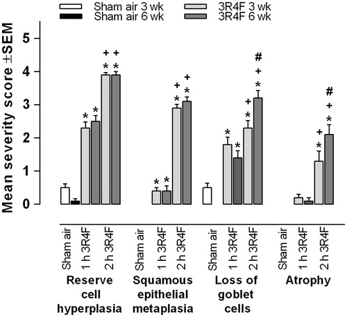
Figure 2. Histopathological responses following 6-week sham air or 3R4F CS exposure (2 h/day). (A–F): 25× magnification, (A′–F′): 200× magnification of sham air exposure group. (A″–F″): 200× magnification of 3R4F CS exposure for 2 h/day. (A″): Atrophy of the nasal olfactory epithelium at dorsal meatus associated with loss of nerve bundles in the lamina propria. (B″): Respiratory epithelium nasal reserve (basal) cell hyperplasia and initial squamous epithelial metaplasia. (C″): Larynx epithelial hyperplasia and cornification at the lower medial region of vocal cords (arytenoid projections). (D″): Reserve (basal) cell hyperplasia of the tracheal epithelium. (E″): Left lung (parenchyma) yellow pigmented and non-pigmented macrophages in the alveolar lumen. (F″): Left lung (main bronchus) bronchial epithelium goblet cell hyperplasia.
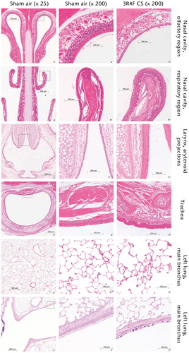
Table 3. Histopathological analysis of the nasal cavity following sham air or 3R4F CS exposure for 3 and 6 weeks.
A dose effect (1 versus 2 h 3R4F CS exposure) was observed for five of six endpoints analyzed at 3 weeks and all endpoints at 6 weeks (). The exception was the level of squamous epithelial metaplasia in the olfactory epithelium of the dorsal meatus: exposure to 3R4F CS for 2 h/day did not induce any significant differences when compared to exposure to 3R4F CS for 1 h/day. For the majority of endpoints, comparison of the 3- and 6-week data showed that continuing exposure for a further 3 weeks (6 weeks total) did not significantly increase the level of histopathological lesions. The loss of respiratory epithelial goblet cells in the nasal septum and atrophy in the olfactory epithelium of the dorsal meatus, following 2 h of 3R4F CS exposure, was the only endpoints where significant increases were observed when comparing the 3- and 6-week data ( and ).
Histopathological evaluation of the larynx
Statistically significant histopathological effects were observed in the larynx following 3 and 6 weeks of 3R4F CS exposure (, ). The exceptions were cornification at the ventral depression (1 h 3R4F CS exposure for 3 and 6 weeks, and 2 h 3R4F CS exposure for 3 weeks) and cornification of lower medial region of the vocal cords (arytenoid projections, 1 h 3R4F CS exposure for 3 weeks). A dose–response effect (1 versus 2 h 3R4F CS exposure) was observed for most of the endpoints examined (); the exceptions were cornification at the ventral depression at the 3-week timepoint, and morphometry at the floor of the larynx at the 3- and 6-week timepoints. For the majority of endpoints, continuing exposure for a further 3 weeks (6 weeks total) did not significantly increase the level of histopathological lesions observed. Statistically significant increases (3 versus 6 weeks) following 1 h of 3R4F CS exposure were detected only in the level of squamous epithelial metaplasia of the ventral depression and floor of the larynx, cornification of the floor of the larynx and morphology of lower medial region. Statistically significant increases (3 versus 6 weeks) following 2 h of 3R4F CS exposure were detected only in the level of squamous epithelial metaplasia of the ventral depression, cornification of the ventral depression and squamous hyperplasia of the lower medial region of the vocal cords (arytenoid projections). Replacement of epithelial hyperplasia with epithelial metaplasia at the base of the epiglottis and at the floor of the larynx was considered an additional epithelial adaptive response to 3R4F CS smoke exposure. This finding was present in all animals exposed to 2 h of 3R4F CS and in a few animals (1–5 rats, depending on the endpoint) exposed to 1 h 3R4F CS/day.
Figure 3. The histopathological response of the larynx following 3- and 6-week sham air or 3R4F CS exposure. Values plotted are mean severity score ± SEM of squamous epithelial metaplasia and cornification (base of epiglottis), epithelial hyperplasia and cornification of lower medial region (LMR) of vocal cords (arytenoid projections). Statistical significance when compared to sham air (*), 1-h 3R4F exposure (+) or 3-week exposure time point (#) are detailed.
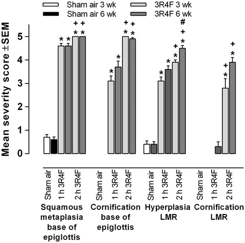
Figure 4. Morphometrical evaluation of the larynx following 3- and 6-week sham air or 3R4F CS exposure. Values are mean severity score ± SEM of mean thickness of epithelium and floor of the larynx, lower medial region (LMR) of vocal cords (arytenoid projections). Statistical significance when compared to sham air (*), 1-h 3R4F exposure (+) or 3-week exposure time point (#) are detailed.
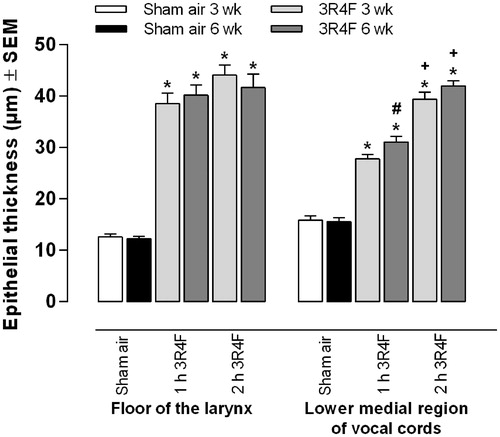
Table 4. Histopathological analysis of the larynx following sham air or 3R4F CS exposure for 3 and 6 weeks.
Histopathological evaluation of the trachea
Statistically significant histopathological effects were observed in the trachea following 3 and 6 weeks of 3R4F CS exposure (, and ). The exceptions were amount of goblet cells and squamous epithelial metaplasia following 1 h of 3R4F CS exposure for 3 and 6 weeks. However, goblet cells were significantly reduced and squamous epithelial metaplasia was detected in animals exposed to 2 h of 3R4F CS for 3 and 6 weeks. Indeed, the level of squamous epithelial metaplasia was significantly increased if 3- and 6-week data were compared. A dose effect (1 versus 2 h) was observed for all endpoints assessed at 3 and 6 weeks.
Figure 5. The histopathological response of the trachea and lung following 3- and 6-week sham air or 3R4F CS exposure. Values plotted are mean severity score ± SEM of the trachea (reserve cell hyperplasia) and left lung lobe (non-pigmented macrophages, yellow pigmented macrophages, bronchial goblet cell hyperplasia). Statistical significance when compared to sham air (*), 1-h 3R4F exposure (+) or 3-week exposure time point (#) are detailed.
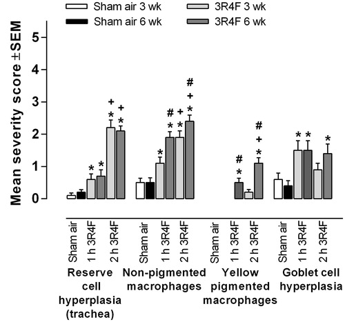
Table 5. Histopathological analysis of the trachea and left lung following sham air or 3R4F CS exposure for 3 and 6 weeks.
Histopathological evaluation of the lung
Statistically significant histopathological effects were observed in the left lung lobe following 3 and 6 weeks of 3R4F CS exposure (, and ). Yellow pigmented macrophages were significantly increased only following 6 weeks of exposure to both 3R4F CS exposure doses (1 or 2 h). Indeed, a dose–response effect (1 versus 2 h) was detected at 6 weeks. Goblet cell hyperplasia was significantly increased following 1 h of 3R4F CS exposure at 3 and 6 weeks, but only following 2 h 3R4F CS exposure at the 6-week timepoint. Non-pigmented macrophages were increased above the level observed in the sham air exposed animals following 1 and 2 h of 3R4F CS exposure at 3 and 6 weeks. A dose–response effect (1 versus 2 h) was observed for non-pigmented macrophages at both timepoints (3 and 6 weeks), and levels were also significantly increased at the 6-week timepoint when compared with the 3-week timepoint. Pigmented macrophage nests were not observed.
Pig-a mutation and micronucleus level following 6 weeks CS exposure
Blood was collected only at the 6-week timepoint from animals exposed to sham air, and 1 and 2 h of 3R4F CS. Values obtained for the Pig-a mutation assay (mean mutant phenotype reticulocyte RETCD59−/106 total reticulocytes [±SD]) were 1.145 [±2.953], 3.130 [±8.839] and 0.968 [±1.677] for exposure to sham air, 1 and 2 h of 3R4F CS, respectively. The values obtained for the level of micronucleus (mean % MN-RET [±SD]) were 0.139 [±0.0456], 0.1485 [±0.064], and 0.184 [±0.076]% for sham air, 1 and 2 h 3R4F CS exposures, respectively. There was no significant difference in the level of RETCD59− (p = 0.566) or % MN-RET (p = 0.078) between the groups.
AEC II viability and purity following 3- or 6-week CS exposure
AEC II isolated from sham air exposed or 3R4F CS exposed rats at both the 3- and 6-week timepoints had >95% viability and average AEC II purity was 60%. These values were comparable to previous studies (Dalrymple et al., Citation2015).
Comet assay in vitro positive controls
Each Comet tank contained both AEC II that were isolated from sham air exposed animals and incubated in vitro with MMS, and NCI-H292 cells that were exposed to 2.0 mM potassium bromate. The level of DNA damage induced by MMS and potassium bromate was 67.28 [± 19.30]% and 53.29 [± 13.79]%, respectively. These values were within historical control values; therefore, all individual experiments were accepted for statistical analysis.
DNA damage as measured by the Alkaline and Modified Alkaline Comet assay following 3- or 6-week CS exposure
DNA damage was statistically significantly increased following 3 and 6 weeks of 3R4F CS exposure. Values obtained for the Alkaline Comet assay (mean % tail DNA [±SD]) were 3.13 [±7.38]%, 10.09 [±14.06]% and 10.56 [±14.36]% for 3-week and 3.19 [±7.29]%, 13.43 [± 14.54]%, and 13.83 [± 16.20]% for 6-week exposure to sham air, 1 h 3R4F, or 2 h 3R4F CS, respectively (). The level of 3R4F CS-induced DNA damage was also statistically significantly increased at 6 weeks when compared to the data obtained at 3 weeks (p < 0.0001). There were no significant differences in the level of DNA damage between the 1 and 2 h 3R4F CS doses at 3 or 6 weeks (p = 0.3306).
Figure 6. The level of DNA damage as measure by the (A) Alkaline Comet assay (single strand breaks) and (B) Modified Alkaline Comet assay (oxidative DNA damage) in isolated AEC II after 3 and 6 weeks exposure to sham air or 3R4F CS for 1 or 2 h/day. Values plotted are mean % DNA tail ± SD. Statistical significance when compared to sham air (*) or 3-week exposure time point (#) are detailed.
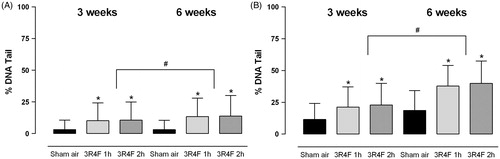
Values obtained for the Modified Alkaline Comet assay (mean % tail DNA [±SD]) were 11.44 [± 12.60]%, 21.20 [± 15.96]% and 22.84 [±16.98]% for 3-week and 18.58 [±15.52]%, 37.75 [± 16.17]% and 39.84 [± 17.56]% for 6-week exposure to sham air, 1 h 3R4F CS, or 2 h 3R4F CS, respectively (). The Modified Alkaline Comet assay also identified that the level of 3R4F CS induced DNA damage was significantly increased at 6 weeks as compared with 3 weeks (p < 0.0001). There were no significant differences in the level of DNA damage between the 1 and 2 h 3R4F CS doses at 3 or 6 weeks (p = 0.0854).
Comparison of basal and MMS induced DNA damage in left and right lung lobes
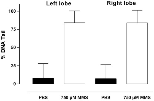
The average viability of isolated AEC II from the left and right lobes was 99.0% and 98.9%, respectively, and AEC II purity was 65.5% and 68.9% from the left and right lobes, respectively. The values obtained were comparable to previous studies (Dalrymple et al., Citation2015). Values obtained for the Alkaline Comet assay (mean % tail DNA [±SD]) for the left lobe was 7.77 [±20.27]% and 84.21 [±16.16]% and right lobe 7.27 [±19.26]% and 83.93 [±17.45]% for PBS or MMS induced DNA damage, respectively, in each lobe ().
Figure 7. The level of basal DNA damage and MMS induced DNA damage as measured by the Alkaline Comet assay in isolated AEC II from the left and right lung lobes. Values plotted are mean % DNA tail ± SD.
Comparison of CS induced DNA damage in left and right lung lobes
The average AEC II viability was 100% regardless of lung lobe or exposure (sham air or 1 h 3R4F CS). The average AEC II purity was 62.5% or 60.7% for the left and right lobes of sham air exposed animals and 64.1% or 66.9% from the left and right lobes of 3R4F CS exposed animals. These values were comparable to previous data (Dalrymple et al., Citation2015). Values obtained for the Alkaline Comet assay (mean % tail DNA [± SD]) for the left lobe was 5.66 [± 16.41]% or 5.90 [± 18.43]% and for the right lobe was 5.89 [± 16.90]% or 5.03 [± 15.92]% for sham air- or 3R4F CS-induced DNA damage, respectively ().
Figure 8. The level of DNA damage as measure by the (A) Alkaline Comet assay (single strand breaks) and (B) Modified Alkaline Comet assay (oxidative DNA damage) in isolated AEC II following 5 days exposure to sham air or 3R4F CS for 1 h/day. Values plotted are mean % DNA tail ± SD. *Statistical significance when compared to sham air exposed corresponding lung lobe.
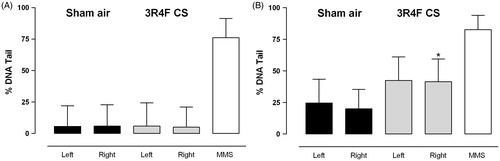
Values for the Modified Alkaline Comet assay (mean % tail DNA [± SD]) for the left lobe were 24.58 [± 18.68]% or 42.37 [± 18.64]% and for the right lobe was 19.94 [± 15.34]% or 41.40 [± 17.90]% for sham air- or 3R4F CS-induced DNA damage, respectively (). The Modified Alkaline Comet assay identified that the level of 3R4F CS induced DNA damage was statistically significantly increased in the right lobe when compared to right lobe of sham air exposed animals (p < 0.05).
Discussion
This study describes a cigarette in vivo inhalation method that has a reduced exposure duration, and details techniques to reduce animal number. A technique to enable separate lung lobes from one animal to be used for histopathology or the Comet assay and the suitability of DNA damage, Pig-a mutation or in vivo mutation assays to measure 3R4F CS induced genotoxicity are detailed.
The inhalation method used in this study is based on the OECD 413 Guideline (OECD, Citation2009), a rodent nose-only sub-chronic inhalation toxicity protocol that has been used previously, with minor modifications, for the testing of cigarettes (Gaworski et al., Citation2009; Schramke et al., Citation2014). Numerous studies have detailed the effect of repeated CS exposure on body weight (Ayres et al., Citation2001; Baker et al., Citation2004; Fujimoto et al., Citation2015; Potts et al., Citation2007; Schramke et al., Citation2014). In this study, only male animal body weight was reduced following 6 weeks of exposure to 3R4F CS for 2 h/day. A reduction in percentage body weight gain was also observed only in male rats; however, differences were noted at both timepoints and both 3R4F CS doses. These data are comparable to studies where animals were exposed to CS for 90 days (Ayres et al., Citation2001; Baker et al., Citation2004; Fujimoto et al. Citation2015; Potts et al., Citation2007; Schramke et al., Citation2014).
In addition to body weight reduction, CS smoke exposure induces a number of characteristic histopathological respiratory tract lesions in the rat. Tissue responses are not permanent: respiratory tract lesions can be regressed or reduced if a recovery phase is included in the smoke inhalation protocol (Ayres et al., Citation2001; Fujimoto et al., Citation2015; Potts et al., Citation2007). Many CS exposure studies in the rat have detailed that histopathological responses do not occur in non-respiratory tract organs (Baker et al., Citation2004; Fujimoto et al., Citation2015; Kogel et al., Citation2014). An exception, however, is a slight increase in thymus atrophy observed in CS-exposed animals (Piade et al., Citation2014; Roemer et al., Citation2014), which is postulated to occur due to smoke irritation and stress induced by restraint during exposure (Gruver & Sempowski, Citation2008). Indeed, those two studies used 6, rather than 1, h of exposure/day, which might have increased the level of stress. In the present study, therefore, histopathology was determined only in respiratory tract organs. In agreement with our data, a recent 35-day tobacco testing study also restricted tissue analysis to respiratory tract organs and heart (Fujimoto et al., Citation2015).
In the present study, histomorphological alterations regardless of the respiratory tract organ analyzed, exposure dose (1 or 2 h 3R4F CS) or length of exposure (3 or 6 weeks) were similar in male and female animals. The responses observed were comparable to other cigarette testing studies that used 3R4F (Fujimoto et al., Citation2015) or 2R4F (Schramke et al., Citation2014) reference cigarettes. A smoke concentration-dependent response was observed for the majority of histopathological endpoints assessed; as expected, responses were significantly higher in animals exposed to 3R4F CS for 2 h/day. The exposure period (3 or 6 weeks) did not appear to affect the severity of the histopathological endpoints analyzed in the nasal cavity or larynx, where the responses observed were comparable. This study also details the use of a preferred tissue sectioning level (Walker, Citation1983) before the first palatine ruga at the incisive papilla, anterior to the level 2 (Young, Citation1981), which is routinely used for cigarette testing. This area of the nasal cavity was selected as it contains the three different types of epithelium (stratified squamous, respiratory and olfactory) that respond to repeated CS exposure (Walker, Citation1983). Although the OECD 413 protocol specifies four tissue levels, the histopathological information obtained from this study suggests that one tissue level is sufficient for cigarette testing. The histopathological responses of the larynx to CS exposure have been extensively studied; indeed, the distal base of the epiglottis is a site of increased sensitivity to inhaled particles (Renne & Gideon, Citation2006; Renne et al., Citation1993). The OECD 413 protocol specifies three larynx tissue levels; two were used in the present study including the base of the epiglottis. For some of the larynx endpoints assessed, a dynamic range of severity scoring was observed, whereas for others, a mean severity score of 5 (saturation) was obtained; these observations demonstrate the sensitivity of this tissue to CS and also that the two tissue levels selected were appropriate for cigarette testing.
Following 1 h of 3R4F CS exposure/day for 3 weeks, minimal or no response was observed in some trachea and lung endpoints; however, responses were observed following 2 h of 3R4F CS exposure per day. The minimal responses obtained in the trachea are possibly due to the reduced exposure duration used in this study, or the transverse tissue level evaluated. Goblet cell quantification is commonly evaluated using a horizontal trachea level at the level of bifurcation (OECD, Citation2009). However, histopathological lesions in the trachea are minimal even after 90 days of exposure (Fujimoto et al., Citation2015; Piade et al., Citation2014). In the lung, yellow pigmented macrophages were observed only following 6 weeks of exposure and pigmented macrophage nests were not observed, again possibly due to the reduced exposure time. Other 90-day CS inhalation studies have reported the formation of pigmented macrophage nests (Ayres et al., Citation2001; Fujimoto et al., Citation2015; Gaworski et al., Citation2009; Walker et al., Citation1978a) and associated incipient alveolar hyperplasia (Walker et al., Citation1978a), which suggests that macrophage accumulation in the lung is proportional to CS dose and days of exposure.
The OECD 413 Guideline (OECD, Citation2009) states that test articles should be tested for 90 days; however, CS induced lesions can be identified within the rat respiratory tract prior to 90 days (Fujimoto et al., Citation2015; Kogel et al., Citation2014; Piade et al., Citation2014; Walker et al., Citation1978a,b). In a recent cigarette testing study using 35 and 90 days of CS exposure (Piade et al., Citation2014), the authors concluded that the histopathology observed after 35 days was in line with data recorded after 90 days. Another study also used 35 days of exposure (Fujimoto et al., Citation2015), and histopathology was observed in all respiratory tract organs. The pathology data from the present study were consistent with these recent publications; we therefore propose that 3 weeks of inhalation, with 2 h CS exposure/day is sufficient to investigate CS responses in the rat respiratory tract. If 1 h/day exposure is to be used, the dose of the WTPM/L should be increased. Indeed, a recent tobacco testing study exposed animals to 1000 μg of 3R4F CS WTPM/L for 2 h/day (Fujimoto et al., Citation2015), a higher dose than was used in the present study.
With the advance of molecular techniques, there is a drive to investigate additional endpoints as part of a rat CS inhalation study. The International Conference on Harmonization guidelines (ICH, 2011) and UK Committee for Mutagenicity test guidelines (UK Committee on Mutagenicity, Citation2011) have recommended that an ex vivo study undertaken at the same time as in vivo work is preferable to a second in vitro study in mammalian cells. The present study investigated the suitability of three additional endpoints to measure CS induced genotoxicity: DNA damage as measured by the Comet assay, the level of Pig-a mutation and the level of in vivo micronuclei. The feasibility/scale-up required to include a Comet endpoint into a regulatory standard inhalation protocol such as OECD 413 (OECD, Citation2009) was also determined. Our recent publication details a method for AEC II isolation (Dalrymple et al., Citation2015) and also the Alkaline Comet assay, which measures DNA single-strand breaks induced by direct acting genotoxic compounds; and the Modified Alkaline Comet assay, which measures indirect DNA damage such as oxidative damage, apurinic/pyrimidinic sites or DNA repair (Smith et al., Citation2006). That study confirmed that CS induced DNA damage can be measured by the Comet assay in isolated rat lung AEC II after single or repeated (5 days) CS exposure and also highlighted the importance of sample handling/processing times to reduce DNA repair (Dalrymple et al., Citation2015).
In the current study, the time for AEC II isolation was set at 45 ± 3 min to limit DNA repair. This follow-up study aimed to determine whether the Comet assay can measure DNA damage following 3 and 6 weeks of 3R4F CS exposure, alongside histopathological analysis. Increased levels of DNA damage were measured following both 3 and 6 weeks of 3R4F CS exposure. The positive control data from each independent Comet experiment, collected over numerous days, were within assay acceptance criteria and did not affect the data, confirming that the Comet assay is a suitable method to measure CS induced DNA in an inhalation study with animal numbers as recommended by OECD 413 (OECD, Citation2009). The Comet data obtained did not concur with histopathological findings: a dose–response was not observed when the 1 and 2 h 3R4F CS exposure data were compared at either timepoint (3 or 6 weeks). Studies have demonstrated that DNA repair mechanisms are rapidly activated in the lung following CS exposure (Dalrymple et al., Citation2015; Tsuda et al., Citation2000). In this study, 2 h 3R4F CS exposure/day was achieved by the following exposure protocol: 1 h 3R4F CS, followed by 1 h fresh air, and then a further 1 h exposure to 3R4F CS. DNA repair mechanisms were possibly activated during the 1 h break and therefore no differences were observed. Interestingly, when the 3-week DNA damage data were compared with the 6-week data, a significant difference was observed. Taken together, these data suggest that DNA repair does occur in the rat lung following CS exposure, but some DNA damage remains and will accumulate in the lung if CS exposure continues.
There is growing interest in Pig-a mutation and in vivo micronucleus assays to determine in vivo genotoxicity (Gollapudi et al., Citation2015; Stankowski et al., Citation2015). The OECD Guideline 474 has also been recently published for the in vivo micronucleus assay (OECD, Citation2014). In the present study, Pig-a gene mutation and in vivo micronucleus levels were not significantly increased following 6 weeks of CS exposure. Others have investigated the in vivo micronucleus assay as part of a rat cigarette inhalation study and have also demonstrated that the level of micronuclei is not increased following CS exposure in peripheral blood or bone marrow samples (Schramke et al., Citation2014; Van-Miert et al., Citation2008). Overall, the present data suggest that at the timepoints and CS doses assessed, CS did not exert any obvious effects on erythropoiesis in the bone marrow. This concurs with the hypothesis that CS does not exert secondary responses in non-respiratory tract tissues in the rat.
CS induced pathology, including 3R4F CS, has been studied for a number of years in the rat lung. We have observed that CS induced pathology is comparable between the two lung lobes (unpublished data). Prior to initiating the CS inhalation study, the level of basal, MMS-induced and 3R4F CS-induced DNA damage was compared between matched right and left lung lobes. The data obtained, in agreement with historical pathology data, confirmed that left and right lobes respond to CS exposure in a comparable manner and justify selecting one lobe for the Comet assay and the other for pathology. Splitting of lung lobes to increase the knowledge obtained from a rat inhalation study has been used in other CS inhalation studies. Histopathology and proteomics have been studied in the right and left lung lobes of Wistar rats (Zhang et al., Citation2008) and bronchoalveolar lavage and histopathology in the right and left lung lobes of Sprague Dawley rats (Kogel et al., Citation2014). The splitting of lung lobes has the potential to reduce animal use significantly. The approach used in this study is aligned to the 3Rs, whereas others have increased animal numbers and included an OECD plus group to expand the information collected from the OECD 413 protocol (Kogel et al., Citation2014; Phillips et al., Citation2015).
Currently, no in vitro models can provide all of the information that an OECD 413 (OECD, Citation2009) CS inhalation study can deliver. There is a wealth of evidence to imply that CS exposure in the rat results in pathology only within the respiratory tract, implying that a surrogate 3D tissue model might be a suitable replacement for in vivo testing. In a drive to reduce and replace the use of animals, there is interest in characterizing 3D models of the lung such as MucilAir™ or EpiAirway™ for use in the testing of CS, plus new products such as e-cigarettes and products that heat tobacco (Baxter et al., Citation2015; Kuehn et al., Citation2015; Neilson et al., Citation2015). Indeed, studies have shown that 3D tissue cultures have gene expression profiles similar to human bronchial cells (Dvorak et al., Citation2011) and are comparable to cells of the respiratory tract (Baxter et al., Citation2015; Mathis et al., Citation2013). Understanding lung 3D tissue cultures further, and comparing such studies with data from an OECD 413 (OECD, Citation2009) CS inhalation study will confirm whether 3D tissue cultures of the lung are a suitable surrogate endpoint for animal inhalation studies.
Numerous studies have identified that cigarette use results in disease in adult smokers (Aoshiba & Nagai, Citation2003; DeMarini, Citation2004; Institute of Medicine, Citation2001); however, the specific mechanisms or pathways that are activated prior to the onset of disease remain unknown. Recent studies have identified the activation of common gene expression signatures in the mouse and human lung following CS exposure (Morissette et al., Citation2014). Understanding fully the gene and protein pathways that are activated by CS might assist in the risk assessment of new products such as e-cigarettes and products that heat tobacco. New tobacco or nicotine products that do not induce these pathways or that activate them at a lower level could be an alternative for people who continue to use tobacco products rather than quit. In addition, if the gene signatures and/or disease pathways that are regulated by CS in the lung are also regulated in vitro in 3D tissue cultures of the lung following CS exposure, it will further confirm the suitability of 3D tissue cultures to replace in vivo tobacco product testing. The present study and others have identified that CS can induce DNA damage in the mouse and rat lung (Dalrymple et al., Citation2015; Tsuda et al., Citation2000) and in cell lines in vitro (Thorne et al., Citation2009; Weber et al., Citation2013); however, it is not known whether CS induced DNA damage can be measured by the Comet assay in a lung 3D tissue culture.
This study was initiated to refine existing in vivo tobacco testing protocols, to align to the 3Rs and to investigate the use of DNA damage, Pig-a mutation and in vivo micronucleus assays as potential endpoints to measure CS induced genotoxicity. It has demonstrated that 3 weeks is a suitable timepoint to investigate pathology induced by CS in the rat respiratory tract and that separate lung lobes can be successfully used to investigate histopathology and the Comet assay concurrently. Pig-a mutation or micronucleus levels were not increased by CS exposure; however, the Comet assay was found to be a suitable method to measure CS induced DNA damage in isolated lung cells collected as part of a regulatory standard inhalation protocol. The method described herein supports a strategy to reduce and refine animal use in tobacco product testing and is aligned to the 3Rs (replacement, reduction or refinement).
Author’s contributions
Annette Dalrymple, Patricia Ordoñez, David Thorne, David Walker, Debbie Dillon and Clive Meredith designed the study. Patricia Ordoñez managed all experimental work at Vivotecnia. Annette Dalrymple drafted the manuscript and prepared the figures. Oscar Camacho analyzed the Comet, Pig-a mutation and micronucleus data. Ansgar Büttner analyzed the histopathology data and prepared the corresponding tables and figures. All authors approved the final manuscript.
Acknowledgements
The authors acknowledge the technical contribution of Ana Belén Sierra, Arantxa Saiz Bautista, Miguel López Angulo and Hernando Servello Ferreiro (AEC II isolation and COMET analysis), Jesús Illán Manero, Elena Fernández Alargunsoro, Ruth Micha Gavilán (inhalation studies), Javier Rivera Trillo, Sara Anca Óvilo, Tamara Domínguez Guerrero and Carlos Martín Llanos (necropsy). We thank Dorothea Torous at Litron Laboratories for support with sample collection, Pig-a mutation and micronucleus analysis data, and review of the manuscript. We would also like to thank Ian Crooks, Marianna Gaḉa and Marc Princivalle for review of the manuscript and Jason Adamson for support with .
Declaration of interest
The authors report no declarations of interest, and are employees of British American Tobacco, were employees of British American Tobacco at the time of study initiation or were contracted by British American Tobacco.
References
- Aoshiba K, Nagai A. (2003). Oxidative stress, cell death, and other damage to alveolar epithelial cells induced by cigarette smoke. Tob Induc Dis 1:219–26
- Ayres PH, Hayes JR, Higuchi MA, et al. (2001). Subchronic inhalation by rats of mainstream smoke from a cigarette that primarily heats tobacco compared to a cigarette that burns tobacco. Inhal Toxicol 13:149–86
- Baker RR, Massey ED, Smith G. (2004). An overview of the effects of tobacco ingredients on smoke chemistry and toxicity. Food Chem Toxicol 42:S53–83
- Baxter A, Thain S, Banerjee A, et al. (2015). Targeted omics analyses, and metabolic enzyme activity assays demonstrate maintenance of key muco-ciliary characteristics in long-term cultures of reconstituted human airway epithelia. Toxicol In Vitro 29:864–75
- Boletín Oficial Del Estado. (2013). Por el que se establecen las normas básicas aplicables para la protección de los animales utilizados en experimentación y otros fines científicos, incluyendo la docencia. Available from: https://www.boe.es/diario_boe/txt.php?id=BOE-A-2013-1337. [Last accessed: 27 Apr 2016]
- Bright J, Aylott M, Bate S, et al. (2011). Recommendations on the statistical analysis of the Comet assay. Pharm Stat 10:485–93
- Dalrymple A, Ordonez P, Thorne D, et al. (2015). An improved method for the isolation of rat alveolar type II lung cells: use in the Comet assay to determine DNA damage induced by cigarette smoke. Regul Toxicol Pharmacol 72:141–9
- DeMarini DM. (2004). Genotoxicity of tobacco smoke and tobacco smoke condensate: a review. Mutat Res 567:447–74
- Dertinger SD, Camphausen K, MacGregor JT, et al. (2004). Three-color labeling method for flow cytometric measurement of cytogenetic damage in rodent and human blood. Environ Mol Mutagen 44:427–35
- Dertinger SD, Phonethepswath S, Avlasevich SL, et al. (2012). Efficient monitoring of in vivo pig-a gene mutation and chromosomal damage: summary of 7 published studies and results from 11 new reference compounds. Toxicol Sci 130:328–48
- Dvorak A, Tilley AE, Shaykhiev R, et al. (2011). Do airway epithelium air–liquid cultures represent the in vivo airway epithelium transcriptome? Am J Respir Cell Mol Biol 44:465–73
- European Union. (2010). Directive 2010/63/EU Of The European Parliament and of the Council on the protection of animals used for scientific purposes. Available from: http://ec.europa.eu/environment/chemicals/lab_animals/legislation_en.htm. [Last accessed: 27 Apr 2016]
- Fujimoto H, Tsuji H, Okubo C, et al. (2015). Biological responses in rats exposed to mainstream smoke from a heated cigarette compared to a conventional reference cigarette. Inhal Toxicol 27:224–36
- Gaworski CL, Schramke H, Diekmann J, et al. (2009). Effect of filtration by activated charcoal on the toxicological activity of cigarette mainstream smoke from experimental cigarettes. Inhal Toxicol 21:688–704
- Gollapudi BB, Lynch AM, Heflich RH, et al. (2015). The in vivo Pig-a assay: a report of the International Workshop On Genotoxicity Testing (IWGT) Workgroup. Mutat Res Genet Toxicol Environ Mutagen 783:23–35
- Gruver AL, Sempowski GD. (2008). Cytokines, leptin, and stress-induced thymic atrophy. J Leukoc Biol 84:915–23
- ICH (International Conference on Harmonization) (2011). Guidance on genotoxicity testing and data interpretation for pharmaceuticals intended for human use S2(R1). International Conference On Harmonization
- Institute of Medicine (2001). Clearing the smoke: the science base for tobacco harm reduction. Washington, DC: National Academic Press
- International Organization for Standardization (1999). ISO 3402:1999 – tobacco and tobacco products – atmosphere for conditioning and testing. Available from: http://www.iso.org/iso/catalogue_detail.htm?csnumber=28324. [Last accessed: 27 Apr 2016]
- International Organization for Standardization (2000a). ISO 3308:2000 – routine analytical cigarette-smoking machine: definitions and standard conditions. Available from: http://www.iso.org/iso/iso_catalogue/catalogue_ics/catalogue_detail_ics.htm?csnumber=28325. [Last accessed: 27 Apr 2016]
- International Organization for Standardization (2000b). ISO 4387:2000 – cigarettes: determination of total and nicotine-free dry particulate matter using a routine analytical smoking machine. Available from: http://www.iso.org/iso/iso_catalogue/catalogue_tc/catalogue_detail.htm?csnumber=28323. [Last accessed: 27 Apr 2016]
- Kamp DW, Greenberger MJ, Sbalchierro JS, et al. (1998). Cigarette smoke augments asbestos-induced alveolar epithelial cell injury: role of free radicals. Free Radic Biol Med 25:728–39
- Kogel U, Schlage WK, Martin F, et al. (2014). A 28-day rat inhalation study with an integrated molecular toxicology endpoint demonstrates reduced exposure effects for a prototypic modified risk tobacco product compared with conventional cigarettes. Food Chem Toxicol 68:204–17
- Kuehn D, Majeed S, Guedj E, et al. (2015). Impact assessment of repeated exposure of organotypic 3D bronchial and nasal tissue culture models to whole cigarette smoke. J Vis Exp 12:96
- Labash C, Avlasevich S, Carlson K, et al. (2016). Mouse Pig-a and micronucleus assays respond to N-ethyl-N-nitrosourea, benzo[a]pyrene, and ethyl carbamate, but not pyrene or methyl carbamate. Environ Mol Mutagen 57:28–40
- Mathis C, Poussin C, Weisensee D, et al. (2013). Human bronchial epithelial cells exposed in vitro to cigarette smoke at the air–liquid interface resemble bronchial epithelium from human smokers. Am J Physiol 304:L489–503
- Morissette MC, Lamontagne M, Berube JC, et al. (2014). Impact of cigarette smoke on the human and mouse lungs: a gene-expression comparison study. PLoS One 9:e92498
- Neilson L, Mankus C, Thorne D, et al. (2015). Development of an in vitro cytotoxicity model for aerosol exposure using 3D reconstructed human airway tissue; application for assessment of e-cigarette aerosol. Toxicol In Vitro 29:1952–62
- OECD (2009). Test guideline 413: subchronic inhalation toxicity: 90-day study. France: OECD Publishing. Available from: http://www.oecd-ilibrary.org/environment/test-no-413-subchronic-inhalation-toxicity-90-day-study_9789264070806-en. [Last accessed: 27 Apr 2016]
- OECD (2014). Test guideline 474: mammalian erythrocyte micronucleus test. France: OECD Publishing
- Phillips B, Esposito M, Verbeeck J, et al. (2015). Toxicity of aerosols of nicotine and pyruvic acid (separate and combined) in Sprague-Dawley rats in a 28-day OECD 412 inhalation study and assessment of systems toxicology. Inhal Toxicol 27:405–31
- Piade JJ, Roemer E, Dempsey R, et al. (2014). Toxicological assessment of kretek cigarettes. Part 3: kretek and American-blended cigarettes, inhalation toxicity. Regul Toxicol Pharmacol 70:S26–40
- Potts RJ, Meckley DR, Shreve WK, et al. (2007). Comparative 13-week inhalation study of cigarette smoke from cigarettes containing cast sheet tobacco. Inhal Toxicol 19:701–24
- Renne RA, Gideon KM. (2006). Types and patterns of response in the larynx following inhalation. Toxicol Pathol 34:281–5
- Renne RA, Gideon KM, Miller RA, et al. (1992). Histologic methods and interspecies variations in the laryngeal histology of F344/N rats and B6C3F1 mice. Toxicol Pathol 20:44–51
- Renne RA, Sagartz JW, Burger GT. (1993). Interspecies variations in the histology of toxicologically-important areas in the larynges of CRL:CD rats and Syrian golden hamsters. Toxicol Pathol 21:542–6
- Rodgman A, Perfetti TA. 2013. The chemical components of tobacco and tobacco smoke. 2nd ed. Boca Raton, FL: CRC Press
- Roemer E, Schramke H, Weiler H, et al. (2014). Mainstream smoke chemistry and in vitro and in vivo toxicity of the reference cigarettes 3R4F and 2R4F. Contrib Tobacco Res 25:316–35
- Schramke H, Roemer E, Dempsey R, et al. (2014). Toxicological assessment of kretek cigarettes. Part 7: the impact of ingredients added to kretek cigarettes on inhalation toxicity. Regul Toxicol Pharmacol 70:S81–9
- Smith CC, O’Donovan MR, Martin EA. (2006). hOGG1 recognizes oxidative damage using the comet assay with greater specificity than FPG or ENDOIII. Mutagenesis 21:185–90
- Stankowski LF Jr, Aardema MJ, Lawlor TE, et al. (2015). Integration of Pig-a, micronucleus, chromosome aberration and Comet assay endpoints in a 28-day rodent toxicity study with urethane. Mutagenesis 30:335–42
- Talikka M, Kostadinova R, Xiang Y, et al. (2014). The response of human nasal and bronchial organotypic tissue cultures to repeated whole cigarette smoke exposure. Int J Toxicol 33:506–17
- Thorne D, Wilson J, Kumaravel TS, et al. (2009). Measurement of oxidative DNA damage induced by mainstream cigarette smoke in cultured NCI-H292 human pulmonary carcinoma cells. Mutat Res 673:3–8
- Tsuda S, Matsusaka N, Ueno S, et al. (2000). The influence of antioxidants on cigarette smoke-induced DNA single-strand breaks in mouse organs: a preliminary study with the alkaline single cell gel electrophoresis assay. Toxicol Sci 54:104–9
- UK Committee on Mutagenicity (2011). Guidance on a strategy for genotoxicity testing of chemical substances. United Kingdom: UK Committee on Mutagenicity. Available from: https://www.gov.uk/government/uploads/system/uploads/attachment_data/file/315800/in_vivo_testing_of_genotoxicity_of_chemicals.pdf. [Last accessed: 27 Apr 2016]
- Van-Miert E, Vanscheeuwijck P, Meurrens K, et al. (2008). Evaluation of the micronucleus assay in bone marrow and peripheral blood of rats for the determination of cigarette mainstream-smoke activity. Mutat Res 652:131–8
- Walker D. (1983). Histopathology of the nasal cavity in laboratory animals exposed to cigarette smoke and other irritants. In: Reznik G, Stinson SF. (eds.) Nasal tumors in animals and man. Boca Raton, FL: CRC Press, 115–35
- Walker D, Wilton LV, Binns R. (1978a). Inhalation toxicity studies on cigarette smoke (VI). 6-Week comparative experiments using modified flue-cured cigarettes: histopathology of the lung. Toxicology 10:229–40
- Walker D, Wilton LV, Binns R. (1978b). Inhalation toxicity studies on cigarette smoke (VII). 6-Week comparative experiments using modified flue-cured cigarettes: histopathology of the conducting airways. Toxicology 10:241–59
- Weber S, Hebestreit M, Wilms T, et al. (2013). Comet assay and air–liquid interface exposure system: a new combination to evaluate genotoxic effects of cigarette whole smoke in human lung cell lines. Toxicol In Vitro 27:1987–91
- Werley MS, Jerome AM, DeSoi DJ, et al. (2013). A comprehensive evaluation of the toxicology of experimental cigarettes manufactured with banded papers. Inhal. Toxicol 25:19–33
- Young JT. (1981). Histopathologic examination of the rat nasal cavity. Fundam Appl Toxicol 1:309–12
- Zhang S, Xu N, Nie J, et al. (2008). Proteomic alteration in lung tissue of rats exposed to cigarette smoke. Toxicol Lett 178:191–6