Abstract
Background: The authors describe a case of unilateral idiopathic acute frosted branch angiitis with premacular hemorrhage.
Case: A 35-year-old woman was referred because of acute vision loss in her left eye during the puerperal period. Her initial visual acuity was 20/20 OD and 20/200 OS. The left eye presented severe sheathing of retinal vessels inferiorly, heavy perivascular intraretinal hemorrhages, and premacular hemorrhage. There was no evidence of vascular leakage in fluorescein angiography. All of the laboratory workup was negative for frosted branch angiitis (FBA). During the follow-up period, FBA resolved spontaneously within a few days, but the amount of premacular hemorrhage increased. Vitrectomy with internal limiting membrane peeling was performed at the third month, resulting in 20/25 vision and no recurrence of the disease during the 13-month follow-up.
Conclusion: This is an idiopathic case of acute FBA that exhibited spontaneous rapid regression of angiitis but was complicated by an unusual premacular hemorrhage.
Frosted branch angiitis (FBA) was first described in 1976 by Ito et al.Citation1 as “bilateral acute uveitis with severe sheathing along all of the retinal vessels.” Since then, FBA has been reported mainly in Japan and occasionally in other countries.Citation2 Here we report a case of idiopathic unilateral FBA with premacular hemorrhage.
CASE REPORT
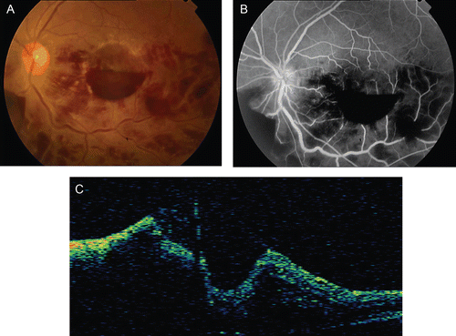
A 35-year-old woman was referred to our hospital with sudden vision loss in her left eye for the last 2 days. Her systemic and ocular history were unremarkable except for giving birth 20 days prior by caesarean section. On her initial examination, her visual acuity was 20/20 OD and 20/200 OS. The intraocular pressures were within normal limits, and there was no abnormal finding in the anterior segment of either eye. The fundus examination was normal OD, but abnormal “frosted” white sheathing was observed around retinal veins from the posterior pole to the inferior periphery with scattered intraretinal hemorrhages. Subhyaloid hemorrhage in premacular and inferonasal areas were found in her left eye (). Some ancillary tests were ordered, and fluorescein angiography (FA) was performed 2 days later. By this time, the appearance of the fundus was considerably altered: the extent of retinal hemorrhage and perivessel sheathing had decreased considerably, the premacular hemorrhage was gravitated inferiorly, and new hard exudates had developed around the macula region (). FA was associated with hemorrhagic blockade and minimal dilation of the inferior retinal veins without any evidence of vascular leakage (). Optical coherence tomography (OCT) revealed the irregular contour of the fovea, which was caused by extensive premacular hemorrhage (). The following parameters were all within normal limits: complete blood cell count, erythrocyte sedimentation rate, C-reactive protein, blood sugar, protein, urea, urine analysis, antinuclear antibody, fluorescent treponemal antibody test, chest X-ray, angiotensin-converting enzyme, serum electrolytes, Mantoux test, and enzyme-linked immunosorbent assay for tuberculosis, human immunodeficiency virus, cytomegalovirus, and herpes zoster and simplex viruses. The patient was evaluated for symptoms of Behçet disease and was considered not to have Behçet disease.
FIGURE 1 Fundus photography of the left eye at the beginning. Note the typical frosted branch angiitis appearance and premacular hemorrhage.
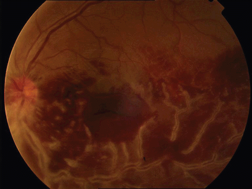
FIGURE 2 (a) Retinal hemorrhages and sheathing were significantly reduced and premacular hemorrhages were gravitated inferiorly within 2 days. (b) Note the absence of vascular leakage in fluorescein angiography. (c) Note irregular contour of fovea caused by extensive premacular hemorrhage.
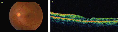
Dramatic spontaneous clinical improvement within the first few days and the breastfeeding status of the patient compelled us to observe her without administering any immunosuppressive therapy. One month later, although the intraretinal hemorrhages were significantly reduced in size, the amount of subhyaloid hemorrhages (premacular and inferonasal) was unchanged or increased. There were new hard exudates in the perifoveal region (). Visual acuity was unchanged (20/200) at that point. The inferonasal subhyaloid hemorrhage had opened into the vitreous cavity, the premacular hemorrhage had decreased in size, and the exudates had resolved spontaneously with a visual acuity improvement to 20/100. During the third month of the disease progression, 23G pars plana vitrectomy with ILM peeling was performed for the persistent premacular hemorrhage, which was considered to be partially intraretinal during the surgery. Visual acuity increased to 20/25 1 month after the surgery and to 20/20 1 year after surgery without any signs of disease recurrence (). OCT images obtained at 1 year after surgery revealed a normal foveal contour ().
FIGURE 3 Fundus picture of the left eye 1 month later. Note new perifoveal hard exudates and subhyaloid hemorrhages in inferonasal and premacular areas.
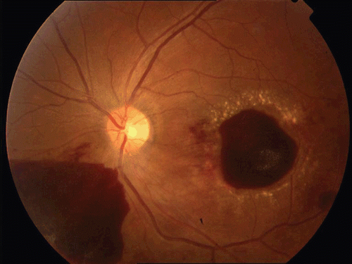
FIGURE 4 (a) Fundus picture of the left eye 1 year postoperatively. (b) OCT of the left eye 1 year postoperatively.
DISCUSSION
The main features of FBA as originally reported by Ito et al.Citation1can be summarized as follows: (1) bilateral acute visual disturbance in a healthy individual; (2) the entire retina is swollen and there is severe sheathing of the retinal vessels, so that they look like frosted branches of a tree, especially at the periphery; (3) fundus FA also shows a normal pattern in the first stage, but leakage of the dye from vessels is seen in later stages; in particular, the sheathed vessels show no sign of occlusion or stasis; (4) visual fields show a marked concentric constriction during the acute stages of the disease but improve with associated reductions in swelling and inflammation; (5) electroretinograms are markedly abnormal; (6) no systemic abnormality was found; (7) high doses of systemic corticosteroids are effective in treating the disorder, and the visual acuity recovers to almost a normal level; (8) the disease does not recur.Citation2
In addition to the primary (idiopathic) cases described originally, FBA may be seen in association with some systemic disorders.Citation2,Citation3 FBA may therefore be a clinical sign that is associated with inflammatory processes rather than a clinical syndrome.Citation3 FBA is observed in viral infections (CMV, HSV, VZV, EBV, or rubella), malignant infiltrations (leukemia, lymphoma), or autoimmune diseases (systemic lupus erythematosus, Crohn disease, Behçet disease).Citation2 The cause of FBA is unknown. The typical onset of FBA after a multifactorial prodromal illness has led to the suggestion of a hypersensitivity reaction to various infective agents, which may initiate FBA via a common pathway that possibly involve immune-complex deposition.Citation2 This characteristic would support the use of systemic corticosteroids for those with substantial visual loss.4 Most patients with acute idiopathic FBA have been treated with systemic corticosteroids with good, rapid visual recovery within weeks.Citation2 There are, however, case reports of patients who recovered without any treatment.4 There is no evidence-based guidance for the treatment of FBA supporting the use of systemic steroids. FBA could even be self-limiting as in our case.
Subhyaloid and vitreous hemorrhages associated with FBA have been reported previously by Agrawal et al.Citation5 We postulate that increased vascular permeability may lead to premacular hemorrhage as well as hard exudates during the course of the disease. Hard exudates often appear in the macula but can be seen elsewhere during the absorption of retinal edema. However, to the best of our knowledge, this is the first case in the literature of a premacular hemorrhage that required vitreoretinal surgery to be cleared.
In conclusion, FBA, a very dramatic clinical picture, may have a favorable prognosis even without treatment.4 However, some cases can be complicated by vitreous hemorrhage, macular epiretinal membrane formation, macular scarring, diffuse retinal fibrosis, and retinal tears.Citation5 Premacular hemorrhage is also a rare and vision-threatening complication, sometimes necessitating surgical intervention.
Declaration of interest: The authors report no conflicts of interest. The authors alone are responsible for the content and writing of the paper.
REFERENCES
- Ito Y, Nakano M, Kyu N., Takeuchi M. Frosted-branch angiitis in a child. Jpn J Clin Ophthalmol. 1976; 30: 797–803.
- Walker S, Iguchi A, Jones NP. Frosted branch angiitis: a review. Eye. 2004;18:524–533.
- Kleiner RC. Frosted branch angiitis: clinical syndrome or clinical sign? Retina. 1997;17:370–371.
- Vander JF, Masciulli L. Unilateral frosted branch angiitis. Am J Ophthalmol. 1991;112:477–478.
- Agrawal S, Agrawal J, Agrawal TP. Unilateral frosted branch angiitis with vitreous haemorrhage. Indian J Ophthalmol. 2001;49(4):269–270.