Abstract
Purpose: To report a case of central serous chorioretinopathy (CSC) as a possible complication of latanoprost treatment.
Methods: A 65-year-old woman presented with a 1-week history of blurred vision and metamorphopsia in her right eye. She was previously diagnosed with unilateral glaucoma, and treatment was initiated with topical latanoprost 0.005% for the right eye. The symptoms occurred 1 month after initiation of glaucoma treatment.
Results: Visual acuity (VA) of the patient had reduced to 20/50 in right eye. Fluorescein angiography revealed a focal RPE leak near to the fovea with neurosensory detachment due to CSC, which was confirmed by optical coherence tomography. After cessation of the therapy, her clinical and morphological status improved spontaneously. The neurosensory detachment resolved almost completely and VA improved to 20/20 simultaneously.
Conclusions: The authors report the first case of CSC associated with latonoprost therapy. This case demonstrates that topical latanoprost may lead to the development of CSC.
Central serous chorioretinopathy (CSC) is characterized by an idiopathic serous detachment of the neurosensory retina at the macula resulting from altered barrier function and deficient pumping function at the level of the retinal pigment epithelium (RPE).Citation1,Citation2 It is usually associated with corticosteroid use, type A personality, organ transplantation, pregnancy, hypertension, and psychopharmacologic medication. The primary pathology of CSC has been altered from the originally proposed level at the retinal pigment epithelium (RPE) to the underlying abnormal choroidal vasculature and blood flow disturbance.Citation2
The once-daily administration of latanoprost (prostaglandin F2α analogue) in patients with glaucoma effectively reduces the intraocular pressure (IOP) and is well tolerated. The reported ocular complications with latanoprost include conjunctival hyperemia, iris discoloration, anterior uveitis and cystoid macular edema (CME) iris cysts, and reactivation of herpes simplex keratitis.Citation3 Although a few studies showing an association between serous macular detachment and CME with latanoprost have been published, there are no previous reports that indicate the possible relationship between latanoprost treatment and CSC. To the best of our knowledge, this is the first case of a patient who developed CSC as a possible complication of latanoprost treatment.
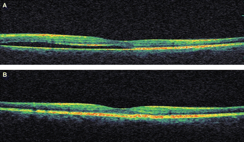
A 65-year-old woman recently diagnosed with unilateral glaucoma was referred to our clinic. Latanoprost 0.005% treatment for the right eye had been initiated once daily as first-line therapy in the referring clinic. The patient reported a 1-week history of blurred vision and metamorphopsia in her right eye that began 1 month after starting latanoprost treatment. According to the records of the previous clinic, visual acuity of the right eye was 20/20 before treatment, and only the right eye was treated. Her ocular history includes uncomplicated cataract surgery with phacoemulsification technique 5 years ago in both eyes. At the time of presentation, best-corrected visual acuity (BCVA) was 20/50 in the right eye and 20/20 in the left eye. IOP was 19 mmHg in the right eye and 16 mm Hg in the left eye. Anterior segment examination of both eyes revealed no evidence of inflammation. Fundoscopic examination showed characteristic appearance of CSC in the right eye. Evidence of smokestack leakage on FA and presence of subretinal fluid on OCT were observed (, ). Central foveal thickness (CFT) was determined as 345 μm. Fundoscopic examination, fluorescein angiography (FA), and optical coherence tomography (OCT) findings of the left eye were unremarkable. The patient had never used topical or systemic steroids and she had no coexisting optic disc pits. Inflammatory, vascular, and infectious causes of CSC were excluded by appropriate investigation. A diagnosis of CSC was made as a possible complication of latanoprost treatment and the treatment was discontinued. Over the next several weeks, the patient’s symptoms and objective clinical findings resolved. BCVA spontaneously improved to 20/20 and the subretinal fluid almost completely resolved. CFT decreased to 198 μm ().
FIGURE 1 (A) At presentation, funduscopic examination of right eye shows evidence of an area of indistinct serous elevation with superiotemporal yellowish pigmentation. (Arrows delineate the limits of serous retinal detachment.) (B) Midphase fluorescein angiography (FA) shows progressive enlargement of a focal leakage point superonasal to the fovea and associated retinal pigment epithelial changes. (C) Four weeks after stopping the latanoprost therapy, FA showing resolution of leakage.
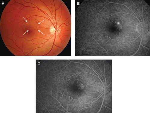
FIGURE 2 (A) Baseline horizontal OCT scan shows serous retinal detachment with a few irregularities on outer neurosensory retina related to central serous chorioretinopathy (CSC). (B) After stopping the therapy, significant resolution of submacular fluid was observed in same section of OCT scan with marked decrease of foveal thickening from 345 to 198 μm and visual acuity improved to 20/20.
Abnormalities in the choroidal circulation and retinal pigment epithelium have been hypothesized to be causative factors in CSC.Citation2 Prostaglandin analogues reduce IOP by enhancing uveoscleral outflow and may also have some effect on vascular permeability as well.Citation3 Prostaglandins, which are metabolic products of arachidonic acid, are the most extensively studied chemical transmitters contributing to general edema in systemic tissues, including eye tissue. It has been suggested that prostaglandins can mimic vascular endothelial growth factor (VEGF) and vascular permeability factor (VPF), inducing vascular permeability.Citation4 We hypothesize that topical latanoprost may increase choroidal vascular permeability and may cause disturbance in choroidal circulation. Although the mechanism is unclear, increase in choroidal vascular permeability induced with latanoprost may be influential in the development of CSC, which is characterized by detachment of the neurosensory retina caused by accumulation of serous fluid between the photoreceptor outer segments and the RPE. Ozdemir et al.Citation5 reported a few cases of serous retinal detachment associated with latanoprost. We believe that this condition is likely to be related to latanoprost treatment because cessation of the drug resulted in the resolution of CSC in our patient. Also, it’s possible that the remission may have been at least in part spontaneous. To the best of our knowledge, this is the first case report of CSC associated with topical latanoprost therapy.
This case demonstrates that topical latanoprost might be responsible for the development of CSC. The patient’s history should be assessed for any previous CSC episodes and clinicians should be cautious for CSC as a possible complication of topical latanoprost therapy.
Declaration of interest: The authors report no conflicts of interest. The authors alone are responsible for the content and writing of the paper.
REFERENCES
- Levy J, Marcus M, Belfair N, Klemperer I, Lifshitz T. Central serous chorioretinopathy in patients receiving systemic corticosteroid therapy. Can J Ophthalmol. 2005;40:217–221.
- Marmor MF. New hypotheses on the pathogenesis and treatment of serous retinal detachment. Graefes Arch Clin Exp Ophthalmol. 1988;226:548–552.
- Alm A, Grierson I, Shields MB. Side effects associated with prostaglandin analog therapy. Surv Ophthalmol. 2008;53:93–105.
- Vinores SA, Sen H, Campochiaro PA. An adenosine agonist and prostaglandin E1 cause breakdown of the blood–retinal barrier by opening tight junctions between vascular endothelial cells. Invest Ophthalmol Vis Sci. 1992;33:1870–1878.
- Ozdemir H, Karacorlu M, Karacorlu SA. Serous detachment of macula in cystoid macular edema associated with latanoprost. Eur J Ophthalmol. 2008;18:1014–1016.