Abstract
The sheep nasal botfly Oestrus ovis is the commonest of several species of dipteran fly whose larvae are obligatory parasites in the nasal cavities and frontal sinuses of sheep but may cause infestation (myiasis) in man, commonly as external infestations and rarely, involves inner coat of the eye (Ocular myiasis interna). We report an imported case of Bilateral Ocular myiasis interna in an Indian boy.
Infection and infestation by insect larvae is common in tropical regions. Among arthropods, flies commonly invade the conjunctival sac (myiasis). The flies that cause conjunctival disease belong to muscid, sarcophacoidae, and oesteriodae groups. The sheep nasal botfly Oestrus ovis is the commonest of several species of dipteran fly whose larvae are obligatory parasites in the nasal cavities and frontal sinuses of sheep but may cause infestation (myiasis) in humans, commonly as external infestations and, rarely, involving inner coat of the eye (ocular myiasis interna). We report an imported case of bilateral ocular myiasis interna in an Indian boy.
CASE REPORT
A 10-year-old Hindu, vegeteranian boy reported to our clinic with history of recurrent redness and diminution of vision of both eyes for 7 days. Patient had a history of an insect bite about 17 days before while holidaying in South Africa and was diagnosed as having a case of preseptal cellulites and was admitted for the same for 3 days. The patient had no other significant past ocular or systemic history.
On his first visit, on examination, his right eye had a best-corrected visual acuity (BCVA) of counting fingers (CF) at 1 m. BCVA in the left eye was 6/12. On slit-lamp examination, both eyes had conjunctival congestion, with the left eye having lid edema, pigments on the endothelium, aqueous cells, and flare 1+ and a sluggish reacting pupil. Intraocular pressures, by Goldmann applanation tonometry, were 18 mmHg in both eyes. Dilated fundus examination by indirect ophthalmoscopy showed a subretinal white larva in the infero-temporal quadrant with subretinal tract all over retina and hemorrhage surrounding the disc in right eye (). The other eye showed preretinal hemorrhage with no sign of any larva but had subretinal tracts like the right eye (). A laser barrage around the larva was planned. The next day, however, the larva was seen in the vitreous cavity in the right eye (). This was confirmed by a B-scan USG of both eyes, which showed a larva in the vitreous cavity in the right eye. However, no larva was seen in the left eye ().
FIGURE 1 Color fundus photograph of the right eye showing hemorrhage nasal to the disc margin and multiple subretinal tracks involving the macula. In the inferotemporal region white subretinal larva is seen.
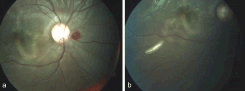
FIGURE 2 Color fundus photograph of the left eye showing large preretinal hemorrhage temporal to disc involving the macula.
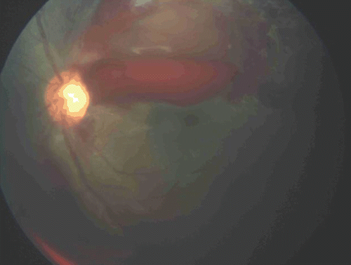
FIGURE 3 Color fundus photograph showing larva in the vitreous cavity along with multiple subretinal tracks.
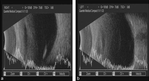
FIGURE 4 USG B scan of right eye showed high reflective echoes corresponding with the larva in the vitreous cavity, while left eye was normal with no evidence of larva.
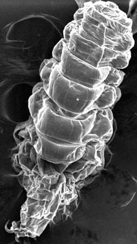
The patient was started on broad-spectrum antibiotics, oral steroid, and ivermectin once daily. A 20-gauge vitrectomy was done in the right eye. The larva was held with end-gripping vitreous forceps and complete vitrectomy was done. The larva was then removed and was sent to the pathology department for identification where it was identified as a botfly larva (, ).
FIGURE 5 Microscopic study of the larva, 1600 μm in size as measured on micrometer and consisting of 11 segments with a pair of oral hooks.
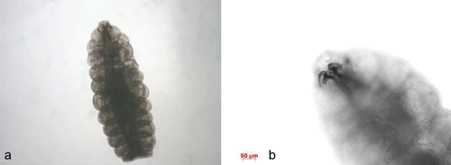
FIGURE 6 Scanning electron microscopy of the whole body of botfly in lateral view with showing multiple segments and a pair of oral hooks (magnification, ×70).
During his last visit, 1 month after the first visit, BCVA in right eye was 6/18 and in the left eye was 6/6. Left eye preretinal hemorrhage had cleared, with a little subretinal blood just above the foveal avascular zone. The subretinal tracts in both eyes, although still present, were resolving. The worm in the left eye was not discovered until the last visit.
DISCUSSION
The larvae of Oestrus ovis are hatched from their eggs in the vagina of the adult female, who darts them into the nostrils of sheepCitation1 in a stream of milky white fluid, possibly without direct contact.Citation2 Migration to the frontal sinuses is followed by maturation for 6–12 months. The larvae are subsequently sneezed out and pupate on the ground for a period of a few weeks. The lifespan of the adult fly is about 4 weeks. Humans are incidental hosts and become involved when larvae are ejected onto the ocular surface instead of the nasal mucosa of sheep. In humans the larvae are unable to mature and survive for up to 10 days. Sudden onset of foreign body symptoms is followed by pain and inflammation. A punctate keratitis is common and small conjunctival hemorrhages may be seen. Single or multiple larvae are observed in the conjunctival sac. The condition is normally benign and self-limiting.Citation3 Invasion of the orbit or globe, more typical of other species, is rarely reported due to O. ovis,Citation4 but the resulting panuveitis may be severe. Treatment may be challenging, as the worms move very quickly and between different compartments and different tissues of the eye. The physician’s imagination and more than one treatment modality, such as argon laser photocoagulation, Yag laser, surgical removal, or even vitrectomy, may be required to destroy the larva Citation13–17. Topical administration of corticosteroids for symptomatic relief and antibiotics to prevent bacterial contamination has been recommended.Citation3 Oestrus ovis is widespread in Africa and the Middle East, where the annual incidence of ocular myiasis is estimated to be 10 per 100,000.Citation5 In this area there is a recognized clinical entity in which patients have been “bitten” by a fly and developed “blindness,” from which they recovered in 2/3 weeks. Although this should be substantiated by medical examination, it is nevertheless an interesting observation, as this would fit the usual benign clinical picture of human infection by the first-stage larvae of Oestrus ovis, i.e., an acute catarrhal conjunctivitis with periorbital edema, which resolves completely in 2 weeks. It is also reported in Australia,Citation6 North America,Citation3,Citation7 India,Citation12 southern Europe,Citation8 and the United KingdomCitation9,Citation10 or may be imported,Citation11 as in this case.
The larva was removed from the vitreous cavity and was examined, photographed, and identified as a first-stage larva of Oestrus ovis. In the absence of any previous ocular disease or injury, it is difficult to explain how the larvae penetrated an intact globe. Presumably, penetration occurred in the posterior region of the eye since the patient originally presented with a severe retinal edema and preretinal haemorrhage. Whether penetration by the larvae was directly through an intact sclera (unlikely) or via the vena vorticosae or at the point of entry of the ciliary and optic nerves can only be speculated upon. Uveitis was probably due to the migration of the larvae within the choroid in an anterior direction (possibly toward the light source shining through the pupil). The involvement of the other eye remained a mystery, as no larva was seen. Possibly, the same larvae had migrated transcutaneously or had a hematogenous spread, or a second larva was present in the left eye but had died because of its short life cycle.
Botfly maggots, very rarely, may invade the sclera and present in the AC, vitreous cavity, or subretinal space. In our patient, aside from impaired VA, the intraocular maggot also caused prominent orbital cellulitis. The prognosis of ophthalmomyiasis interna varies greatly, from vision loss to excellent prognosis, as in our case. A history of recent travel to endemic areas should prompt a high index of suspicion and careful ocular examination for larvae.
Declaration of interest: The authors report no conflicts of interest. The authors alone are responsible for the content and writing of the paper.
REFERENCES
- Zumpt F. Myiasis in Man and Animals in the Old World. London: Butterworth; 1965.
- Kcan BN, Sun T, Elisworth W. Colour Atlas/Text of Ophthalmic Parasitology. New York: Igaku-Shoin;1991.
- Reingold WJ, Robin JB, Leipa D, Kondra L, Schanzh DJ, Smith RE. Oestrus ovis ophthalmo-myiasis externa. Am J Ophthalmol. 1984;97:7–10.
- Rakusin W. Ocular myiasis interna caused by the sheep nasal botfly (Oestrus ovis L.). S Afncan Med J. 1970;44:1155–1157.
- Dar MS, Arncr MB, Dar FK, Papazotos V. Ophthalmomyiasis caused by the sheep nasal bot, Oestrus ovis (Oestridae) larvae, in the Bcnghazi area of Eastern Libya. Trans R Soc Tmp Med Hyg. 1980;74:303–306.
- Harrington NN. External ophthalmomyiasis in Australia. Med J Aust. 1968;1:152–153.
- Brown HS Jr, Hitchcmk JC Jr, Foos RY. Larval conjunctivitis in California caused by Oestrus ovis L. Calif Med. 1969;111:272–274.
- Le Fjchoux Y, Mariy P, Denis G, Couturier P, Dellamomca P. A case of extemal ophthalmomyiasis by Oestrus ovis, Lie, 1758 caught on the Nice beach [author’s translation]. Acta Tropica. 1981;38:461–468.
- Romanes GJ. Ocular myiasjs [letter]. Br J Ophthalmol. 1983;67:332.
- Smith JH. Ophthalmomyiasis in England. Br J Ophthalmol. 195135:242–243.
- Wong D. External ophthalmomyiasis caused by the sheep bot Oestrus ovis. Br J Ophthalmol. 1982;66:786–787.
- Narayanan S, Jayaprakash K. Incidence of ocular myiasis due to infection with the larva of Oestrus ovis (Oestridae Diptera). Indian J Ophthalmol. 1991;39:176–178
- Mason GI. Bilateral ophthalmomyiasis interna. Am J Ophthalmol. 1981; 91: 65–70.
- Saraiva Vda S, Amaro MH, Belfort R Jr, Burnier MN Jr. A case of anterior internal ophthalmomyiasis: case report. Arq Bras Oftalmol. 2006; 69: 741–743.
- Sharifipour F, Feghhi M. Anterior ophthalmomyiasis interna: an ophthalmic emergency. Arch Ophthalmol. 2008; 126: 1466–1467.
- Syrdalen P, Nitter T, Mehl R. Ophthalmomyiasis interna posterior: report of case caused by the reindeer
- “Intraocular safari: ophthalmomyiasis internal” Clin Exp Ophthalmol. 2011 Jan;39(1):84–5. doi: 10.1111/j.1442-9071.2010.02407.x.