Abstract
Purpose: To show that diffuse unilateral subacute neuroretinitis (DUSN) may masquerade as multiple evanescent white dot syndrome, and to describe structural and functional recovery following treatment in DUSN.
Design: Case report.
Methods: Baseline and serial optical coherence tomography (OCT) and perimetry following photocoagulation of nematode.
Results: After laser treatment, the inner segment-outer segment (IS–OS) junction was restored and the visual field was improved.
Conclusions: DUSN, early on, can be mistaken for other inflammatory white dot syndromes. OCT and perimetry, in combination, provide strong support for success of treatment with photocoagulation.
Keywords::
CASE
A 41-year-old woman was referred for management of multiple evanescent white dots syndrome (MEWDS). She reported a 2-week history of visual obscurations in the right eye, described specifically as a “spot” in her vision. She had no medical or ophthalmic history and was taking no medications.
Visual acuity was 20/20 in each eye. Pupils were reactive without an afferent pupillary defect. Slit-lamp examination was normal. Funduscopy revealed granular pigment epithelial changes in the macula of the right eye and a normal retina in the left eye ().
FIGURE 1 Fundus photographs (A) and fluorescein angiogram (FA) (B). Retinal pigment epithelial changes and foveal granularity are seen in the right eye (left), and the left fundus (right) is normal (A). FA of the right eye reveals pinpoint hyperfluorescent spots that stain late (B).
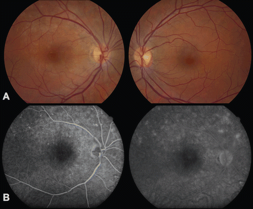
Fluorescein angiography (FA) revealed pinpoint hyperfluorescent spots in the right eye (), and optical coherence tomography (OCT) showed disruption of the inner segment–outer segment (IS–OS) layer (). Visual field testing revealed an enlarged blind spot (). Our diagnosis was MEWDS, and the plan was to observe closely for resolution.
FIGURE 2 Spectral Domain Optical Coherence Tomography ((SD-OCT) Spectralis; Heidelberg Engineering, Germany), right eye. Baseline OCT shows disruption of the inner segment-outer segment junction (arrow) (A). OCT five months following treatment shows restoration of the inner segment-outer segment junction (arrowhead) (B).
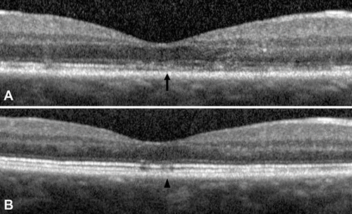
FIGURE 3 Humphrey visual field 30-2, right eye. Baseline visual field shows an enlarged blind spot and temporal depression (A). The visual field three months after treatment is improved (B).

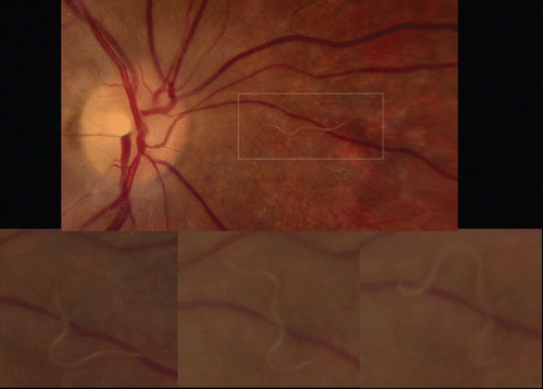
One week later, repeat funduscopy revealed a motile subretinal nematode nasal to the optic nerve (), and the diagnosis of diffuse unilateral subacute neuroretinitis (DUSN) was made. The nematode was immediately photocoagulated. On further questioning, the patient reported recent visits to Colorado where she had exposure to raccoons.
FIGURE 4 Fundus photographs, right eye. One week after initial presentation, a peri-papillary nematode is identified (top), and its movement through the subretinal space is observed (bottom).
On follow-up, there was mild improvement in the patient’s symptoms, and retinal exam revealed photocoagulation scars without nematode motility. Additional confirmation of presumed worm death included recovery of the IS–OS junction on OCT (), as well as improvement in the visual field (). The residual temporal depression of the visual field may relate to underlying permanent structural damage incurred by the motile nematode in the subretinal space.
DISCUSSION
DUSN, a progressively blinding retinal disorder, is due to migration of a nematode worm in the subretinal space. Baylisascaris procyonis, a raccoon roundworm endemic to the northern midwestern United States and Canada has been implicated in this condition. Findings in the acute phase include successive crops of evanescent white outer retinal lesions in different parts of the fundus, reflecting nematode motility through the subretinal space, as well as mild vitritis and papillitis. Toxic injury from the nematode eventually causes retinal pigment epithelium degeneration, disc pallor, and retinal vessel narrowing, seen in the later stages. Photocoagulation of the worm definitively arrests visual deterioration, whereas treatment with antihelminthics has had variable reports of success.Citation1,Citation2,Citation3 Improvement of the visual field has been reported in cases of DUSN treated with either albendazole or laser.Citation4,Citation5 MEWDS typically presents with unilateral vision changes, including photopsias and scotomata, and is characterized by small outer retinal white spots, which are transitory and leave behind granular macular pigmentary changes. The hyperfluorescent lesions on FA in MEWDS patients may resemble those of DUSN, although MEWDS lesions classically stain late in a perifoveal wreathlike configuration. Indocyanine green angiography in MEWDS, typically showing hypofluorescent dark spots up to the midperipheral fundus, have also been reported in DUSN, thereby pointing to choroidal involvement in both these entities.Citation6,Citation7 Additionally, the field abnormalities of MEWDS mimic those of DUSN, including paracentral scotomata and blind spot enlargement. Unlike DUSN, prognosis for MEWDS is excellent with visual recovery within weeks.Citation8
Our patient’s presentation highlights the ability of DUSN to masquerade as a white dot syndrome, as formerly demonstrated.Citation9 Since prompt diagnosis of DUSN may prevent vision loss, it is important to consider this diagnosis early in the management of patients with presumed inflammatory white dot syndromes.
This case also emphasizes the utility of serial OCT and perimetry, in combination, for helping confirm death of the nematode following laser photocoagulation. In the short term after laser, it can be challenging to ascertain treatment success given the uncertainty of the presence of eradicated nematode beneath the laser scar, and the ability of a live nematode to remain clinically undetected. A recent report demonstrated restoration of photoreceptor architecture following treatment with both photocoagulation and albendazole.10 Our case highlights the point that OCT can be followed for evidence of retinal anatomical restoration after laser treatment alone. Moreover, OCT and perimetry, used together, provide powerful support for nematode death following photocoagulation, by demonstrating recovery of both retinal structure and function.
ACKNOWLEDGMENTS
We would like to acknowledge Dr. Richard Weber, MD, of Stamford, CT, for his input to this case and for providing us with the fluorescein angiogram images.
Declaration of interest: The authors report no conflicts of interest. The authors alone are responsible for the content and writing of the paper.
REFERENCES
- Gass JD, Braustein RA. Further observations concerning the diffuse subacute neuroretinitis syndrome. Arch Ophthalmol. 1983;101(11):1689–1697.
- Goldberg MA, Kazacos KR, Boyce WM, Ai E, Katz B. Diffuse unilateral subacute neuroretinitis: morphometric, serologic, and epidemiologic support for Baylisascaris as a causative agent. Ophthalmology. 1993;100(11): 1695–1701.
- Raymond LA, Gutierrez Y, Strong LE, Wander AH, Buten R, Cordan D. Living retinal nematode (filarial-like) destroyed with photocoagulation. Ophthalmology. 1978;85(9):944–949.
- Souza EC, Casella AM, Nakashima Y, Monteiro ML. Clinical features and outcomes of patients with diffuse unilateral subacute neuroretinitis treated with oral albendazole. Am J Ophthalmol. 2005; 140(3): 437–445.
- De Souza EC, Abujamra S, Nakashima Y, Gass JD. Diffuse bilateral subacute neuroretinitis: first patient with documented nematodes in both eyes. Arch Ophthalmol. 1999;117(10):1349–1351.
- Cimino L, Auer C, Herbort CP. Sensitivity of indocyanine green angiography for the follow-up of active inflammatory choriocapillaropathies. Ocul Immunol Inflamm. 2000;8(4):275–283.
- Vianna RN, Onofre G, Ecard V, Muralha L, Muralha A, de A Garcia CA. Indocyanine green angiography in diffuse unilateral subacute neuroretinitis. Eye. 2006;20(9):1113–1116.
- Jampol LM, Sieving PA, Pugh D, Fishman GA, Gilbert H. Multiple evanescent white dot syndrome, I: clinical findings. Arch Ophthalmol. 1984;102(5):671–674.
- Barbazetto IA, Lesser RL, Tom D, Freund KB. Diffuse unilateral subacute neuroretinitis masquerading as a white-dot syndrome. Br J Ophthalmol. 2009;93(5):574–576, 655.
- Tarantola RM, Elkins KA, Kay CN, Folk JC. Photoreceptor recovery following laser photocoagulation and albendazole in diffuse unilateral subacute neuroretinitis. Arch Ophthalmol. 2011;129(5):669–671.