Abstract
We present a case of a patient with bilateral posterior uveitis HLA-A29 positive, masquerading intraocular lymphoma. A 43 year-old woman presented with bilateral vitritis and chorioretinal lesions compatible with “birdshot lesions”. The patient was initially diagnosed with birdshot retinochoroidopathy and later on, during follow up, the occurrence of neurologic involvement and the lack of response to systemic immunosuppression led us to re-evaluate the diagnosis. A definite diagnosis of intraocular lymphoma with central nervous system involvement was made. This case is presented to highlight the importance of careful follow-up of patients with chronic uveitis and re-evaluation of systemic symptoms and signs, in particular when ocular findings are highly suggestive for masquerade syndrome.
We present the case of a patient with bilateral posterior uveitis, HLA-A29 positive, and chorioretinal lesions compatible with “birdshot lesions.” The patient was initially diagnosed with birdshot chorioretinopathy, but later during follow-up the occurrence of neurologic involvement and the lack of response to systemic immunosuppression led us to reevaluate the diagnosis. A definite diagnosis of intraocular lymphoma with central nervous system involvement was made.
A 43-year-old Italian woman with a diagnosis of bilateral uveitis and progressive bilateral visual loss was referred to our Uveitis Service for a second diagnostic opinion. The patient complained of visual impairment from the beginning of 2007 and had been previously treated with 25 mg/day of systemic prednisone for about 30 days without visual improvement. On February 2007, at our first evaluation, her visual acuity was 20/32 in the right eye and 20/25 in the left eye. Anterior segment and intraocular pressure were normal. Fundus examination showed the presence of bilateral 2+ vitreous cells with a grade 2 vitreous haze and small, round, yellowish chorioretinal lesions at the posterior pole in both eyes. Optical coherence tomography revealed the presence of cystoid macular edema. The patient’s past medical history was positive only for the presence of kidney stones. Uveitis questionnaire and review of system were unremarkable. We performed extensive laboratory tests for autoimmune and infectious diseases, including the serology for syphilis (MHATP and RPR) and ACE and lysozyme associated with a high-resolution chest CT scan to rule out sarcoidosis. Results were all negative, except for the presence of HLA-A 29 antigen. Magnetic resonance imaging was negative for the presence of brain abnormalities. A diagnosis of birdshot chorioretinopathy was then made and the patient was started on higher doses of systemic prednisone (50 mg/day) and cyclosporine 200 mg/day.
Two months later, despite a visual acuity improvement (visual acuity 20/20 in both eyes) the patient had a subjective impression of visual disturbances and the ophthalmoscopic examination showed resolution of macular edema but persistence of bilateral 1+ vitreous cells with an improvement of vitreous haze to grade 1. We decided to increase the dose of systemic cyclosporine to 300 mg/day to adequately immunosuppress the patient. In June 2007, 4 months after our first evaluation, there was a complete resolution of intraocular inflammation and at fundus examination there was a complete absence of haze and vitreitis. A pigmentation of the chorioretinal lesion was evident (). Unfortunately, the patient began to have some psychological disturbances, such as severe depression and “behavioral changes.”
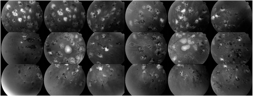
FIGURE 1 Fundus red free photography showing the bilateral small fundus spots on the retina, scattered in a pattern like birdshot. The photo corresponds to the July ophthalmologic examination and shows the resolution of intraocular inflammation and the pigmentation of the lesions.
We decided to hospitalize the patient to perform additional investigations and to reassess the diagnosis. During hospitalization the patient had an acute visual loss in the left eye to hand motion and the visual field showed a dense central scotoma. Ophthalmoscopically, a bilateral, very active relapse of disease was evident, characterized by bilateral 3+ vitreous cells, vitreous haze grade 3, and new yellowish chorioretinal lesions. Visual symptoms were rapidly followed by the development of aphasia and confusion. The magnetic resonance imaging of the brain showed multiple and diffuse periventricular lesions compatible with central nervous system lymphoma. Therefore, the patient was followed by the Lymphoma Unit, who decided, in accordance with the patient, to perform a diagnostic stereotactic biopsy that confirmed the clinical suspicion. The cytopathology revealed a hypercellular specimen with numerous degenerated cells and scattered viable atypical lymphoid cells with prominent nucleoli compatible with large B-cell lymphoma. Immunocytochemical staining was positive for CD19 and CD20 and flow cytometry confirmed a monoclonal B-cell population. Therefore, the patient was started on systemic chemo- and radiotherapy. The patient unfortunately died after 9 months of follow-up from a pulmonary mycetoma.
The diagnosis of intraocular lymphoma requires a high clinical suspicion and it can be suspected in the presence of retinal or chorioretinal lesions similar to “birdshot lesions.” However, the definite diagnosis is only established with demonstration of malignant lymphocytes in the vitreous, retina, or choroid.Citation1,Citation2 On the other hand, the diagnosis of birdshot chorioretinopathy is only clinical and relies on the recognition of round or oval hypopigmented chorioretinal lesions at the level of the choroid, most often one-quarter to one-half optic disk diameter in size and clustered around the optic disc.Citation3
Our patient fulfilled the criteria for birdshot chorioretinopathy, because of the presence of yellowish chorioretinal lesions. Moreover, she was characterized by the HLA-A29 gene positivity, which is not a diagnostic criterion but is present in a high percentage of patients affected by birdshot chorioretinopathy.Citation4 The absence of neurologic symptoms and normal magnetic resonance findings at the time of first evaluation, prompted us to exclude the diagnosis of intraocular lymphoma in favor of a diagnosis of birdshot chorioretinopathy. The lack of response to adequate immunosuppressive therapy and the later occurrence of “behavioral changes” raised a suspicion of a masquerade syndrome.Citation2 Reevaluation of clinical diagnosis led to the definite diagnosis of intraocular lymphoma with central nervous system involvement. MRI negativity at the first evaluation cannot, in fact, exclude that central lesions subsequently develop or small lesions previously undetectable become evident. The lack of response to adequate steroid and immunosuppressive treatment should always alert the ophthalmologist to reassess the first diagnosis. The absence of neuroradiologic imaging findings may not be a sufficient criterion to exclude a definite diagnosis of intraocular lymphoma. Although the majority of patients with birdshot chorioretinopathy present with the positive HLA-A29 gene, this laboratory finding should not be considered as a major diagnostic criterion for diagnosis of the disease.
In conclusion, with the presented case we would like to highlight the importance of differential diagnosis and reevaluation of systemic symptoms and signs in patients presenting with ocular findings suggestive of birdshot chorioretinopathy. In particular, when intraocular lymphoma can be suspected, it is important to investigate for the presence of neurologic symptoms and signs with careful history and close monitoring with neuroimaging.
Declaration of interest: The authors report no conflicts of interest. The authors alone are responsible for the content and writing of the paper.
REFERENCES
- Zaldivar RA, Martin DF, Holden JT, Grossniklaus HE. Primary intraocular lymphoma: clinical, cytologic and flow cytometric analysis. Ophthalmology. 2004; 9:1762–1767.
- Read RW, Zamir E, Rao NA. Neoplastic masquerade syndromes. Surv Ophthalmol. 2002; 2:81–124.
- Levinson RD, Brezin A, Rothova A, Accorinti M, Holland GN. Research criteria for the diagnosis of birdshot chorioretinopathy: results of an International consensus conference. Am J Ophthalmol. 2006; 1:185–187.
- Shah KH, Levinson RD, Yu F, et al. Birdshot chorioretinopathy. Surv Ophthalmol. 2005;6:519–541.