Abstract
Purpose: To describe cases of pars planitis associated with retinoschisis, in which laser photocoagulation was carried out.
Methods: Retrospective review.
Results: Three pars planitis cases were associated with retinoschisis and underwent laser photocoagulation. All cases were idiopathic. Retinoschisis was located in the inferior retinal quadrants in all cases and all of them were in bullous formation. None of the cases developed retinal detachment.
Conclusion: As well as posterior vitreous detachment, or peripheral retinal tears, retinoschisis may accompany pars planitis. Laser photocoagulation of the pars plana is effective in these cases both as a treatment and to prevent sight-threatening complications like retinal detachment.
Intermediate uveitis was first introduced by the International Uveitis Study Group in 1987. The entity was defined due to the anatomical location of uveitis. The inflammatory process is primarily limited to the anterior vitreous, peripheral retina, and pars plana. The term pars planitis is used by some authors for idiopathic intermediate uveitis, which shows no association with a systemic disorder.Citation1,Citation2 Besides well-known sight-threatening, chronic complications like band keratopathy, epiretinal membrane, retinal detachment, neovascularization, and vitreous hemorrhage, retinoschisis associated with pars planitis was also reported.Citation3,Citation4
Herein, we report 3 cases of pars planitis associated with bullous retinoschisis in which laser photocoagulation was applied to limit the extent of the schisis.
PATIENTS AND METHODS
Data of 3 consecutive cases of pars planitis aged between 9 and 17, which were associated with retinoschisis, were reviewed. Diagnosis of pars planitis was based on a quiet or minimally reactive anterior chamber, cells in the anterior vitreous, and snowball opacities in the inferior vitreous. Bullous retinoschisis was accompanying pars planitis in all 3 cases, and prophylactic laser photocoagulation was performed to make a barrier to extend of the schisis. The diagnosis of retinoschisis was made upon clinical appearance by 90 diopters non-contact lens in slit-lamp and confirmed by three-mirror contact lens. Retinoschisis, especially bullous forms, features peripheral smooth elevation and a uniformly convex posterior border that entends anteriorly to or very close to the ora serrata. Indirect opthalmoscopy with peripheral indentation was also performed to rule out retinal holes and confirm the diagnosis.
To exclude the association with a systemic disorder, tests for Lyme disease, sarcoidosis, and syphilis were performed and found to be negative. For a possible vasculitis association, serological tests for ANA and ANCA were also negative. Neurological examination revealed no sign of multiple sclerosis (MS). Magnetic resonance imaging (MRI), which was performed in cases 2 and 3, demostrated no features of MS.
CASE REPORTS
Case 1
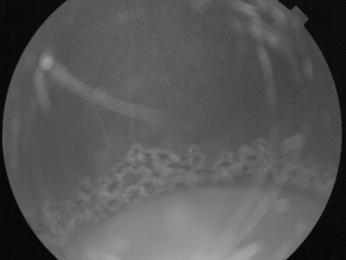
A 9-year-old boy was referred for further evaluation with a diagnosis of uveitis. Best-corrected visual acuities were 18/20 and 16/20 in his right and left eyes, respectively. Slit-lamp anterior segment findings were normal. Both eyes revealed bullous retinoschis, and snowball opacities in the fundus examination. Optical coherence tomography (OCT) scans of both eyes were normal. In fundus fluorescein angiography (FFA), late-phase leakage from the optic disc and peripheral retinal vessels was detected; cystoid macular edema (CME) was not evident. The patient underwent prophylactic laser photocoagulation to make a barrier around the bullous retinoschisis (). Additional corticosteroid therapy was not needed.
FIGURE 1 Fundus fluorescein angiogram of patient 1 following photocoagulation.
He is under follow-up for 4 months and the eye is quiescent and the bullous retinoschisis shows no further progression.
Case 2
A 17-year-old girl had a complaint of blurry vision in her left eye. The visual acuity was 20/20 in both eyes. Slit-lamp findings were normal. In the fundus examination, bullous retinoschisis in the inferior quadrant of the right eye and vitreous hemorrhage in the left eye were detected; both eyes showed mild to moderate vitritis and vascular sheeting. Fundus fluorescein angiography revealed a hyperfluorescent optic disc as well as a, late-phase leakage from the optic disc and peripheral retinal vessels of both eyes, but did not demonstrate CME.
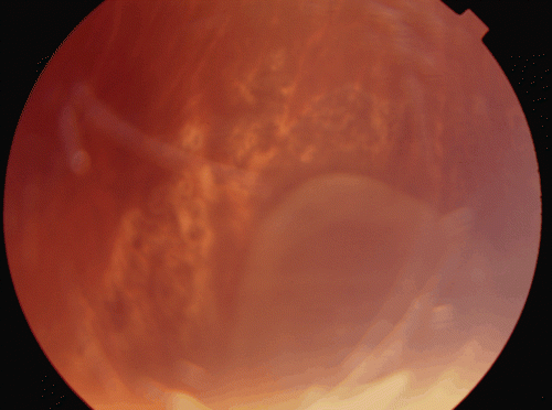
Prophylactic laser photocoagulation was applied around the bullous retinoschisis (). Additional corticosteroid therapy was not needed. She is under follow-up for 5 months and the eye is quiescent.
FIGURE 2 Fundus picture of patient 2 following photocoagulation. Note that laser spots form a barrier around the bullous retinoschisis.
Case 3
A 17-year-old girl had a complaint of blurry vision in her both eyes. Her visual acuity was 20/20 in both eyes. In the slit-lamp examination, diffuse posterior synechia and mild to moderate reaction in the anterior chamber and the anterior vitreous were detected in both eyes. Fundus examination revealed optic disc and macula edema in both eyes and snowball opacities in the right eye.
While under follow-up she occasionally developed recurrences that responded well to topical and oral corticosteroid therapy, until bullous retinoschisis was detected in the inferior temporal quadrant, 2 years after the first examination.
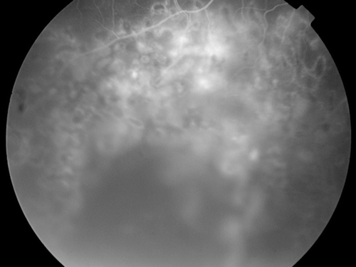
Prophylactic laser photocoagulation was carried out around the bullous retinoschisis (). Following photocoagulation she was under follow-up for 3 years. Visual acuity was 20/20, and the anterior chambers were normal except posterior synechia in both eyes. The retinoschisis showed no further extent or progression.
FIGURE 3 Fundus fluorescein angiogram of patient 3 following photocoagulation.
DISCUSSION
The inflammatory process involving the vitreous is considered to associated with posterior vitreous detachment and retinal tear formation. It is proposed that an increase of inflammatory cells in the vitreous induces vitreous shrinkage, thus causing early PVD.Citation5,Citation6 Particularly, snowbanks in pars planitis can cause vitreo-retinal traction that can lead to retinoschisis.Citation7,Citation8 Sungur and co-workers reported that Fuchs uveitis is also associated with an increase in retinal break formation of 6.1%, whereas this rate is 3.3% in the general population.Citation9
Jalil and co-workers, reported the rate of retinal elevation in their intermediate uveitis series to be 4.3% (11 of 256), 6 of these 11 being retinoschisis, all located at the inferior quadrants. They proposed the traction to result from snowbanking or glial fibrosis over the pars plana and peripheral retina, not the vitreous. Except the therapy for suppression of the uveitis, the authors carried out no further treatment for the retinoschisis and during follow-up they reported that all cases were stable.Citation10 We carried out laser photocoagulation around the schisis in our cases to prevent retinal detachment.
Pollack and co-workers reported 10 cases of retinoschisis in 13 eyes with intermediate uveitis; 3 of these cases were accompanied by rhegmatogenous/tractional retinal detachment. Of the remaining 7 cases that were not accompanied by retinal detachment, 1 underwent cryotherapy, whereas the remaining 6 did not receive any treatment. These retinoschisis cases were associated with various vascular disorders, including peripheral neovascularization, telangiectasies, and retinal capillary angiomas. The authors postulated that, besides peripheral retinal traction, vasoproliferation based inflammatory processes plays role in the pathogenesis of retinoschisis formation.Citation3 Retinal detachment occurs in 2.5% of retinoschisis cases of different etiologies.Citation11
We did not detect any vascular abnormality except vascular sheeting in our cases. Currently, there is very small number of studies reporting the association of uveitis and peripheral retinal changes—particularly retinoschisis. We believe that laser photocoagulation also helped to inhibit vascular endothelial growth factor (VEGF) production of the retinal pigment epithelium. Similarly, Pulido and co-workers found that laser photocoagulation helped to diminish inflammation in pars planitis; they hypothesized this effect to be related to the decreased release of angiogenic factors following photocoagulation.Citation12
In conclusion, peripheral vitreous and retinal changes, including bullous retinoschisis, may occur as a result of prolonged inflammation in intermediate uveitis. The physician should be aware of these complications and all patients should be carefully examined. Laser photocoagulation of the pars plana might be effective as a treatment of pars planitis and might also play a role in preventing sight-threatening complications like retinal detachment.
Declaration of interest: The authors report no conflicts of interest. The authors alone are responsible for the content and writing of the paper.
REFERENCES
- Bloch ME, Nussenblatt RB. International Uveitis Study Group recommendations for the evaluation of intraocular inflammatory disease. Am J Ophthalmol. 1987;103:234–235.
- Bonfioli AA, Damico FM, Curi ALL, Orefice F. Intermediate uveitis. Semin Ophthalmol. 2005;20:147–154.
- Pollack AL, McDonald R, Johnson RN, et al. Peripheral retinoschisis and exudative retinal detachment in pars planitis. Retina. 2002;22:719–724.
- Brockhurst RJ. Retinoschisis. Complications of peripheral uveitis. Arch Ophtalmol. 1981;99:1988–1999.
- Akova YA, Yilmaz G, Aydin P. Retinal tears associated with panuveitis and Behçet’s disease. Ophthalmic Surg Lasers. 1999;30:762–765.
- Sebag J. Classifying posterior vitreous detachment: a new way to look at the invisible. Br J Ophthalmol. 1997;81:521–522.
- Zimmerman PL, Boyle TM. Pars planitis and other intermediate uveitis. In Yanoff M, Duker JS, eds. Ophthalmology. St. Louis: CV Mosby; 2004: 1213–1218.
- Green WR, Sebag J. Vitreoretinal interface. In Ryan SJ, eds. Retina. Philadelphia: Elsevier Mosby; 2006:1921–1989.
- Sungur G, Hazirolan D, Duman S, Kasim R. Fuchs’ heterochromic uveitis associated with retinal break or dialysis. Can J Ophthalmol. 2008;43:109–110.
- Jalil A, Dhawair-Scala FE, Jones NP. Nonprogressive tractional inferior retinal elevation in intermediate uveitis. Ocul Immunol Inflamm. 2010;18:60–63.
- Pecold K, Czaplicka E, Bernardczyk J. Retinoschisis vs. retinal detachment—diagnosis and treatment. Klin Oczna. 1993;95:32–34.
- Pulido JS, Mieler WF, Walton D, et al. Results of peripheral laser photocoagulation in pars planitis. Tr Am Ophth Soc. 1998;96:127–141.