Abstract
Purpose: To report a case of cytomegalovirus (CMV) retinitis in an immunocompetent patient with proliferative diabetic retinopathy (PDR). Design: Case report.
Methods: A 69-year-old man presented with a 44-year history of diabetes mellitus and 4 years of PDR. Fundus of left eye could not be visualized because of vitreous hemorrhage. Laboratory tests indicated normal immunological status.
Results: Yellowish white retinal exudative lesion and whitening inside vascular arcades were observed during vitrectomy. Multiplex PCR using vitreous sample detected CMV DNA at 4.37 × 10⁴ copies/mL. CMV retinitis was diagnosed.
Conclusions: If atypical findings of PDR are observed, a multiplex PCR test should be performed for further investigation.
Cytomegalovirus (CMV) infection is a frequent cause of posterior uveitis in human immunodeficiency virus (HIV)-infected individuals with CD4+ T lymphocyte counts less than 100/µL or immunosuppressed individuals.Citation1,Citation2 Diabetic patients with impaired immune states are susceptible to various infectious diseases.Citation3 Especially, diabetes mellitus is a risk factor for bacterial or mycotic endophthalmitis, but it has not been associated with CMV retinitis.Citation4 We describe a case of CMV retinitis in an HIV-negative, CMV Ag-, IgM-, and IgG-negative patient with proliferative diabetic retinopathy (PDR) causing vitreous hemorrhage but no evidence of immunological dysfunction, and we consider diabetes mellitus as a risk factor for CMV infection.
A 69-year-old man was referred to our hospital because vitreous hemorrhage in the left eye had occurred 2 weeks previously. He had a 44-year history of diabetes mellitus that was poorly controlled (hemoglobin A1c was 9.6%) and had been diagnosed with PDR 4 years before. He had never received any steroid medication. At presentation, the left eye had best-corrected visual acuity of hand motion, elevated intraocular pressure (36 mmHg) because of rubeosis, and vitreous hemorrhage due to PDR, which obscured the fundus. The right eye had nonproliferative retinopathy. Laboratory tests indicated a white blood cell count of 8500/dL, CD4+ T-lymphocyte count of 593/µL, and CD8+ T-lymphocyte count of 776/µL; serum HIV was negative.
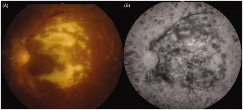
During vitreous surgery, a yellowish white retinal exudative lesion involving the posterior pole and whitening arcade vessels were observed, both of which were atypical findings of PDR (). A multiplex PCR test for human herpes virus (HHV) 1–8, 16S rRNA, and 28S rRNA using the vitreous sample was performed, and detected only CMV (HHV-5) DNA at 4.37 × 10⁴ copies/mL, although serum CMV IgM, CMV IgG, and CMV antigen were negative. Since the ocular fundus lesions were compatible with findings of CMV retinitis, CMV retinitis concurrent with PDR was diagnosed. Intravenous ganciclovir therapy (600 mg/day) for 3 weeks achieved successful remission of the retinal lesion. However, visual acuity of the left eye did not improve since macular atrophy had already progressed (). CMV IgM and CMV IgG became positive 4 months later.
Figure 1. Fundus of left eye during vitrectomy. During vitreous surgery, a yellowish white retinal exudative lesion involving the posterior pole and whitening inside vascular arcades are observed, both of which are atypical findings of PDR. Proliferative membrane was around the disc.
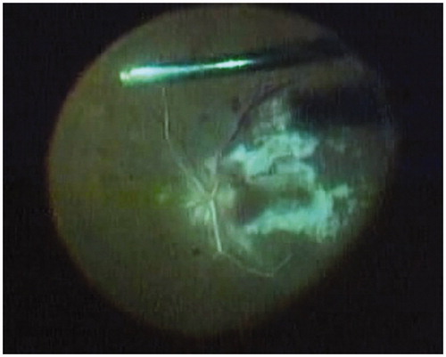
Figure 2. Fundus of left eye after treatment. (A) Macular color photograph and (B) macular fluorescein angiography. Ganciclovir therapy (600 mg/day) for 3 weeks achieved successful remission of the retinal lesion, but visual acuity of the left eye did not improve since macular atrophy had already progressed.
Although CD4+ T lymphocytopenia is associated with occurrence of CMV retinitis, the number of CD4+ T lymphocytes was within normal range in this patient. Since vitreous hemorrhage due to PDR obstructed observation of the fundus, CMV retinitis would not have been detected if multiplex PCRCitation5 using the vitreous sample was not performed, and the diagnosis would remain only PDR. Interestingly, it is possible that this patient had not been infected with CMV previously, because of the initial negative CMV IgM and CMV IgG findings and later seroconversion, although CMV retinitis usually occurs by reactivation of previous infection.Citation4 If multiplex PCR becomes more generally available and simpler to perform, more cases of primary or secondary CMV retinitis might be detected regardless of the visibility of the fundus. Nevertheless, ophthalmologists should be aware that if atypical features of PDR are observed, the multiplex PCR test using vitreous sample should be performed to investigate the real disease condition.
Declaration of interest
The authors report no conflicts of interest. The authors alone are responsible for the content and writing of the paper.
References
- Kelkar A, Kelkar J, Kelkar S, et al. Cytomegalovirus retinitis in a seronegative patient with systemic lupus erythematosus on immunosuppressive therapy. J Ophthalmic Inflamm Infect. 2011;1:129–132
- Wiwanitkit V. Recurrent CMV retinitis in non HIV. Graefe’s Arch Clin Exp Ophthalmol. 2011;249:147; author reply 149
- Leegaard A, Riis A, Kornum JB, et al. Diabetes, glycemic control, and risk of tuberculosis: a population-based case-control study. Diabetes Care. 2011;34:2530–2535
- Carmichael A. Cytomegalovirus and the eye. Eye (Lond). 2012;26:237–240
- Sugita S, Shimizu N, Watanabe K, et al. Use of multiplex PCR and real-time PCR to detect human herpes virus genome in ocular fluids of patients with uveitis. Br J Ophthalmol. 2008;92:928–932