Abstract
Purpose: Intraocular TB is usually known to present as granulomatous uveitis and presence of hypopyon is extremely rare. The authors describe a case of intraocular tuberculosis presenting as hypopyon uveitis.
Design: Case report.
Methods: A 40-year-old immunosuppressed woman presented with acute granulomatous uveitis with hypopyon and cervical lymphadenopathy. She underwent aqueous tap for polymerase chain reaction, lymph node biopsy, and PET scan.
Results: Molecular diagnostic procedures provided a definite diagnosis of ocular tuberculosis. Antitubercular treatment and steroids led to improvement.
Conclusion: Intraocular TB can present as hypopyon uveitis and a high index of suspicion is needed, especially in the endemic areas.
Intraocular tuberculosis is known to have diverse manifestations, and the phenotypic expression of disease depends on the epidemiological, bacteriological, and bacterial variables.Citation1 The anterior segment presentation usually comprises granulomatous uveitis, while the presence of hypopyon is associated with the nongranulomatous form of uveitis. The disease, however, may have atypical presentation in immunosuppressed patients. We report a case of intraocular TB presenting as hypopyon uveitis in a woman who had been on immunosuppressive treatment (methotrexate 7.5 mg per week) for rapidly proliferative glomerlunephritis for 18 months.
Case Report
A 40-year-old woman, who was suffering from hypertension and renal failure and was receiving hemodialysis, complained of blurred vision in her left eye of 15 days duration. General physical examination showed pallor and right cervical lymphadenopathy. Her best-corrected visual acuity was 6/36 in the right eye and hand motions in the left eye. Intraocular pressure was 4 mmHg in the right and 10 mmHg in the left eye.
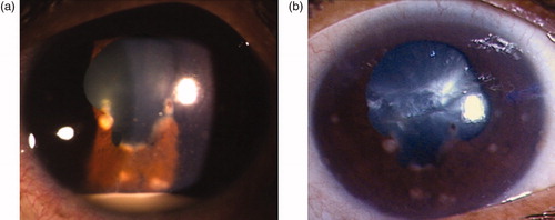
Slit-lamp biomicroscopy of the left eye showed granulomatous uveitis with cells (4+ grade), intense flare (4+ grade),Citation2 hypopyon of 2 mm in the anterior chamber with mutton fat keratic precipitates, iris nodules, posterior subcapsular cataract, and broad-based posterior synechiae (). Fundus details were not clear. The anterior segment of the right eye was normal. Dilated funduscopy of the right eye revealed narrowing of vessels and hard exudates suggestive of hypertensive changes. Ultrasound B scan of the left eye showed few vitreous echoes.
FIGURE 1. (a) Anterior segment photograph of left eye at presentation showing the presence of Koeppe nodules at the pupillary border, broad-based posterior synechiae, and hypopyon. (b) Anterior segment photograph showing resolution of hypopyon.
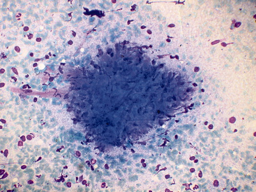
The patient was diagnosed with acute anterior granulomatous uveitis in the left eye and hypertensive retinopathy in the right eye, and a detailed workup was done to investigate the cause. Mantoux skin test, interferon gold test for TB, and ELISA for HIV were negative. She had no history of any infectious disease and was not on rifabutin therapy. Chest x-ray showed hilar lymphadenopathy with normal lung fields. Laboratory examination revealed elevated sedimentation rate (70 mm/h), low Hb (5 g%), and rise in serum creatinine (15.53 mg%) and blood urea (251.23 mg%). Ultrasonography of the abdomen was suggestive of end stage renal disease. Aqueous tap for TB PCR was positive for Mycobacterium tuberculosis. Biopsy from a right-side cervical lymph node stained positive for acid-fast bacilli (), suggestive of tuberculous cervical adenitis along with the presence of granuloma, multinucleate giant cells, and degenerated inflammatory cells with necrosis (). A PET scan showed an intense uptake in the right cervical and supraclavicular lymph nodes and mildly increased uptake in the inferior aspect of the globe.
FIGURE 2. Histopathology section from cervical lymph node showing acid-fast bacilli (arrows).
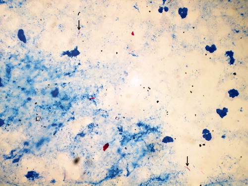
FIGURE 3. Cervical node biopsy showing granuloma with multinucleate giant cells, degenerated inflammatory cells, and necrosis.
The patient received antituberculosis therapy with concomitant systemic and topical corticosteroids. One week later, the anterior chamber showed improvement with disappearance of hypopyon and media clarity improved to grade 2 (). The fundus at this stage showed little vitreous debris inferiorly in pars plana region. Long-term follow-up was not possible because the patient succumbed to renal failure.
Discussion
Tubercular anterior uveitis commonly presents as chronic iridocyclitis, complicated inevitably by extensive broad-based posterior synechiae and cataract.Citation1,Citation3,Citation4 Presence of hypopyon is generally not associated with a clinical impression of intraocular TB and thus may lead to investigating the patient in a different direction. In a clinocopathological report, Rathinam and RaoCitation5 reported a case of pigmented hypopyon where the eye had to be enucleated and the diagnosis of Tb confirmed. Ni et al.Citation6 reported turbid, hemorrhagic, grayish yellow exudate in the anterior chamber in 1 patient and fibrinous hypopyon in 3 patients with presumed TB. However, the hypopyon in our case was nonmobile, 2 mm in height, white associated with granulomatous inflammation, and not fibrinous, hemorrhagic, or pigmented, which makes it a unique presentation. TB may have developed in this case due to prolonged immunosuppression. The documentation of acid-fast bacilli from the cervical nodes and molecular biology confirmed the diagnosis of TB. This case illustrates that TB may rarely present with hypopyon uveitis.
Declaration of interest
The authors report no conflicts of interest. The authors alone are responsible for the content and writing of the paper.
References
- Gupta V, Gupta A, Rao NA. Intraocular tuberculosis—an update. Surv Ophthalmol. 2007;52:561–587
- Jabs DA, Nussenblatt RB, Rosenbaum JT. Standardization of Uveitis Nomenclature (SUN) Working Group. Am J Ophthalmol. 2005;140:509–516
- Gupta A, Bansal R, Gupta V, et al. Ocular signs predictive of tubercular uveitis. Am J Ophthalmol. 2010;149:562–570
- Gupta A, Gupta V, Bansal R, et al. (2009). Ocular tuberculosis in endemic areas. In: Gupta A, Gupta V, Herbort CP, Khairallah M (eds.), Uveitis Text and Imaging (pp 563–577). New Delhi: Jaypee Brothers
- Rathinam SR, Rao NA. Tuberculous intraocular infection presenting with pigmented hypopyon: a clinicopathological case report. Br J Ophthamol. 2004;88:721–722
- Ni C, Palpale JJ, Robinson JL, et al. Ocular tumors and other ocular pathology: a Chinese-American collaborative study. Int Ophthalmol Clin. 1982;22:103–124