Pemphigus vulgaris (PV) is an autoimmune bullous disease characterized by blistering of the skin and mucous membranes, which usually precede former.Citation1 The most common presenting sign of PV is oral mucosal ulceration, but all stratified squamous epithelial mucosal surfaces can be involved in the disease process. Ocular involvement in patients with PV has rarely been reported.Citation2,Citation3 The most common ocular finding is bilateral conjunctivitis, which can be associated with conjunctival hyperemia, ocular irritation, and mucus secretions. In the advanced stage of the disease rupturing of blisters can give rise to conjunctival erosions of the bulbar/palpebral conjunctiva or at the eyelid margin.Citation4 In all reported cases, ocular involvement is limited to the conjunctiva and eyelids and has no effect on visual acuity.Citation3–7 As an addition to the current knowledge we herein report an unusual case of PV who presented with a conjunctival mass with corneal involvement confirmed by histopathological evaluation.
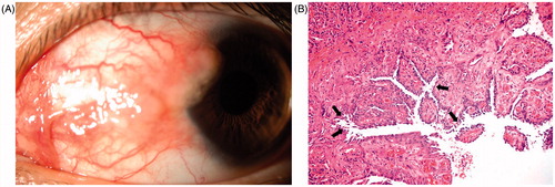
A 22-year-old Caucasian male was referred to our department due to hyperemia and swelling of conjunctiva on his left eye. His medical history was remarkable for a 13-month history of blisters and erosions on his trunk and extremities and erosions on the buccal mucosa. At that time, histology and immunofluorescence findings together with clinical presentation were diagnostic for PV and he was put on prednisone at 1 mg/kg/day and azathioprine at 100 mg/day treatment by the attending dermatologist. Mucosal and cutaneous lesions were healed with postinflammatory hyperpigmentation. The doses of prednisone and azathioprine were tapered gradually. Ophthalmologic examination was normal in the right eye. Entering uncorrected visual acuities were 20/20 in both eyes, and external examination was unremarkable. Slit-lamp examination of the left eye revealed localized conjunctival hyperemia with lobulated, 12 × 6 mm in size tumoral mass encroaching on the cornea on the nasal conjunctiva (). Based on the understanding of the current diagnosis and atypical presentation, an incisional biopsy was performed. Routine histopathologic examination of the specimen revealed suprabasal acantholytic separation in the conjunctival epithelium, which is characteristic of PV (). In some areas of the specimen, subepithelial inflammatory cells and granulation tissue-like congested vessels were also observed. Topical prednisolone acetate 4 times daily was initiated. Healing occurred within 3 weeks with little scar formation. At 6-month follow-up, the patient remained asymptomatic with no evidence of recurrence.
FIGURE 1. (A) Clinical picture shows solitary, lobulated conjunctival tumoral mass associated with conjunctival hyperemia encroaches on the cornea nasally. (B) Histopatology demonstrates suprabasal acantholitic separation (arrows) in the conjunctival epithelial cells and subepithelial congested vessels giving a papillary configuration to mucosa (hematoxylin–eosin stain × 200).
In PV, autoantibodies directed against keratinocyte cell surfaces bind to the keratinocyte desmosomal proteins (desmogleins 1 and 3) and to desmosome-free areas of the keratinocyte cell membrane, resulting in acantholysis, which is a diagnostic clue sought by pathologists. ELISA and direct and indirect immunofluorescence techniques may be used to confirm or enhance definitive diagnosis.Citation8 However, as stated by some authors, diagnosis based on medical history, clinical findings, and histopathology seemed satisfactory in the current case.Citation9 Usually histopathology is not needed in cases with ocular PV associated with classical presentation, including conjunctivitis, blepharitis, mucous discharge, blisters, and eventually erosions. However, mass lesion on the bulbar or palpebral conjunctiva (with or without corneal involvement) in a patient with PV warrants histopathologic confirmation to differentiate it particularly from conjunctival tumors. Avisar et al. reported two PV cases associated with multiple conjunctival papillomas of eyelid margins, which were associated with human papilloma virus infection.Citation10 Fiore et al. reported a case with bilateral vegetative plica semilunaris involvement.Citation11 However, conjunctival tumoral lesion encroaching on the cornea in PV to our knowledge has not been reported before. Though bilateral involvement is almost a rule, unilateral and asymmetric cases have been reported.Citation3 Contrary to the previous understanding, severity of ocular involvement does not necessarily indicate severity of systemic disease.Citation7 Accordingly, the current case presented with unilateral involvement and controlled systemic PV. Topical or systemic corticosteroid therapy is the mainstay treatment for ocular PV, depending on the degree of ocular and systemic involvement. While mild ocular cases can be controlled with topical corticosteroid therapy, severe cases, especially those with an erosive form of the disease, may require further treatment options, including subconjunctival or systemic corticosteroids, topical or systemic immunosuppresants or immunomodulators, and intravenous immunoglobulins.Citation12,Citation13 Surprisingly, our patient responded well to topical corticosteroid therapy, probably due to the additional therapeutic effect of surgery. Similarly, Fiore et al. found excision of the vegetative plica semilunaris lesion as beneficial in alleviating symptoms. On the other hand, surgery would be the cause of little scar formation left on the nasal conjunctiva after treatment of ocular PV, which typically recover without leaving a scar.
In conclusion, PV may present with a conjunctival inflammatory mass without associated typical findings, such as conjunctivitis, mucous discharge, blisters, and erosions. Incisional or excisional biopsy is required in differential diagnosis as they resemble conjunctival tumors. Topical corticosteroid therapy with the additional therapeutic effect of surgery seems satisfactory in the treatment of ocular PV associated with conjunctival inflammatory mass lesion. However, further studies are needed to confirm this observation.
Declaration of interest
The authors report no conflicts of interest. The authors alone are responsible for the content and writing of the paper.
References
- Casiglia J, Woo SB, Ahmed AR. Oral involvement in autoimmune blistering disease. Clin Dermatol. 2001;19:737–741
- Ahmed AR, Sami N. Uncommon manifestations of pemphigus vulgaris. J Eur Acad Dermatol Venereol. 2002;16:313–315
- Daoud YJ, Cervantes R, Foster CS, et al. Ocular pemphigus. J Am Acad Dermatol. 2005;53:585–590
- Brackley R, Pagani JM. Conjunctival erosions associated with pemphigus vulgaris. Optom Vis Sci. 2011;88:1010–1013
- Smith RJ, Manche EE, Mondino BJ. Ocular cicatricial pemphigoid and ocular manifestations of pemphigus vulgaris. Int Ophthalmol Clin. 1997;37:63–75
- Lifshitz T, Levy J, Cagnano E, et al. Severe conjunctival and eyelid involvement in pemphigus vulgaris. Int Ophthalmol. 2004;25:73–74
- Palleschi GM, Giomi B, Fabbri P. Ocular involvement in pemphigus. Am J Ophthalmol. 2007;144:149–152
- Elchahal S, Kavosh ER, Chu DS. Ocular manifestations of blistering diseases. Immunol Allergy Clin North Am. 2008;28:119–136
- Mokhtari M, Rasolmali R, Kumar PV. Pemphigus vulgaris of skin: cytological findings and pitfalls. Acta Cytol. 2012;56:310–314
- Avisar I, Yassur I, Kremer I. Multiple conjunctival papillomas of eyelid margins in pemphigus vulgaris. Case Rep Ophthalmol Med. 2011;2011:174912
- Fiore JM, Perry HD, Donnenfeld ED, et al. Pemphigus vulgaris: bilateral plica semilunaris involvement. Cornea. 2011;30:357–359
- Olszewska M, Komor M, Mazur M, et al. Response of ocular pemphigus vulgaris to therapy. Case report and review of literature. J Dermatol Case Rep. 2008;29:1–3
- Kozeis N, Tyradellis S, Dragiotis E, et al. Triamcinolone acetonide for rare ocular manifestations of pemphigus vulgaris: a case report. Clin Ophthalmol. 2010;26:365–368