Abstract
Purpose: To report the detection of Toxoplasma gondii cysts in intraocular aspirates of patients with necrotizing retinitis following periocular/intraocular corticosteroid injection.
Design: Case report.
Methods: Two patients (2 eyes) with widespread necrotizing retinitis in a steroid-exposed eye posed a diagnostic challenge and underwent pars plana vitrectomy (PPV). Intraocular samples (vitreous fluid, retinal tissue, and subretinal aspirate in case 1, and vitreous fluid in case 2) were subjected to cytological examination.
Results: The subretinal aspirate (case 1) revealed encysted bradyzoites of Toxoplasma gondii. Vitreous fluid (case 2) tested positive for anti-toxoplasma antibodies and the smear showed encysted forms of Toxoplasma gondii on cytology.
Conclusion. Toxoplasma gondii cysts were detected in eyes with necrotizing retinitis that developed secondary to injudicious use of corticosteroids.
Toxoplasma gondii is an intracellular parasite with a predilection for multiple tissues, including the eye. Ocular toxoplasmosis characteristically displays focal necrotizing retinitis, occurring from the activation of cysts in the retina. Within the necrotic retina, cysts of Toxoplasma gondii can be found.Citation1 Yet, direct confirmation by demonstrating organisms in ocular samples is extremely difficult.Citation2 Its diagnosis is usually clinical, confirmed by presence of anti-toxoplasma antibodies. Direct evidence from vitreous fluid is increasingly becoming possible by polymerase chain reaction (PCR) and, rarely, by cytology.Citation3 More invasive retinal/subretinal biopsies are performed following a failure to confirm a diagnosis.Citation4
Diagnosis of ocular toxoplasmosis presents a challenge in immunocompromised individuals when it causes diffuse necrotizing retinitis, mimicking viral retinitis. We report two patients who developed widespread necrotizing retinitis following exposure to corticosteroids, and did not respond to empiric therapy for viral retinitis. Cytological examination revealed the presence of Toxoplasma gondii cysts in intraocular samples.
Case 1
A 71-year-old male presented to us with right eye panuveitis, having received elsewhere a posterior subtenon injection of triamcinolone acetonide 9 months before. The best-corrected visual acuity (BCVA) was 6/60 and 6/6 in the right and left eyes, respectively. The intraocular pressure (IOP) was 30 and 13 mm Hg in the right and left eyes, respectively. The right eye biomicroscopy showed ++ flare and + cells in the anterior chamber, posterior synechiae, and significant vitritis. Fundus examination revealed a patch of retinitis in the macula with scattered retinal hemorrhages (). An extensive area of retinal atrophy was noted inferotemporal to the macula. The left eye was normal. He was seronegative for human immunodeficiency virus infection. The clinical picture and a positive serum IgG for HSV was suggestive of acute retinal necrosis and was unsuccessfully treated with acyclovir and oral corticosteroid therapy. In view of the persistent inflammation at 6 months, he underwent pars plana vitrectomy (PPV). Vitreous fluid revealed no organisms or malignant cells on cytology. At 10 months, retinal tissue and subretinal aspirate were obtained during repeat PPV for cytology, followed by silicon oil tamponade. Retinal tissue was normal but the subretinal aspirate revealed encysted bradyzoites, structures consistent with the encysted bradyzoites of Toxoplasma gondii cysts (). After 3 days of antitoxoplasma drugs, BCVA was counting fingers, IOP 10 mmHg, anterior chamber showed cells +, flare +, and retina was attached (). The patient was lost to follow up thereafter.
Figure 1. Fundus photograph (a) of right eye of the patient (case 1) at initial visit with visual acuity 6/60. There was significant vitritis with a yellowish placoid lesion in posterior pole and retinal hemorrhages in periphery.
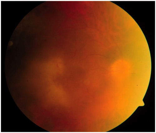
Figure 2. Subretinal aspirate (case 1) showing structures consistent with Toxoplasma gondii cysts with numerous bradyzoites, and a pigment-laden macrophage (May-Grumwald Giemsa, ×1000).
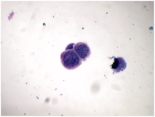
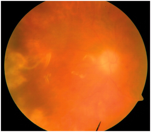
Figure 3. Fundus photograph (case 1) 3 days after surgery (pars plana vitrectomy + partial retinectomy + endolaser photocoagulation + silicon oil tamponade) showing attached retina in a silicon-oil filled eye.
Case 2
A 29-year-old female presented with decreased vision in the right eye since 8 months. She had received intravitreal triamcinolone acetonide, oral corticosteroids, and antitubercular therapy elsewhere. The BCVA was counting fingers and 6/6 in the right and left eyes, respectively. The IOP was 15 and 13 mmHg in the right and left eyes, respectively. The right eye anterior segment examination was unremarkable. Fundus examination revealed a widespread area of necrotizing retinitis in the posterior pole with a healed choroiditis scar along inferotemporal arcade (). The left eye was normal. Serum antitoxoplasma IgG antibodies were positive. Vitreous aspirate from diagnostic PPV was positive for antitoxoplasma IgG and IgM antibodies. Vitreous cytology smear showed structures consistent with Toxoplasma gondii cysts (). One month after initiating antitoxoplasma therapy, BCVA was counting fingers and lesions were resolving ().
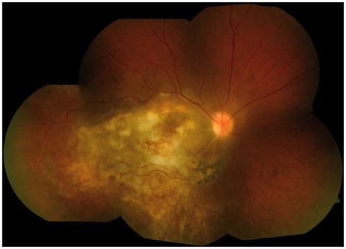
Figure 4. Fundus photograph of right eye (case 2) shows a widespread necrotizing retinitis with a healed choroiditis scar along the lower temporal arcade.
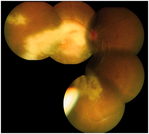
Figure 5. Vitreous smear (case 2) showing structures consistent with Toxoplasma gondii cysts in the main picture and in the inset (May-Grumwald Giemsa, ×1000).
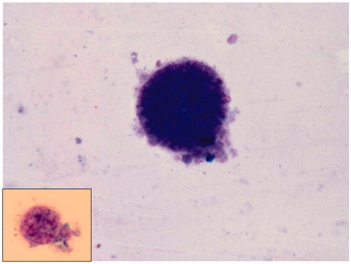
Figure 6. Fundus photograph (case 2) at 1 month follow-up showing healing lesions after anti-toxoplasma therapy was initiated.
Discussion
There are occasional reports of demonstration of toxoplasma organisms in tissue biopsies/cytology,Citation3,Citation4, necropsy material,Citation5 or enucleated eyes.Citation6,Citation7 Within the eye, the parasite has an affinity for retina, particularly the posterior pole. Leucocyte taxis and direct interaction with the vascular endothelium have been proposed mechanisms. An in vitro study has demonstrated that tachyzoites are capable of independent migration across human vascular endothelium.Citation8
Our patients had been extensively treated with local (periocular/intravitreal) and oral corticosteroids that triggered widespread retinal necrosis facilitating the growth and migration of toxoplasma parasites leading to their subsequent detection on cytology of subretinal and vitreous aspirates. In the first case, failure to detect them in the vitreous aspirate or retinal tissue highlights the difficulty in detecting these organisms on cytology of intraocular samples. The parasite may harbor in selected areas of the eye, and may not be diffusely present in the uvea and retina. Their isolation from the subretinal aspirate in this case, as also reported by Adan et al. recently,Citation4 may suggest their preferential colonization in the deeper layers in an immunocompromised state. Adan et al. reported an atypical toxoplasmic retinochoroiditis in an elderly male with post-transfusional hepatitis C who also received high-dose systemic corticosteroids.Citation4 The vitreous aspirate did not reveal toxoplasma in PCR or on cytology. The subretinal aspirate revealed toxoplasma organisms on cytology. Their isolation from the vitreous aspirate (as in our second case, and also reported previouslyCitation3,Citation7) may suggest a more extensive necrosis of the retina, releasing the cysts into the vitreous cavity.Citation9
While PCR is an easier technique and preferred in certain infectious uveitis (viral) over the Goldmann-Witmer coefficient (GWC), which detects intraocular antibody production, the latter has shown superior results in diagnosing ocular toxoplasmosis when compared to PCR of aqueous humor.Citation10 The slow release of Toxoplasma gondii tachyzoites from the cysts into the ocular fluid may explain the absence of positive PCR results in early stages.Citation10 Further, their subsequent accumulation within the vitreous cavity may explain their detection on cytological examination, which is usually performed much later following an initial negative vitreous biopsy report of PCR. In an immunocompromised individual, PCR seems preferable to GWC analysis since the antibody production is unpredictable. However, in case of ocular toxoplasmosis, GWC analysis has contributed to its diagnosis considerably more than PCR in immunosuppressed patients.Citation11
Due to the atypical clinical presentations, an initial noncontributory vitreous biopsy report, a poor response to therapy, and a high clinical suspicion of toxoplasmosis in an immunocompromised eye, we resorted to more invasive tests, such as retinal or subretinal biopsy. In our cases, the long period of inadequate treatment before definite cytological examination might have contributed to the diagnostic yield of these tests. The cytopathologist needs to be vigilant for a meticulous search for organisms in these samples. With advancement in sampling techniques, it may be possible to demonstrate the protozoan in a greater proportion of ocular samples sent to the cytology laboratory.
Differentiation of infectious from noninfectious etiology is of utmost importance in the treatment of uveitis as they differ largely in terms of treatment and prognosis. Inadvertent use of corticosteroids should be avoided in an inflamed eye, particularly in the absence of a definitive diagnosis.
Declaration of interest
The authors report no conflicts of interest. The authors alone are responsible for the content and writing of the paper.
References
- Wilder HC. Toxoplasma chorioretinitis in adults. Arch Ophthalmol. 1952;48:127–136
- Brezin AP, Kasner L, Thulliez P, et al. Ocular toxoplasmosis in the fetus: immunohistochemistry analysis and DNA amplification. Retina. 1994;14:19–26
- Greven CM, Teot LA. Cytologic evaluation of Toxoplasma gondii from vitreous fluid. Arch Ophthalmol. 1994;112:1086–1088
- Adan A, Sole M, Mateo C, et al. Cytologic identification of Toxoplasma gondii from subretinal aspirate. Acta Ophthalmol. 2012;90:392–393
- Yeo JH, Jakobiec FA, Iwamoto T, et al. Opportunistic toxoplasmic retinochoroiditis following chemotherapy for systemic lymphoma: a light and electron microscopic study. Ophthalmology. 1983;90:885–898
- Rao NA, Font RL. Toxoplasmic retinochoroiditis: electron microscopic and immunofluorescence studies of formalin-fixed tissue. Arch Ophthalmol. 1977;95:273–277
- Belfort RN, Rasmussen S, Kherani A, et al. Bilateral progressive necrotizing retinochoroiditis in an immunocompromised patient: histopathological diagnosis. Acta Ophthalmol. 2010;88:614–615
- Furtado JM, Bharadwaj AS, Chipps TJ, et al. Toxoplasma gondii tachyzoites cross retinal endothelium assisted by intercellular adhesion molecule-1 in vitro. Immunol Cell Biol. 2012 Apr 24. doi: 10.1038/icb.2012.21. [Epub ahead of print]
- Moshfeghi DM, Dodds EM, Couto CA, et al. Diagnostic approaches to severe, atypical toxoplasmosis mimicking acute retinal necrosis. Ophthalmology. 2004;111:716–725
- De Groot-Mijnes JDF, Rothova A, Van Loon AM, et al. Polymearse chain reaction and Goldmann-Witmer coefficient analysis are complimentary for the diagnosis of infectious uveitis. Am J Ophthalmol. 2006;141:313–318
- Westeneng AC, Rothova A, De Boer JH, De Groot-Mijnes JDF. Infectious uveitis in immunocompromised patients and the diagnostic value of polymerase chain reaction and Goldmann-Witmer coefficient in aqueous analysis. Am J Ophthalmol. 2007;144:781–785