
We read with great interest the article entitled “Divided nevus of the eyelid: successful treatment with CO2 laser” (Citation1) and commend the author on the good result. Reconstruction of eyelid and periorbital defects has always been a challenge for plastic surgeons. Aesthetic and functional repair requires donor tissue highly matched in color, elasticity and thickness. The skin of the eyelid is the thinnest in the body, meaning that its matching tissue is not easy to find. Various minimally invasive approaches have been proposed with the purpose of avoiding surgeries with donor site morbidity. CO2 laser has been reported to be effective in divided eyelid nevus with the advantages of simplicity and precision, but is it really a perfect choice for eyelid nevus?
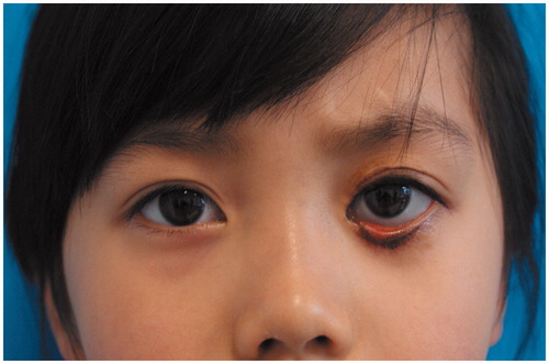
In this study, we present a case with both upper and lower eyelids ectroption after CO2 laser treatment of divided eyelid nevus. The patient was a four-year-old girl. CO2 laser was performed when she was two years old in another hospital. One month after the treatment, most of the crusts have fallen off, exposing erythematous wound base. The ectroption of lower eyelid was noticed by her parents at the same time (). It can be deduced from the photo that the area of the nevus was more than two-thirds of the upper and lower eyelids, respectively. Conservative treatment with ointment and anti-cicatricial drugs was then provided. She presented to our department for help 2 years after the laser treatment. The ectroption persisted. There were residual nevus and hyperpigmentation on both eyelids with asymmetric double eyelid height ().
Figure 1. A two-year-old girl with congenital divided eyelid nevus. One month after CO2 laser therapy, most crusts have fallen off, exposing an erythematous wound. Lower eyelid ectropion was also noted.
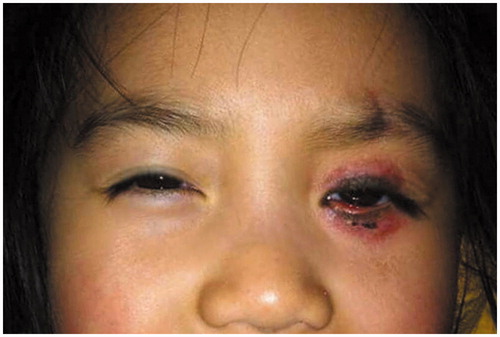
Figure 2. Two years after the laser treatment, ectropion of left lower eyelid persisted, with residual nevus, hyperpigmentation and asymmetric double eyelid height.
Possible reasons of the ectroption are as follows: (1) the area of both the upper and lower lesions was large, so there was extensive scar contracture after wound healing; (2) the patient was a child, which means more skin tissue was needed during her later growth; and (3) the CO2 laser treatment might be too radical. The wound healing time was 30 days, which was much longer than usual.
For young patients and large lesions, we strongly suggest that laser therapy of eyelid nevus should be deliberate. Possible complications include ectroption, scar, recurrence and pigmentation. Various surgical approaches of eyelid reconstruction have been proposed, lots of which have produced satisfactory results (Citation2–4). The treatment consists of full-thickness excision followed by repair with skin grafts or flaps. According to our practice, surgical treatment is more reliable in these cases for nevus removal and functional and aesthetic reconstruction.
Declaration of interest
None of the authors has a financial interest in any of the products, devices or drugs mentioned in this manuscript.
References
- Zeng Y. Divided nevus of the eyelid: successful treatment with CO2 laser. J Dermatolog Treat. 2014;25:358–9
- Glat PM, Longaker MT, Jelks EB, et al. Periorbital melanocytic lesions: excision and reconstruction in 40 patients. Plast Reconstr Surg. 1998;102:19–27
- Margulis A, Adler N, Bauer BS. Congenital melanocytic nevi of the eyelids and periorbital region. Plast Reconstr Surg. 2009;124:1273–83
- Zhu L, Qiao Q, Liu Z, et al. Treatment of divided eyelid nevus with island skin flap: report of ten cases and review of the literature. Ophthal Plast Reconstr Surg. 2009;25:476–80