
ABSTRACT
Since the 1970s, computational modeling has been used to investigate the fundamental mechanisms of cochlear implant stimulation. Lumped parameter models and analytical models have been used to simulate cochlear potentials, as well as three-dimensional volume conduction models based on the Finite Difference, Finite Element, and Boundary Element methods. Additionally, in order to simulate neural responses, several of these cochlear models have been combined with nerve models, which were either simple activation functions or active nerve fiber models of the cochlear auditory neurons. This review paper will present an overview of the ways in which these computational models have been employed to study different stimulation strategies and electrode designs. Research into stimulation strategies has concentrated mainly on multipolar stimulation as a means of achieving current focussing and current steering, while modeling work on electrode design has been chiefly concerned with finding the optimal position and insertion depth of the electrode array. Finally, the present and future of computational modeling of the electrically stimulated cochlea is discussed.
1. Introduction
Cochlear implants stimulate the auditory nerve electrically, thereby providing an audible sound percept to the hearing impaired implant user. Since both the electrical fields generated by the implant as well as the dynamic responses of the auditory neurons can be described in terms of electromagnetic physics, the functioning of cochlear implants is an ideal subject for computational modeling. Indeed, the earliest computational model relevant to the study of the electrically stimulated cochlea dates back to the 1970s, when Strelioff (Citation1973) used a so-called lumped parameter model to describe electrical properties of cochlear structures (). Computational models from the early years of cochlear implant research used this type of lumped parameter approach to describe electrical stimulation of the cochlea (Black and Clark Citation1980; Black et al. Citation1983; Suesserman and Spelman Citation1993; Rodenhiser and Spelman Citation1995; Spelman et al. Citation1995; Jolly et al. Citation1996; Kral et al. Citation1998), in addition to employing analytical functions (O’Leary et al. Citation1985; Spelman et al. Citation1995; Jolly et al. Citation1996). These lumped parameter and analytical models were not yet coupled to computational models of the auditory nerve, so they were restricted to examining the electric potentials and current distributions inside the cochlea.
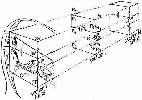
Figure 1. Illustration of the lumped parameter model from Strelioff (Citation1973).
Meanwhile, in the field of neural science, computational models of electrically stimulated nerve fibers were being developed, starting with the pioneering work of Frankenhæuser and Huxley (Citation1964) on the myelinated nerve fiber of toads. Subsequent researchers developed the principle of neurons modeled as electrical networks (McNeal Citation1976; Reilly et al. Citation1985; Rattay Citation1987), leading to the first electrical cable model of the mammalian auditory neuron (Colombo and Parkins Citation1987).
Although earlier studies had already used three-dimensional volume conduction models to investigate electric potentials in the cochlea (Girzon Citation1987; Sapozhnikov Citation1990), Finley et al. were the first to publish simulations of a three-dimensional volume conduction model combined with a (preliminary) cable model of the auditory nerve (Finley et al. Citation1990). They used the Finite Element Method (FEM) to calculate the electric potential distribution in their unrolled human cochlear geometry and then coupled the results to their version of an auditory nerve fiber model, which was based on the neural modeling works referenced above. From there on, several research groups have used volume conduction models to simulate electric potentials in increasingly sophisticated geometries of implanted cochleae. First as unrolled cochleae (), then with rotationally symmetric geometries (), and finally as increasingly realistic spiraling structures () (Finley et al. Citation1990; Frijns et al. Citation1995; Briaire and Frijns Citation2000; Frijns et al. Citation2001; Hanekom Citation2001; Rattay et al. Citation2001b; Choi et al. Citation2004; Choi et al. Citation2005; Choi et al. Citation2006; Tognola et al. Citation2007; Whiten Citation2007; Nogueira et al. Citation2014; Pau et al. Citation2014; Kalkman et al. Citation2015; Malherbe et al. Citation2015b; Wong et al. Citation2016). Despite this tendency to move to more detailed cochlear geometries, simpler mathematical models of unrolled cochleae have retained their usefulness and are still employed in specific situations (Litvak et al. Citation2007; Bonham and Litvak Citation2008; Goldwyn et al. Citation2010).
Figure 2. Illustration of the unrolled human cochlea geometry used by Finley et al. (Citation1990). Figure a shows a close-up view of how the cross section of the cochlea and electrode is segmented; Figure b shows the full geometry.
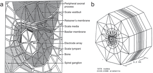
Figure 3. Rotationally symmetric geometry of the guinea pig cochlea from Frijns et al. (Citation1995, Citation1996).
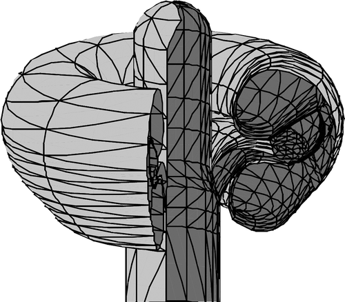
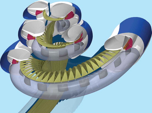
Figure 4. Spiraling tapered geometry of the implanted human cochlea from Kalkman et al. (Citation2015).
Additionally, electrical models of the auditory nerve have been further refined by incorporating data from electrophysiological single fiber experiments on mammalian neurons and morphological details of the human auditory nerve (Frijns et al. Citation1995; Rattay et al. Citation2001a; Briaire and Frijns Citation2005; Dekker et al. Citation2014). However, not all of the developed cochlear models have incorporated active neural models; instead, some studies employ the so-called activation function to estimate neural responses (Finley et al. Citation1990; Litvak et al. Citation2007; Bonham and Litvak Citation2008; Choi and Hsu Citation2009; Goldwyn et al. Citation2010; Wong et al. Citation2016). This activation function is equal to the second spatial derivative of the electric potential along the nerve fibers (Rattay Citation1986). Although relatively simple to implement, the activation function only gives an indication of neural thresholds, and cannot be used to model more complex aspects of neural stimulation. For instance, simulating neural responses to pulse trains require either active nerve fiber models or stochastic nerve models; the latter of which have mainly been implemented as models of single nodes or fibers (Bruce et al. Citation1999b; Bruce et al. Citation1999a; Rubinstein et al. Citation1999; Imennov and Rubinstein Citation2009; Woo et al. Citation2010). Furthermore, active nerve models also enable the simulation and validation of electrically evoked compound action potentials (eCAPs) (Briaire and Frijns Citation2005; Briaire and Frijns Citation2006; Whiten Citation2007; Smit et al. Citation2009; Westen et al. Citation2011; Choi and Wang Citation2014).
Computational models are well suited to provide insight into the underlying mechanisms of cochlear stimulation. Furthermore, they can be used to simulate various types of experiments that are impractical or impossible to perform in cochlear implant patients or animal models. For example, new types of electrode arrays can be tested and experimental stimulation paradigms can be evaluated iteratively, without the need for human or animal test subjects, or any of the practical requirements for performing in vivo tests. The aim of the present paper is to review the modeling studies that have been performed over the years to gain insight into stimulation strategies and electrode designs.
2. Multipolar stimulation
Multipolar stimulation has been a major theme throughout cochlear implant modeling research. In the years before the now commonly used Continuously Interleaved Sampling (CIS) strategy (Wilson et al. Citation1991), there was much interest in multipolar stimulation as a way of reducing the extensive electrical interaction inherent in simultaneous monopolar stimulation, which was hampering cochlear implant performance at the time. However, even though the majority of modern clinical stimulation strategies avoid simultaneous activation of cochlear implant electrode contacts, multipolar stimulation has continued to be of interest in the research field, particularly as a means of producing more localized regions of neural excitation in order to increase spatial selectivity (current focussing).
In this section, we will be reviewing modeling studies which have investigated various forms of multipolar stimulation. As will be shown, the most commonly investigated multipolar configurations have been bipolar and tripolar stimulation. In bipolar stimulation, two intracochlear electrode contacts are stimulated in opposite polarity, which causes no net current to leave or enter the cochlea (a). In tripolar stimulation, three intracochlear contacts are stimulated, one of which is considered the center contact and the other two flanking/inhibiting contacts that stimulate at a polarity opposite to that of the center contact (b). The current amplitudes injected on the flanking contacts are half that of the center contact, so that, as in bipolar stimulation, the net current in the cochlea is zero.
Figure 5. Schematic illustration of different multipolar strategies: bipolar stimulation (a1&a2), tripolar stimulation (b1&b2), partial tripolar stimulation with σ = 0.5 (c1&c2), and phased array stimulation for an electrode array with five contacts (d1&d2). The top figures (a1–d1) show the stimulus current amplitudes used for each multipolar strategy, and bottom figures (a2–d2) show the resulting electrical potentials along the electrode array for each individual contact (blue, red, green, purple, and orange curves), as well as their combined potential (black curve).
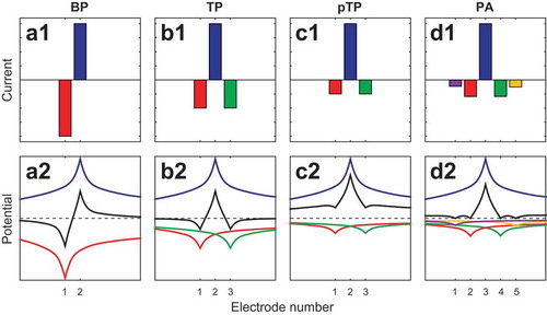
Other multipolar configurations have simulated as well, one of which is called the partial tripole (c). Partial tripolar stimulation is, as the name implies, essentially a mitigated version of tripolar stimulation, where the current amplitude of the flanking contacts is multiplied by a fraction, usually denoted as σ. This means that the net current injected into the cochlea is not zero, but that part of the current, equal to (1 − σ) times the amplitude of the center contact, leaves or enters the cochlea to or from the return electrode. Note that by this definition, σ = 1 results in a normal tripole, while σ = 0 simply amounts to monopolar stimulation.
Another, relatively new, multipolar configuration is referred to as phased array stimulation (d), which was proposed in a study by Van den Honert and Kelsall (Van den Honert and Kelsall Citation2007), though its concept traces back to the work of Van Compernolle (Citation1985). In phased array stimulation, all contacts of the array are stimulated in such a way that the electrode potentials are zero everywhere except at one specific contact, which we will designate the center contact (though it need not be located in the center of the array). The currents required to achieve this are computed using an impedance matrix, determined by recording electrode potentials for each stimulating contact of the array.
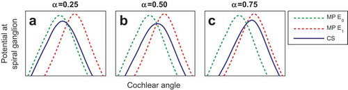
The aforementioned multipolar configurations are all used as a means of current focussing; however, other applications for multipolar stimulation exist. In this section, we will also encounter the so-called current steering paradigm (), also referred to as simultaneous dual electrode stimulation. In current steering, two electrode contacts are stimulated at equal polarity, with the current amplitudes of both controlled by a parameter that is usually denoted as α. The current amplitude on one of the contacts is then equal to a base value multiplied by α, while the amplitude of the other contact is equal to a base value multiplied by (1 − α). This means that increasing the value of α from 0 to 1 will gradually shift the potential field from one electrode to the other, with the intent of creating “virtual channels” that lie in between the two stimulating contacts.
Figure 6. Schematic illustration of the current steering strategy. The curves show the electrical potential along the spiral ganglion generated by two electrode contacts, labeled E0 and E1, stimulated individually in monopolar mode (green and red dotted curves), and stimulated together as a current steered electrode pair for different values of α (blue curves). Figure a, b, and c show the potentials at α = 0.25, α = 0.5, and α = 0.75, respectively. Since the monopolar curve for E0 (green) is essentially the current steered potential for α = 0 and the curve of E1 (red) is that of α = 1, it is clear that the current steered curve (blue) gradually shifts from the monopolar field of E0 to that of E1 as the value of α increases. Note that although the peak of current steered potential is lower for intermediate values of α, this does not necessarily imply that the neural threshold is also lower.
2.1. Multipoles in simple mathematical models
The earlier lumped parameter modeling studies examined electric potentials from monopolar and bipolar stimulation in the cochlea and compared them to electrophysiological experiments in cats (Black and Clark Citation1980; Black et al. Citation1983; O’Leary et al. Citation1985). They came to the conclusion that bipolar stimuli produced sharper, more localized potentials than monopolar ones did. Furthermore, Black and Clark observed that the spread of current through the cochlear scalae could be quite different than the current spread in other parts of the cochlea, such as the organ of Corti (Black and Clark Citation1980), which underlined the usefulness of modeling as a way of estimating electrical field distributions in cochlear locations that are difficult or impossible to access clinically or electrophysiologically.
Spelman et al. performed several modeling studies which explored the possibilities of multipolar stimulation. In the first, Suesserman and Spelman examined potentials at the organ of Corti induced by parallel stimulation of bipolar and (partial) tripolar configurations in a lumped parameter model of the first turn of the guinea pig cochlea, with the goal of determining independent channels that could safely be stimulated simultaneously (Suesserman and Spelman Citation1993). Their results showed that bipolar stimuli produced localized potential peaks at the organ of Corti, and suggested that tripolar configurations would be capable of generating peaks that were even sharper than those of bipolar stimuli.
Since it was apparent that multichannel cochlear implants were able to shape potential fields by parallel stimulation of channels, Rodenhiser and Spelman investigated the possibility of creating focussed electrical fields calculated from impedance data of individual cochlear implant electrode contacts (Rodenhiser and Spelman Citation1995). Based on the work of Van Compernolle (Citation1985), they used their lumped parameter model to calculate potentials induced along the organ of Corti by each electrode contact and used them to define an impedance matrix. This impedance matrix was then used to find the optimal combination of driving currents necessary to generate potentials that were similar to a desired potential curve, by employing the least-squares method. The results indicated that current focussing using impedance data was a promising technique, but the authors noted that the smaller potential peaks induced by focussed stimuli might make them too electrically inefficient or even unsafe to use in clinical practice.
In their third modeling study, Spelman et al. (Citation1995) combined insights from their lumped parameter model with an analytical model of the neural activation function and electrophysiological measurements performed in monkeys and guinea pigs. The study used monopolar and bipolar stimuli, in addition to what they referred to as “quadrupolar” stimulation, which is nowadays known as tripolar stimulation (Spelman et al. reasoned that it can be seen as two dipoles, with one of the polarities from both dipoles physically overlapping each other). They found that, when comparing tripolar to monopolar stimulation, the model did not agree very well with psychophysical thresholds measured in monkeys. They hypothesized that this disagreement was due to separate areas of excitation caused by the two inhibiting flanking electrodes of the tripolar configuration. This hypothesis was further elaborated on by Clopton and Spelman in an accompanying neural modeling study (Clopton and Spelman Citation1995). Results from guinea pig measurements from Spelman et al. (Citation1995) also supported the idea that current steering and current focussing were feasible with cochlear implants, although with certain constraints.
The final modeling study from Spelman et al. investigated tripolar stimulation once more (which was again referred to as quadrupolar stimulation), for which they used both an analytical model of point sources located in an infinite homogeneous isotropic medium as well as their lumped parameter model (Jolly et al. Citation1996). Their results reiterated the idea that tripolar stimulation has reduced current spread and electrical channel interaction, making parallel stimulation feasible and possibly improving pitch and electrode pair discrimination. However, their results again suggested the possible presence of “side lobes”: secondary areas of excitation near the flanking contacts.
In 2007, Litvak et al. published a joint modeling and psychophysical study on loudness growth with partial tripolar stimulation (Litvak et al. Citation2007). Their model had a straightforward approach similar to the analytical part of Jolly et al.’s model, describing point sources located in an infinite homogeneous medium at a parametric distance from neuronal elements. They calculated neural activation functions for partial tripolar stimulation, while varying the parameter σ, the electrode–neuron distance, and changing the spacing between the center contact and flanking contacts. Their psychophysical experiments consisted of loudness balancing of partial tripolar stimuli in seven subjects for different values of σ and flanking electrode spacing. They found that for increasing values of σ more current was needed to achieve comfortable loudness, to the point where in some subjects the compliance limits of the implant were reached. This effect was generally greater when there was more electrical interaction between the stimulating contacts, such as for larger electrode–neuron distances or smaller electrode spacing. However, they also found that in some cases the increase in required current diminished at higher values of σ, which their modeling results suggested was due to the occurrence of side lobes. They concluded that (partial) tripolar stimulation reduced spatial selectivity and that choosing an optimal value of σ could help avoid possible side-lobe excitation and keep stimulation within compliance limits.
The next year, Bonham and Litvak presented another study, which not only looked at dipoles and (partial) tripoles, but also at current steering (Bonham and Litvak Citation2008). The study contained data from modeling, electrophysiology, and psychophysics, in addition to reviewing earlier studies. They used an FEM model with a simple geometry of a conductive tube representing the scala tympani which contained several spherical electrodes, and which was located in an infinite homogeneous medium. Neural activation functions were determined along rudimentary neural trajectories located outside the tube, orthogonal to its axis. Concerning (partial) tripoles, Bonham and Litvak’s observations were in agreement with Litvak et al.’s findings; however, the novelty of the study was in its findings on current steering. Modeling results showed that it was possible to steer the electric potential and the region of neural excitation, which was confirmed by electrophysiological data in the inferior colliculus. Bonham and Litvak concluded that a combination of current focussing and steering might improve cochlear implant perception.
Goldwyn et al. used a similar geometrical setup for their modeling study on partial tripolar stimulation; however, instead of using the FEM, they derived an analytical solution of the electric potential distribution generated by point sources in an infinite tube, and used it to determine activation functions (Goldwyn et al. Citation2010). Goldwyn et al. looked at partial tripoles at different electrode positions and included localized degeneration in their neural distribution. Results were in general agreement with previous studies, and they additionally showed that (partial) tripoles were sensitive to dead neural regions, as spatially restricted excitation region of a tripole could conceivably overlap with an area of neural degeneration. This led Goldwyn et al. to speculate that (partial) tripolar stimulation could be used clinically to locate possible neural dead regions in cochlear implant patients.
2.2. Multipoles in volume conduction models of the cochlea
Finley et al. investigated four bipolar configurations in their three-dimensional volume conduction model of the unrolled human cochlea (Finley et al. Citation1990). First they examined a “pure radial” bipolar setup, where two stimulating plate contacts were located at the same insertion depth, in other words in the same mid-modiolar cross section. Second they used a “pure longitudinal” setup, where the two contacts were separated along the length of the scala tympani. Their third configuration was the “offset radial” setup, which was a combination of radial and longitudinal spacing of the two contacts. Their fourth configuration was bipolar stimulation of two banded contacts spaced longitudinally. In the banded configuration, the electrode array was located along the lateral wall; in the other three configurations, the array was located along the modiolar wall. Their results suggested that a pure radial setup was the most capable of localized neural stimulation, but they noted that their results were strongly dependent on the presence of the neural peripheral processes. The main conclusion from Finley et al. was that, while the basic principles involved in electrical stimulation of the auditory nerve were simple, combining electrical field generation with neural activation could cause complex results. Electrical fields depended heavily on electrode configuration and the neural responsiveness depended on the morphological and electrophysiological details of the nerve fibers. Nonetheless they expressed confidence that an understanding of the mechanisms behind cochlear implant functioning would lead to better electrode designs and stimulation strategies.
In 1995, Frijns et al. investigated longitudinal bipolar stimulation in a rotationally symmetric representation of the guinea pig cochlea (Frijns et al. Citation1995). Their model used the Boundary Element Method (BEM) to compute electric potentials generated by bipolar current sources; these potentials were then used as input for a deterministic active nerve fiber model of the auditory neurons of the guinea pig, which was a generalized version of the Schwarz–Eikhof–Frijns (SEF) model, called gSEF. Excitation patterns showed two distinct areas of excitation near the current sources, and that this excitation was largely occurring at the neural peripheral processes, especially at lower stimulus levels. Furthermore, the rotational nature of the model had introduced the so-called ectopic or cross-turn stimulation into the model; at high current levels, fibers belonging to cochlear turns that did not contain current sources were being stimulated, due to the relatively close spacing of the neural axons in the cochlear modiolus.
Frijns et al. expanded on the study by examining radial dipoles as well as longitudinal dipoles in the same cochlear geometry (Frijns et al. Citation1996), showing that radial dipoles excite neurons at lower thresholds than longitudinal dipoles. They also concluded that, unlike longitudinal dipoles, radial dipoles do not generate separated regions of excitation near the current sources. However, experiments with different pulse shapes revealed that the use of monophasic pulses could reduce one of the two regions of excitation in longitudinal bipolar stimulation, due to the fact that neural excitation thresholds for anodic pulses were generally higher than for cathodic pulses; this increased spatial selectivity and could potentially double the number of nonoverlapping stimulation sites. Since monophasic pulses are considered unsafe for long-term in vivo stimulation, Frijns et al. suggested mimicking monophasic stimulation by using charge-balanced asymmetric biphasic pulses (nowadays referred to as pseudo-monophasic pulses), which was shown to produce equivalent results in the model. It should be noted, however, that in Frijns et al.’s model, the neurons were more sensitive to cathodic pulses than to anodic pulses, while later experiments in human subjects have shown the opposite effect (Macherey et al. Citation2008; Macherey et al. Citation2010); this discrepancy in the computational model has not yet been fully explained.
A subsequent paper updated their guinea pig cochlea to a tapered spiraling geometry (Briaire and Frijns Citation2000) and found comparable results, including the presence of ectopic stimulation. The tapered spiraling geometry allowed the cochlear scalae to act as a transmission line along the entire length of the cochlea, and resulted in asymmetric potential distributions. The study also looked more closely at the near-field potentials of current sources in the scala tympani and found that while scalar potentials roughly followed an exponential decay in the far field (as was widely assumed in preceding lumped parameter models), cochlear potentials near the current source contained an additional spherical component. The cochlear geometry of Frijns et al. would be further updated to human anatomy and a geometrical representation of an electrode array a year later (Frijns et al. Citation2001).
Around the same time, Hanekom published a model of the implanted human cochlea (Hanekom Citation2001). Hanekom’s model contained a spiraling (but not tapered) FEM geometry of the first one and a half turn of the human cochlea, complete with electrode arrays modeled in either lateral or medial position, and was coupled with a neural model which was based on the gSEF model of Frijns et al. (Citation1995, Citation1996). The electrode configurations modeled by Hanekom were reminiscent to those of Finley et al. (Citation1990); longitudinal bipolar stimulation, radial stimulation, offset radial stimulation, and pseudo-monopolar stimulation (i.e., monopolar stimulation with a distant, but intracochlear, reference contact) were simulated, at different electrode spacing and using plate contacts as well as banded contacts. As in the studies above, ectopic stimulation and asymmetric potential distributions were observed, and results showed that longitudinal dipoles generated two areas of excitation, while radial dipoles did not. Additionally, threshold levels for bipolar stimulation were higher than those of pseudo-monopolar stimulation, and increased as the spacing between the two stimulating contacts decreased.
Also in that year, Rattay et al. presented another cochlear implant model containing a tapered spiraling FEM geometry of the implanted human cochlea (Rattay et al. Citation2001b). Neural simulations were performed using a deterministic nerve fiber model presented in a companion study (Rattay et al. Citation2001a). The study examined monopolar, bipolar, and tripolar stimulation; their results were consistent with the idea that bipolar and tripolar stimulation has higher thresholds, but narrower spread of excitation. They also concluded that the spatial trajectories of auditory nerve fibers are of importance when modeling electrically induced neural excitation in the cochlea, as strongly curved parts of the neurons were found to be easier to excite.
More insights would follow from the Ph.D. thesis of Whiten, which presented comprehensive modeling work of the implanted cochlea (Whiten Citation2007). Unlike the geometries of previous volume conduction models, which were created by interpolating single histological slices, Whiten’s geometry was based on high resolution imaging and three-dimensional segmentation of the temporal bones of two implanted patients. Electrical field distributions were calculated using the Finite Difference Method (FDM), and were used in conjunction with a deterministic gSEF-based nerve fiber model. Whiten also had access to a wealth of objective and psychophysical data obtained from the two temporal bone subjects, which were used to validate the model. The study suggested that the electrical resistivity value of the temporal bone surrounding the human cochlea was about 10 times higher than the value used in previous models, which was derived from electrophysiological measurements in guinea pigs (Suesserman Citation1992). Later modeling studies would also re-evaluate the electrical resistivity of temporal bone based on clinical intracochlear potential recordings, and arrived at the same conclusion, with comparable resistivity values (Kalkman et al. Citation2014; Malherbe et al. Citation2015a). Because of the 10-fold increase in temporal bone resistivity, Whiten found much wider spread of excitation for monopolar and bipolar stimulation than previous modeling studies, due to the increased electrical insulation of the cochlear scalae. Whiten also presented data for partial tripolar stimulation (referred to as “hybrid-quadrupolar”), and observed that it was successful at focussing the electrical current density, but that side lobes would emerge at (nearly) full tripolar stimulation.
A 2011 modeling study by Frijns et al. investigated phased array stimulation in a human cochlear geometry (Frijns et al. Citation2011), which showed neural excitation patterns that were considerably more narrow than monopolar stimulation patterns, albeit at higher threshold levels. The results also suggested that phased array channels could maintain their spatial selectivity during parallel stimulation, and that it was less prone to producing side lobes than tripolar stimulation. The study concluded that phased array stimulation, while requiring more electrical power than monopolar stimulation, was more energy efficient than tripolar stimulation and was therefore less likely to exceed compliance limits.
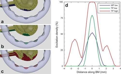
A subsequent modeling study from the same group examined various current focussing strategies in an updated version of their model (Kalkman et al. Citation2015). The model had previously been updated to include four different human cochlear geometries, two of which were based on µCT imaging, as well as using more realistic nerve fiber trajectories and modified tissue conductivities, which were derived from patient-specific modeling of intracochlear potentials (Kalkman et al. Citation2014). The 2015 study on current focussing strategies added a realistic spatial distribution of the auditory neurons’ cell bodies, which meant that they were not linearly aligned, as they had been in all previously published modeling studies, but were spread out in the spiral ganglion, essentially filling up Rosenthal’s canal in the modiolus. Spatial selectivity of stimuli was expressed in terms of excitation density: the percentage of neurons excited at a specific length along the spiral ganglion (). This showed that current focussing strategies, such as (partial) tripolar stimulation and phased arrays, are capable of penetrating the spiral ganglion more deeply than monopolar stimulation, when exciting the same number of auditory neurons (a, b, and d). Previous modeling studies had also shown current focussing strategies exciting a larger number of neurons at a location close to the stimulating contacts, but had expressed it in terms of stochastic response of linearly aligned neurons (Litvak et al. Citation2007; Goldwyn et al. Citation2010), while Kalkman et al. presented it as a purely spatial effect, using deterministic nerve fibers. Additionally, as in the modeling studies before it, the side-lobe effect for tripolar stimulation was demonstrated (c and d) and it was again shown, in both modeling results as well as (preliminary) psychophysical loudness growth curves, that current focussing strategies require more power to achieve sufficient loudness levels than monopolar stimulation. Furthermore, the study reiterated the idea that, for current focussing to be effective, a sufficient level of electrical channel interaction was needed at the site of neural stimulation.
Figure 7. Illustration of excitation patterns and excitation density plots from Kalkman et al. (Citation2015). Figures a–c show neural excitation patterns in auditory neurons with degenerated peripheral processes for three different situations: (a) monopolar stimulation at low amplitude, (b) tripolar stimulation at low amplitude, exciting the same number of neurons as the monopolar stimulus above, and (c) tripolar stimulation at high amplitude. Blue, green, and red fibers in Figures a, b, and c indicate excited neurons. In Figure d, the corresponding excitation density curves are plotted, which show the percentage of neurons that are being excited along the cochlea. The blue curve corresponds to the monopolar excitation pattern shown in Figure a, the green curve to the low amplitude tripolar excitation pattern shown in Figure b, and the red curve corresponds to the excitation pattern generated by high-amplitude tripolar stimulation shown in Figure c. Comparing Figures a and b and their curves in Figure d, it is clear that tripolar stimulation excites neurons in a more spatially restricted pattern than monopolar stimulation does with the same number of excited neurons. In Figure c and the red curve in Figure d, the excitation pattern produced by a high-amplitude tripolar stimulus reveals the presence of side lobes on either side of the main excitation region, close to the flanking contacts.
Aside from current focussing, there have also been several modeling studies that have investigated the subject of current steering. Choi and Hsu presented a study that examined current steered potential fields and activation functions in a half-turn rotational FEM geometry of the human cochlea (Choi and Hsu Citation2009). The results of the study showed gradually shifting activation function curves when varying the value of the current steering parameter α.
In the same year, Frijns et al. published a modeling study on current steering, which they referred to as dual electrode stimulation (Frijns et al. Citation2009a). The study looked at excitation patterns generated by current steered stimuli, using both simultaneous stimulation of the current steering electrode pair, as well as rapid sequential stimulation of the contacts. The study identified two current steering modalities; the first consisted of a single region of neural excitation that shifted gradually from one electrode contact to another, which could be considered the intended goal of current steering. In the second modality, there were two areas of excitation, one of which would diminish as the other expanded while increasing or decreasing α. The first modality mainly occurred for high stimulus levels, using simultaneous stimulation of electrode contacts with a large degree of electrical interaction, for example, closely spaced or lateral wall contacts. The second modality often occurred at low stimulus levels, for sequential stimulation and in cases where the peripheral processes were the main sites of excitation. Furthermore the study showed that for sequentially stimulated current steering loudness correction was needed at intermediate values of α, whereas simultaneous current steering required almost no correction of injected current to maintain constant loudness across the total range of α.
A later study used the same model in conjunction with psychophysical testing in 12 implanted subjects, involving loudness balancing for current steered stimuli near threshold level, rather than at maximum comfortable loudness (MCL) level (Snel-Bongers et al. Citation2013). Psychophysical loudness correction factors were plotted against α, which were triangular in shape for most subjects, comparable to correction factors determined at MCL for sequentially stimulated current steering in earlier experiments (Frijns et al. Citation2009a); in other words, for α = 0.5 more current was needed to achieve threshold than for other values of α. For some subjects however, the loudness balancing curve showed a dip around α = 0.5; modeling results indicated that this was consistent with the dual excitation region modality described in Frijns et al. (Citation2009a). Modeling results also suggested that the occurrence of the dip could be related to the degree of degeneration of the peripheral neural processes, especially the unmyelinated terminal, which is suspected of being the primary site of neural degeneration after, for example, noise trauma (Kujawa and Liberman Citation2009; Lin et al. Citation2011).
3. Electrode design
Design of the electrode array itself has been another important subject of modeling research, with the main points of interest being determining the optimal distance from the modiolar wall and insertion depth of the electrode array. By their nature, the early lumped parameter model were not suitable for investigating the effects of array design, so it was not until the advent of three-dimensional volume conduction modeling that electrode design aspects could be tested in computational models.
The first three-dimensional modeling study that looked at the effect of electrode position was that of Finley et al. (Citation1990). They had placed the banded electrodes in their study along the lateral wall of the cochlear geometry, while their array with plate electrode contacts was in a perimodiolar position. Though this meant that the results conflated the effects of electrode size, shape, and location, they did observe that the laterally placed banded contacts produced more broadly spreading electrical fields and lower activation function amplitudes than the medially placed array, which suggested higher neural thresholds and larger spread of excitation.
In the rotationally symmetric cochlea models of Frijns et al., point sources were placed inside the scala tympani in lateral, midscalar, and medial position, as well as underneath the osseous spiral lamina (Frijns et al. Citation1995; Frijns et al. Citation1996), similar to cat experiments performed by Shepherd et al. (Citation1993). Consistent with the physiological cat data, neural excitation thresholds decreased as the current sources were placed close to the modiolus; the lowest thresholds were found for the current source underneath the osseous spiral lamina, provided the neural peripheral process was present. A later study investigated the effect of lateral and medial placement of an electrode array in spiraling geometries of the guinea pig and human cochlea (Frijns et al. Citation2001). Instead of using ideal point current sources, a geometrical representation of the Clarion HiFocus electrode array both with and without a positioner was used. Monopolar stimulation in the basal turn of the human cochlear geometry showed lower thresholds for medial arrays, with higher spatial selectivity. However, after the basal turn this reduction of threshold was considerably smaller, while at the same time the model predicted that apical contacts close to the modiolus were more likely to produce ectopic stimulation than those located along the lateral wall. This led Frijns et al. (2001) to conclude that a perimodiolar placement of the array was beneficial in the basal turn, but that it should be avoided in more apical parts of the cochlea. Furthermore, it was observed that the positioner was able to electrically insulate apical regions of the cochlea from current injected in the basal turn.
In the same paper, comparison of the results from the human geometry to those from the guinea pig geometry revealed that differences in anatomical features between the two cochleae, specifically the size and shape of the basal turn, had notable effects on the neural excitation patterns. In particular, ectopic stimulation was affected, having a higher chance of occurring in the basal turn of the guinea pig cochlea than in that of the human cochlea. These differences underlined the importance of using species-specific cochlear geometries in models, and urged caution in interpreting data from animal studies when applying them to human situations.
Hanekom’s Citation2001 study found similar results in their spiraling geometry, and concluded that medial placement of the array was preferable to lateral placement, due to lower thresholds, better spatial selectivity, and a smaller reliance on the presence of the neural peripheral processes (Hanekom Citation2001). In a subsequent study, Hanekom also investigated the effect of adding encapsulation tissue around the electrode array, modeled as a 50-µm layer of fibrous tissue, which surrounded the electrode array either directly, or with a 50-µm layer of perilymph in between the array and its encapsulation (Hanekom Citation2005). Results showed that without a layer of perilymph, the encapsulation tissue caused lower thresholds, and a reduction in spread of excitation, while with the layer of perilymph the effects on threshold and spread of excitation were small and inconsistent. Furthermore, Hanekom found that changes in threshold due to encapsulation were stronger for medial arrays than for lateral ones.
Briaire and Frijns followed with a study on neural degeneration in the human cochlea, where neural excitation for lateral and medial electrodes was simulated for intact neurons and for neurons with completely degenerated peripheral processes (Briaire and Frijns Citation2006). The study reiterated the results of Frijns et al. (Citation2001), and the idea that lateral arrays are more likely to excite neurons at their peripheral processes, which makes thresholds and spatial selectivity of lateral contacts more sensitive to neural degeneration than those of perimodiolar electrodes.
Though the modeling work of Whiten (Citation2007) also predicted lower thresholds and increased spatial selectivity for medial electrodes, the effect was much smaller than it was for previous modeling studies. Whiten attributed this difference to using a higher temporal bone resistivity, which caused more current to flow in longitudinal direction along the scala tympani compared to other models. In most modeling studies that followed, however, the electrode’s distance from the modiolar wall or neural elements remained an important parameter that affected modeling outcomes. In particular, the results of the subsequent studies on multipolar stimulation, described in Sections 2.1 and 2.2, generally agreed that increasing the electrode–neuron distance caused more electrical interaction to occur at the excitable elements, which was considered beneficial for current focussing and steering purposes (Litvak et al. Citation2007; Frijns et al. Citation2009a; Choi and Hsu Citation2009; Goldwyn et al. Citation2010; Frijns et al. Citation2011; Kalkman et al. Citation2015).
In a series of five papers published in 2009, Cohen presented a stochastic model of electrical stimulation in a population of single node fibers (Cohen Citation2009a; Cohen Citation2009b; Cohen Citation2009c; Cohen Citation2009d; Cohen Citation2009e). In the second paper of the series, Cohen simulated electrical potentials in a rotationally symmetric FEM model of a single turn of the human cochlea, containing either a nucleus straight array or a nucleus contour array (Cohen Citation2009b). Results showed that, at the neural elements, the contour array produced electrical potential distributions that were higher and sharper than those of the more laterally located straight array. Despite its relatively simple representation of both the cochlea and the nerve fibers, Cohen’s model was able to describe data from eCAP masking experiments and loudness growth functions of individual patients quite well.
Another 2009 modeling study investigated the effect of otosclerosis on facial nerve stimulation by cochlear implants, using Frijns et al.’s computational model (Frijns et al. Citation2009b). Thresholds were determined for auditory nerve and facial nerve stimulation, for lateral and medial electrodes, with either plate contacts, half-banded contacts or full-banded contacts. To model the effects of otosclerosis, the conductivity of the temporal bone surrounding the cochlea was varied. Results of the study reinforced the intuitively obvious clinical observations that lateral wall electrodes are more likely to stimulate the facial nerve than medially placed electrodes, and that full banded contacts are more likely to produce facial nerve stimulation than plate contacts or half-banded contacts that faced the modiolus. Furthermore, lowering the conductivity of temporal bone in the model increased the likelihood of facial nerve stimulation; the novel insight, however, was that this was not so much due to the lowering of facial nerve thresholds (as was commonly thought) as it was due to the raising of auditory MCL levels.
More recently, the updated Frijns et al. model was used to predict pitch percepts in cochlear implant induced hearing (Kalkman et al. Citation2014; Van der Marel et al. Citation2016). Instead of using radial nerve fiber trajectories, as in earlier studies, the nerve fibers in this study were given realistically curved trajectories which took the length difference between the organ of Corti and the spiral ganglion into account, based on histological data (Stakhovskaya et al. Citation2007). Pitch percepts were predicted based on neural excitation patterns in four different cochlear geometries, induced by lateral and medial electrode contacts of up to 805° insertion depth; nerve fibers were modeled both with and without peripheral processes. The study found that simulated pitch percepts in the first cochlear turn were more or less the same for lateral and medial contacts, and followed the pitch predicted by direct application of the Greenwood function to the electrodes’ positions in the cochlea. Furthermore, Kalkman et al. found that beyond the first cochlear turn, medial electrodes tended to produce lower pitch percepts than lateral ones when the peripheral processes were intact. Intuitively this made sense, considering the closer proximity of the medial electrode to the spiral ganglion. However, predicted pitch of the medial electrodes generally did not reach values expected from direct spiral ganglion stimulation as long as the peripheral processes were present. Without peripheral processes, lateral and medial pitch percepts were very similar, and more closely followed the pitch expected from direct spiral ganglion stimulation. Electrode contacts located deeper than approximately 540° from the round window (which was near the end of the spiral ganglion in the model) produced unpredictable pitch percepts that were spectrally broad and showed considerable overlap with their neighboring contacts, which called their usefulness into question.
A 2010 psychophysical study by Carlyon et al. used an earlier iteration of the same computational model to compare data from pitch matching experiments to model predictions, and found that they were in good agreement (Carlyon et al. Citation2010). However, the test subjects’ electrodes that were used in the pitch matching experiments were all located in the first cochlear turn, so modeling results in apical regions of the cochlea could not be validated.
Finally, while the above studies have all examined the influence of electrode design on cochlear electrical fields and neural excitation, cochlear implant modeling studies that investigate the fundamental processes occurring at the electrode contacts themselves are surprisingly rare. Additionally, all of the previously described volume conduction models employ the quasi-static approximation of assuming that the electrode array and all of the cochlear structures are purely resistive. In reality, there is an electrochemical interface impedance at the boundary between stimulating contacts and the scalar perilymph, which has a capacitive component (Vanpoucke et al. Citation2004). Lai and Choi proposed a method to implement this interface impedance into FEM models of the implanted cochlea (Lai and Choi Citation2007). In a spiraling geometry of an implanted human cochlea, they modeled the interface as a thin 50-µm layer on the surface of the electrode contacts, which had a complex permittivity value. The study used geometrical representations of half-banded, full-banded, and flat plate contacts, and showed that the interfacial layer produced the same potentials as their equivalent electrical circuits. Plots of the neural activation functions suggested that the electrochemical interface impedance could have an influence on neural excitation, though the nature and extent of this influence was not clear.
To assess the current density distribution on platinum electrodes used for electrical stimulation of neurons, Rubinstein et al. (Citation1987) developed a model of disk-shaped electrode contacts, both surface mounted and recessed into the electrode carrier. The model numerically solved Laplace’s equation for the quasi-static electrical fields using a Green’s function approach, and found that the current density across the electrode contact was not uniform, with the current density increasing as one gets closer to the edge. The model also showed that recessing the electrode would make the current density distribution more uniform, which was deemed advantageous, since nonuniform current distribution could locally exceed electrochemical safety limits near the edge of the electrode.
Recently, Sue et al. have presented modeling work on the effects of electrochemical processes and electrode recession on current distributions across half-banded platinum contacts embedded in an electrode carrier, located in a perilymph-filled cylindrical geometry representing the cochlear scalae (Sue et al. Citation2013; Sue et al. Citation2015). Their results showed that faradaic current density was dependent on time, and that, in accordance with Rubinstein et al. (Citation1987), it was not uniform across the electrode contact, with current densities being higher at the corners of the half-banded contacts. However, Sue et al. again showed that these concentrations of current could be mitigated by recessing the contact into the carrier, though the improvement was more modest than it was for Rubinstein et al.’s model.
4. Discussion
4.1. Computational modeling of stimulation strategies and electrode design
It has become clear that most of the general properties of multipolar stimulation can be understood readily using simple mathematical models of unrolled cochleae. These models can successfully simulate electrical field interaction of electrode contacts, and thereby describe sharpening of the fields due to current focussing techniques quite well (Black and Clark Citation1980; Black et al. Citation1983; O’Leary et al. Citation1985; Suesserman and Spelman Citation1993; Rodenhiser and Spelman Citation1995; Spelman et al. Citation1995; Jolly et al. Citation1996; Kral et al. Citation1998; Litvak et al. Citation2007; Bonham and Litvak Citation2008; Goldwyn et al. Citation2010). When including an estimate of neural excitation through the activation functions, these models can even predict certain features of multipolar stimuli, such as increased localization of the excitation patterns and the presence of side lobes due to neural excitation near the flanking contacts in tripolar stimulation. Using a more realistic volume conduction model will create a more detailed view of intracochlear potentials and neural excitation; a rotationally symmetric geometry will reveal the possibility of ectopic stimulation at high stimulus amplitudes (Frijns et al. Citation1995). A spiraling geometry will additionally show asymmetrical potential distributions through the cochlea (Briaire and Frijns Citation2000). However, while realistic volume conduction models are quantitatively more accurate, qualitatively speaking mathematical models are sufficient for a basic understanding of multipolar stimulation strategies. This also strengthens confidence that modeling predictions on multipolar stimulation can be trusted, as they generally agree with each other and make sense from an electrophysical point of view, making it unlikely that such predictions are a result of modeling artifacts.
For matters concerning electrode design, the applicability of simple mathematical models is more limited; while simple models can shed light on the basic consequences of moving the current sources toward the neural elements (i.e., lower thresholds and less spread of excitation), other aspects of electrode design require a realistic three-dimensional volume conduction model of the implanted cochlea. For instance, investigating the effects of changing electrode shape and size in a computational model requires the actual electrode geometry to be represented accurately and not simply reduced to point sources in the scala tympani; this has previously allowed for the investigation of the insulating effects of positioner systems (Frijns et al. Citation2001) and the probability of facial nerve stimulation by different electrode contact geometries (Frijns et al. Citation2009b). Furthermore, understanding the effect insertion depth has on neural excitation requires not only a realistic cochlear geometry, but also an accurate modeling of neural trajectories, since these vary considerably over the length of the cochlea, which, in turn, was shown to have serious implications for neural recruitment by electrical stimulation (Kalkman et al. Citation2014).
It should be noted that, while there are many modeling studies that have shown the effect of electrode position in the scala tympani, there have not been many investigations on the shape and size of electrode arrays. Most modeling studies only use one type of electrode array, and the few that use multiple geometries have not made an extensive comparison of different arrays. It is unclear what the reason for this could be; possibly, the electrode geometry is thought to have little effect on neural excitation, or at least not as much as other factors. Alternatively, the modeling of different electrode geometries could be considered too laborious and time-consuming to be worth the effort. Whatever the case, it is conceivable that there are still some insights to be gained from computational modeling of different electrode array designs.
4.2. Present and future of cochlear implant modeling
Many of the past cochlear implant modeling studies, particularly the early lumped parameter models, have coupled their results with electrophysiological or psychophysical experiments. This was essential, because there is an unavoidable need to validate modeling results by comparing them to data from live subjects. After all, any computational model contains a number of assumptions and simplifications; if these are inappropriately chosen, the model’s output will be misleading or outright wrong. In short, a computational model always produces results, but in order to know if one can trust these results, validation is necessary.
The most straightforward way of validation a computational model of an implanted cochlea is by comparing simulated electrical fields to electrophysiological data, since it concerns only directly measurable physical quantities and does not require interpretation of neural signals. While this type of validation is necessary and gives confidence in a model’s ability to simulate the electrical properties of the cochlea, electrical field distributions in the cochlea only give limited information about how cochlear implants function. Therefore, to meaningfully investigate the subject of electrical stimulation of the cochlea, one cannot avoid including a neural model.
Most of the models discussed in this review have contained some estimate of neural activity, either by determining the activation function, or by incorporating an electrical cable model of the auditory neurons. However, validation of simulated auditory neural activity is substantially more difficult. The only objective data at one’s disposal are eCAPs or electrically evoked auditory brainstem responses (EABRs), and inferior colliculus measurements in animal subjects. Even though eCAPs have been modeled successfully (Briaire and Frijns Citation2005; Briaire and Frijns Citation2006; Whiten Citation2007; Smit et al. Citation2009; Westen et al. Citation2011; Choi and Wang Citation2014), these types of neurophysiological data only offer indirect evidence of model validity, and can usually only verify that the model is producing realistic output in broad strokes. The only other tool available is psychophysical testing, but it has the additional drawback of involving central neural processing, while current cochlear implant models only simulate neural stimulation at the peripheral level. This means that validation will remain the primary challenge that researchers will face when enhancing their computational models in the future.
These future model enhancements will likely include adding more detail to the cochlear geometries. Advances in imaging techniques have made it relatively easy to acquire high-resolution three-dimensional anatomical scans, which can be used to create new model geometries. Recent models have already used (micro-)CT data to create more realistically shaped model cochleae with individual anatomical characteristics (Malherbe et al. Citation2013; Kalkman et al. Citation2014; Malherbe et al. Citation2015b), while researchers at the University of Sydney are currently working on a highly detailed model based on scanning thin-sheet laser imaging microscopy (sTSLIM) of the guinea pig cochlea (Wong et al. Citation2016). In addition to improving the level of detail of the cochlea itself, modeling work from the past years has also seen the inclusion of detailed geometries of the human head (Malherbe et al. Citation2015a; Tran et al. Citation2015). These enhancements in anatomical detail may lead to new insights regarding electrical field distributions in or around the cochlea and possibly add more realism to simulated neural excitation patterns.
Aside from purely anatomical improvements, several other more fundamental changes to cochlear implant models are possible. First, all of the models thus far have restricted themselves to using isotropic conductivity values in their geometries. This is likely sufficient for most of the cochlear structures, but neural tissue cannot be expected to conduct electrical currents equally well in all directions. Since neural tissue takes up a considerable amount of space in the cochlear modiolus and is obviously the target of electrical stimulation, adding anisotropy to volume conduction models of the cochlea could have significant impact.
Next, the majority of the models presented have employed a quasi-static approximation when calculating electrical field distributions, and have assumed that the electrical fields can be determined by simply multiplying a normalized static field with the time-dependent amplitude of the electrode stimulus. This assumption was based on work by Spelman and coworkers, who showed that cochlear potentials in the scala tympani were largely independent of stimulation frequencies of up to 12.5 kHz (Spelman et al. Citation1982); unpublished data from their group has shown that the assumption of frequency independency is even valid up to 100 kHz (F.A. Spelman, personal communication). However, the electrochemical interface is not completely resistive, and it is possible that a small frequency dependency of cochlear structures exists. Some investigation has been done into the electrode interface impedance (Lai and Choi Citation2007; Sue et al. Citation2013; Sue et al. Citation2015), and initial work concerning frequency dependency in the implanted cochlea has been presented (Inguva et al. Citation2015), but an extensive analysis of the subject has not yet been performed in a full volume conduction model coupled with an active neural model.
The neural models themselves are also still improving, as there is much that is unknown concerning auditory nerve fiber morphology and kinetics. Additionally, the discussed neural models have an ongoing problem of being unable to reasonably predict realistic neural threshold levels, which are invariably higher in the models than they are in clinical reality. It is unclear what the cause of this discrepancy is, since the models do actually simulate electrophysiological experiments on single fibers quite well. To understand this, more input from the field of neurophysiology is necessary.
One obvious addition that can be made to the cochlear implant models is the inclusion of neural stochasticity. While stochasticity has been implemented in models of single nodes or fibers (Bruce et al. Citation1999a; Bruce et al. Citation1999b; Rubinstein et al. Citation1999; Imennov and Rubinstein Citation2009; Cohen Citation2009d; Cohen Citation2009e; Woo et al. Citation2010), few of the presented cochlear implant models have included it, instead relying on activation functions or deterministic electrical cable models of the auditory neurons. The research groups of Hanekom et al. and Frijns et al. recently presented preliminary work on extensions of their models with relatively simple models of stochasticity (Badenhorst et al. Citation2015; Frijns et al. Citation2015). Badenhorst et al. investigated the addition of stochasticity to the Hanekom model; the results of this suggested that stochastic fibers will predict more realistic neural thresholds. Frijns et al. used the thresholds calculated by their volume conduction and deterministic neural models as input for a simulation of stochastic variability, which, apart from stochastic thresholds, includes neural adaptation, refractoriness, and neural accommodation; the future goal of this extension is to make predictions on the functioning and effectiveness of speech processing strategies.
Another subject of interest is the exact nature of neural degeneration in the cochlea. Several cochlear implant modeling studies have included neural degeneration in their simulations, but most have modeled it by the complete removal of all peripheral processes from the auditory neurons. Snel-Bongers et al. discussed the possibility of gradually degenerating peripheral processes (Snel-Bongers et al. Citation2013), in line with findings from the Liberman group (Kujawa and Liberman Citation2009; Lin et al. Citation2011), and only Goldwyn et al. have so far investigated the consequences of hypothetical neural dead regions (Goldwyn et al. Citation2010). These methods of modeling neural degeneration are somewhat speculative, as the precise mechanisms of neural degeneration in humans are still a subject of research (Kujawa and Liberman Citation2009; Lin et al. Citation2011; Wan and Corfas Citation2015).
Tangentially related to neural degeneration is the effect of electrode insertion trauma on cochlear implant stimulation. All of the modeling studies discussed in this review have exclusively used the scala tympani as the location of the stimulating electrodes, even though it is known that many cochlear implant electrode arrays end up perforating cochlear structures such as the basilar membrane and Reissner’s membrane, thereby ending up in the scala vestibuli, rather than the scala tympani (Aschendorff et al. Citation2005; Skinner et al. Citation2007; Aschendorff et al. Citation2007; Finley et al. Citation2008; Holden et al. Citation2013; Wanna et al. Citation2014). The unintended position of the electrode and the potential damage to the neural elements caused by this insertion trauma could have noteworthy consequences for cochlear implant stimulation; however, no cochlear implant modeling studies have investigated this subject thus far.
In conclusion, computational modeling of the electrically stimulated cochlea has had a long history. It has been a valuable tool for the study of stimulation techniques and electrode design, enabling types of experimentation that would be difficult or impossible to achieve in clinical or laboratory conditions. There are still many issues left to be investigated and improvements to be made to existing models; the increasing availability of high resolution imaging techniques and open sourced or commercial modeling software mean that the threshold for developing new models has never been lower, making sure that computational modeling of the electrically stimulated cochlea has a long future ahead. A crucial factor in the further application of models in clinical practice will be the validation of their outcomes and predictions.
References
- Aschendorff A, Kromeier J, Klenzner T, Laszig R. 2007. Quality control after insertion of the nucleus contour and contour advance electrode in adults. Ear Hear. 28:75S–79S.
- Aschendorff A, Kubalek R, Turowski B, Zanella F, Hochmuth A, Schumacher M, Klenzner T, Laszig R. 2005. Quality control after cochlear implant surgery by means of rotational tomography. Otol Neurotol. 26:34–37.
- Badenhorst W, Malherbe TK, Hanekom T, Hanekom JJ. 2015. Development of a voltage dependent current noise algorithm for conductance based stochastic modelling of auditory nerve fibre populations in compound models. Conference on Implantable Auditory Prostheses; 2015; Pacific Grove, CA.
- Black RC, Clark GM. 1980. Differential electrical excitation of the auditory nerve. J Acoust Soc Am 67:868–874.
- Black RC, Clark GM, Tong YC, Patrick JF. 1983. Current distributions in cochlear stimulation. Ann NY Acad Sci. 405:137–145.
- Bonham BH, Litvak LM. 2008. Current focusing and steering: modeling, physiology, and psychophysics. Hear Res. 242:141–153.
- Briaire JJ, Frijns JHM. 2000. Field patterns in a 3D tapered spiral model of the electrically stimulated cochlea. Hear Res. 148:18–30.
- Briaire JJ, Frijns JHM. 2005. Unraveling the electrically evoked compound action potential. Hear Res. 205:143–156.
- Briaire JJ, Frijns JHM. 2006. The consequences of neural degeneration regarding optimal cochlear implant position in scala tympani: a model approach. Hear Res. 214:17–27.
- Bruce IC, Irlicht LS, White MW, O’Leary SJ, Dynes S, Javel E, Clark GM. 1999b. A stochastic model of the electrically stimulated auditory nerve: pulse-train response. IEEE Trans Biomed Eng. 46:630–637.
- Bruce IC, White MW, Irlicht LS, O’Leary SJ, Dynes S, Javel E, Clark GM. 1999a. A stochastic model of the electrically stimulated auditory nerve: single-pulse response. IEEE Trans Biomed Eng. 46:617–629.
- Carlyon RP, Macherey O, Frijns JHM, Axon PR, Kalkman RK, Boyle P, Baguley DM, Briggs J, Deeks JM, Briaire JJ, et al. 2010. Pitch comparisons between electrical stimulation of a cochlear implant and acoustic stimuli presented to a normal-hearing contralateral ear. J Assoc Res Otolaryngol. 11:625–640.
- Choi CTM, Hsu CH. 2009. Conditions for generating virtual channels in cochlear prosthesis systems. Ann Biomed Eng. 37:614–624.
- Choi CTM, Lai WD, Chen YB. 2004. Optimization of cochlear implant electrode array using genetic algorithms and computational neuroscience models. IEEE Trans Magn. 40:639–642.
- Choi CTM, Lai WD, Chen YB. 2005. Comparison of the electrical stimulation performance of four cochlear implant electrodes. IEEE Trans Magn. 41:1920–1923.
- Choi CTM, Lai WD, Lee SS. 2006. A novel approach to compute the impedance matrix of a cochlear implant system incorporating an electrode-tissue interface based on finite element method. IEEE Trans Magn. 42:1375–1378.
- Choi CTM, Wang SP. 2014. Modeling ECAP in cochlear implants using the FEM and equivalent circuits. IEEE Trans Magn. 50:7001004.
- Clopton BM, Spelman FA. 1995. Electrode configuration and spread of neural excitation: compartmental models of spiral ganglion cells. Ann Otol Rhinol Laryngol Suppl. 166:115–118.
- Cohen LT. 2009a. Practical model description of peripheral neural excitation in cochlear implant recipients: 1. Growth of loudness and ECAP amplitude with current. Hear Res. 247:87–99.
- Cohen LT. 2009b. Practical model description of peripheral neural excitation in cochlear implant recipients: 2. Spread of the effective stimulation field (ESF), from ECAP and FEA. Hear Res. 247:100–111.
- Cohen LT. 2009c. Practical model description of peripheral neural excitation in cochlear implant recipients: 3. ECAP during bursts and loudness as function of burst duration. Hear Res. 247:112–121.
- Cohen LT. 2009d. Practical model description of peripheral neural excitation in cochlear implant recipients: 4. Model development at low pulse rates: general model and application to individuals. Hear Res. 248:15–30.
- Cohen LT. 2009e. Practical model description of peripheral neural excitation in cochlear implant recipients: 5. Refractory recovery and facilitation. Hear Res. 248:1–14.
- Colombo J, Parkins CW. 1987. A model of electrical excitation of the mammalian auditory-nerve neuron. Hear Res. 31:287–311.
- Dekker DMT, Briaire JJ, Frijns JHM. 2014. The impact of internodal segmentation in biophysical nerve fiber models. J Comput Neurosci. 37:307–315.
- Finley CC, Holden TA, Holden LK, Whiting BR, Chole RA, Neely GJ, Hullar TE, Skinner MW. 2008. Role of electrode placement as a contributor to variability in cochlear implant outcomes. Otol Neurotol. 29:920–928.
- Finley CC, Wilson BS, White MW. 1990. Models of neural responsiveness to electrical stimulation. In: Miller JM, Spelman FA, editors. Cochlear implants: models of the electrically stimulated ear. New York (NY): Springer; p. 55–96.
- Frankenhaeuser B, Huxley AF. 1964. The action potential in the myelinated nerve fiber of xenopus laevis as computed on the basis of voltage clamp data. J Physiol. 171:302–315.
- Frijns JHM, Briaire JJ, Grote JJ. 2001. The importance of human cochlear anatomy for the results of modiolus-hugging multichannel cochlear implants. Otol Neurotol. 22, 340–349.
- Frijns JHM, de Snoo SL, Schoonhoven R. 1995. Potential distributions and neural excitation patterns in a rotationally symmetric model of the electrically stimulated cochlea. Hear Res. 87:170–86.
- Frijns JHM, de Snoo SL, ten Kate JH. 1996. Spatial selectivity in a rotationally symmetric model of the electrically stimulated cochlea. Hear Res. 95:33–48.
- Frijns JHM, Dekker DMT, Briaire JJ. 2011. Neural excitation patterns induced by phased-array stimulation in the implanted human cochlea. Acta Otolaryngol. 131:362–370.
- Frijns JHM, Kalkman RK, Briaire JJ. 2009b. Stimulation of the facial nerve by intracochlear electrodes in otosclerosis: a computer modeling study. Otol Neurotol. 30:1168–1174.
- Frijns JHM, Kalkman RK, Vanpoucke FJ, Bongers JS, Briaire JJ. 2009a. Simultaneous and non-simultaneous dual electrode stimulation in cochlear implants: evidence for two neural response modalities. Acta Otolaryngol. 129:433–439.
- Frijns JHM, Van Gendt MJ, Kalkman RK, Briaire JJ. 2015. Modeled neural response patterns from various speech coding strategies. Conference on Implantable Auditory Prostheses; 2015; Pacific Grove, CA.
- Girzon G. 1987. Investigation of current flow in the inner ear during electrical stimulation of intracochlear electrodes [M.Sc. thesis]. Cambridge (MA): Massachusetts Institute of Technology.
- Goldwyn JH, Bierer SM, Bierer JA. 2010. Modeling the electrode-neuron interface of cochlear implants: effects of neural survival, electrode placement, and the partial tripolar configuration. Hear Res. 268:93–104.
- Hanekom T. 2001. Three-dimensional spiraling finite element model of the electrically stimulated cochlea. Ear Hear. 22:300–315.
- Hanekom T. 2005. Modelling encapsulation tissue around cochlear implant electrodes. Med Biol Eng Comput. 43:47–55.
- Holden LK, Finley CC, Firszt JB, Holden TA, Brenner C, Potts LG, Gotter BD, Vanderhoof SS, Mispagel K, Heydebrand G, Skinner MW. 2013. Factors affecting open-set word recognition in adults with cochlear implants. Ear Hear. 34, 342–60.
- Imennov NS, Rubinstein JT. 2009. Stochastic population model for electrical stimulation of the auditory nerve. IEEE Trans Biomed Eng. 56:2493–2501.
- Inguva C, Wong P, Sue A, McEwan A, Carter P. 2015. Frequency-dependent simulation of volume conduction in a linear model of the implanted cochlea. Conference proceedings: 7th International IEEE/EMBS Conference on Neural Engineering; 2015; Montpellier. 426–429.
- Jolly CN, Spelman FA, Clopton BM. 1996. Quadrupolar stimulation for Cochlear prostheses: modeling and experimental data. IEEE Trans Biomed Eng. 43:857–865.
- Kalkman RK, Briaire JJ, Dekker DMT, Frijns JHM. 2014. Place pitch versus electrode location in a realistic computational model of the implanted human cochlea. Hear Res. 315:10–24.
- Kalkman RK, Briaire JJ, Frijns JHM. 2015. Current focussing in cochlear implants: an analysis of neural recruitment in a computational model. Hear Res. 322:89–98.
- Kral A, Hartmann R, Mortazavi D, Klinke R. 1998. Spatial resolution of cochlear implants: the electrical field and excitation of auditory afferents. Hear Res. 121:11–28.
- Kujawa SG, Liberman MC. 2009. Adding insult to injury: cochlear nerve degeneration after “temporary” noise-induced hearing loss. J Neurosci. 29:14077–14085.
- Lai WD, Choi CTM. 2007. Incorporating the electrode-tissue interface to cochlear implant models. IEEE Trans Magn. 43:1721–1724.
- Lin HW, Furman AC, Kujawa SG, Liberman MC. 2011. Primary neural degeneration in the Guinea pig cochlea after reversible noise-induced threshold shift. J Assoc Res Otolaryngol. 12:605–616.
- Litvak LM, Spahr AJ, Emadi G. 2007. Loudness growth observed under partially tripolar stimulation: model and data from cochlear implant listeners. J Acoust Soc Am. 122:967–81.
- Macherey O, Carlyon RP, van WA, Deeks JM, Wouters J. 2008. Higher sensitivity of human auditory nerve fibers to positive electrical currents. J Assoc Res Otolaryngol. 9:241–251.
- Macherey O, van WA, Carlyon, RP, Dhooge I, Wouters J. 2010. Forward-masking patterns produced by symmetric and asymmetric pulse shapes in electric hearing. J Acoust Soc Am. 127:326–338.
- Malherbe TK, Hanekom T, Hanekom JJ. 2013. Can subject-specific single-fibre electrically evoked auditory brainstem response data be predicted from a model? Med Eng Phys. 35:926–936.
- Malherbe TK, Hanekom T, Hanekom JJ. 2015a. The effect of the resistive properties of bone on neural excitation and electric fields in cochlear implant models. Hear Res. 327:126–135.
- Malherbe TK, Hanekom T, Hanekom JJ. 2015b. Constructing a three-dimensional electrical model of a living cochlear implant user’s cochlea. Int J Numer Method Biomed Eng. [Epub ahead of print].
- McNeal DR. 1976. Analysis of a model for excitation of myelinated nerve. IEEE Trans Biomed Eng. 23:329–337.
- Nogueira W, Würfel W, Büchner A. 2014. Development of a model of the electrically stimulated auditory nerve. Biomed Eng-Biomed Tech. 59:S786–S789.
- O’Leary SJ, Black RC, Clark GM. 1985. Current distributions in the cat cochlea: a modelling and electrophysiological study. Hear Res. 18, 273–281.
- Pau HW, Grunbaum A, Ehrt K, Dahl R, Just T, van Rienen U. 2014. Would an endosteal CI-electrode make sense? Comparison of the auditory nerve excitability from different stimulation sites using ESRT measurements and mathematical models. Eur Arch Otorhinolaryngol. 271:1375–1381.
- Rattay F. 1986. Analysis of models for external stimulation of axons. IEEE Trans Biomed Eng. 33:974–947.
- Rattay F. 1987. Ways to approximate current–distance relations for electrically stimulated fibers. J Theor Biol. 125:339–349.
- Rattay F, Lutter P, Felix H. 2001a. A model of the electrically excited human cochlear neuron. I. Contribution of neural substructures to the generation and propagation of spikes. Hear Res. 153:43–63.
- Rattay F, Leao RN, Felix H. 2001b. A model of the electrically excited human cochlear neuron. II. Influence of the three-dimensional cochlear structure on neural excitability. Hear Res. 153:64–79.
- Reilly JP, Freeman VT, Larkin WD. 1985. Sensory effects of transient electrical-stimulation—evaluation with a neuroelectric model. IEEE Trans Biomed Eng. 32:1001–1011.
- Rodenhiser KL, Spelman FA. 1995. A method for determining the driving currents for focused stimulation in the cochlea. IEEE Trans Biomed Eng. 42:337–342.
- Rubinstein JT, Spelman FA, Soma M, Suesserman MF. 1987. Current-density profiles of surface mounted and recessed electrodes for neural prostheses. IEEE Trans Biomed Eng. 34:864–875.
- Rubinstein JT, Wilson BS, Finley CC, Abbas PJ. 1999. Pseudospontaneous activity: stochastic independence of auditory nerve fibers with electrical stimulation. Hear Res. 127:108–118.
- Sapozhnikov A. 1990. Computer modelling of the implanted cochlea [B.Sc. thesis]. Parkville, Victoria, Australia: University of Melbourne.
- Shepherd RK, Hatsushika S, Clark GM. 1993. Electrical stimulation of the auditory nerve: the effect of electrode position on neural excitation. Hear Res. 66:108–120.
- Skinner MW, Holden TA, Whiting BR, Voie AH, Brunsden B, Neely JG, Saxon EA, Hullar TE, Finley CC. 2007. In vivo estimates of the position of advanced bionics electrode arrays in the human cochlea. Ann Otol Rhinol Laryngol Suppl 197:2–24.
- Smit JE, Hanekom T, Hanekom JJ. 2009. Estimation of stimulus attenuation in cochlear implants. J Neurosci Methods 180:363–73.
- Snel-Bongers J, Briaire JJ, Van der Veen EH, Kalkman RK, Frijns JHM. 2013. Threshold levels of dual electrode stimulation in cochlear implants. J Assoc Res Otolaryngol. 14:781–790.
- Spelman FA, Clopton BM, Pfingst BE. 1982. Tissue impedance and current flow in the implanted ear. Implications for the cochlear prosthesis. Ann Otol Rhinol Laryngol Suppl. 98:3–8.
- Spelman FA, Pfingst BE, Clopton BM, Jolly CN, Rodenhiser KL. 1995. Effects of electrical current configuration on potential fields in the electrically stimulated cochlea: field models and measurements. Ann Otol Rhinol Laryngol Suppl. 166:131–136.
- Stakhovskaya O, Sridhar D, Bonham BH, Leake PA. 2007. Frequency map for the human cochlear spiral ganglion: implications for cochlear implants. J Assoc Res Otolaryngol. 8:220–233.
- Strelioff D. 1973. A computer simulation of the generation and distribution of cochlear potentials. J Acoust Soc Am. 54:620–629.
- Sue A, Tran P, SP Wang, Li Q, Carter P. 2013. Time-domain finite element models of electrochemistry in intracochlear electrodes. Conference proceedings: 35th Annual International Conference of the IEEE Engineering in Medicine and Biology Society; 2013; Osaka, Japan. 1554–1557.
- Sue A, Wong P, Tran P, Li Q, Carter P. 2015. Modeling the effects of electrode recessing on electrochemical safety in cochlear implant electrodes. Conference proceedings: 7th International IEEE/EMBS Conference on Neural Engineering; 2015; France: Montpellier; 490–493.
- Suesserman MF. 1992. Noninvasive microelectrode measurement technique for performing quantitative, in vivo measurements of inner ear tissue impedances [Ph.D. thesis]. Seattle (WA): University of Washington.
- Suesserman MF, Spelman FA. 1993. Lumped-parameter model for in vivo cochlear stimulation. IEEE Trans Biomed Eng. 40:237–245.
- Tognola G, Pesatori A, Norgia M, Parazzini M, Di Rienzo L, Ravazzani P, Burdo S, Grandori F, Svelto C. 2007. Numerical modeling and experimental measurements of the electric potential generated by cochlear implants in physiological tissues. IEEE Trans Instrum Meas. 56,:87–93.
- Tran P, Sue A, Wong P, Li Q, Carter P. 2015. Development of heather for cochlear implant stimulation using a new modeling workflow. IEEE Trans Biomed Eng. 62:728–35.
- Van Compernolle D. 1985. Speech processing strategies for a multichannel cochlear prosthesis [PhD thesis]. Stanford (CA): Stanford University.
- Van den Honert C, Kelsall DC. 2007. Focused intracochlear electric stimulation with phased array channels. J Acoust Soc Am. 121:3703–3716.
- Van der Marel KS, Briaire JJ, Wolterbeek R, Verbist BM, Frijns JHM. 2016. Development of insertion models predicting cochlear implant electrode position. Ear Hear. (in press).
- Vanpoucke FJ, Zarowski AJ, Peeters SA. 2004. Identification of the impedance model of an implanted cochlear prosthesis from intracochlear potential measurements. IEEE Trans Biomed Eng. 51:2174–2183.
- Wan G, Corfas G. 2015. No longer falling on deaf ears: mechanisms of degeneration and regeneration of cochlear ribbon synapses. Hear Res. 329:1–10.
- Wanna GB, Noble JH, Carlson ML, Gifford RH, Dietrich MS, Haynes DS, Dawant BM, Labadie RF. 2014. Impact of electrode design and surgical approach on scalar location and cochlear implant outcomes. Laryngoscope. 124(Suppl 6):S1–S7.
- Westen AA, Dekker DMT, Briaire JJ, Frijns JHM. 2011. Stimulus level effects on neural excitation and eCAP amplitude. Hear Res. 280:166–76.
- Whiten DM. 2007. Electro-anatomical models of the cochlear implant [PhD thesis]. Cambridge (MA): Massachusetts Institute of Technology.
- Wilson BS, Finley CC, Lawson DT, Wolford RD, Eddington DK, Rabinowitz WM. 1991. Better speech recognition with cochlear implants. Nature. 352:236–238.
- Wong P, George S, Tran P, Sue A, Carter P, Li Q. 2016. Development and validation of a high-fidelity finite-element model of monopolar stimulation in the implanted guinea pig cochlea. IEEE Trans Biomed Eng. 63:188–198.
- Woo J, Miller CA, Abbas PJ. 2010. The dependence of auditory nerve rate adaptation on electric stimulus parameters, electrode position, and fiber diameter: a computer model study. J Assoc Res Otolaryngol. 11:283–296.