Abstract
During infection, the enteric pathogen Vibrio cholerae encounters a bile-containing environment. Previous studies have shown that bile and/or bile acids exert several effects on the virulence and physiology of the bacterial cells. These observations have led to the suggestion that bile acids may play a signaling role in infection. We have previously reported that the bile component deoxycholic acid blocks the general diffusion porin OmpT in a dose-dependent manner, presumably as it transits through the pore. V. cholerae colonizes the distal jejunum and ileum, where a mixture of various conjugated and unconjugated bile acids are found. In this work, we have used patch clamp electrophysiology to investigate the effects of six bile acids on OmpT. Two bile acids (deoxycholic and chenodeoxycholic acids) were found to block OmpT at physiological concentrations below 1 mM, while glycodeoxycholic acid was mildly effective and cholic, lithocholic and taurodeoxycholic acids were ineffective in this range. The block was also voltage-dependent. These observations suggest the presence of a specific binding site inside the OmpT pore. Since deconjugation is due to the activity of the endogenous flora, the preferential uptake of some unconjugated bile acids by OmpT may signal the presence of a hospitable environment. The results are also discussed in terms of the possible molecular interactions between the penetrating bile acid molecule and the channel wall.
Introduction
Vibrio cholerae is a motile, Gram-negative, facultative pathogenic organism responsible for the disease cholera. The bacteria are a part of the natural population of aquatic environments and infection is typically acquired through the ingestion of contaminated water. Once in the host, V. cholerae colonizes the small intestine, specifically the distal jejunum and ileum (Butler and Camilli Citation2005), where it produces the cholera toxin which is mostly responsible for the symptoms of severe diarrhea and dehydration. In the host, the bacteria encounter a number of changing environments and host antimicrobial defences such as stomach acidic pH, proteolytic enzymes, and bile. Bile is a heterogeneous mixture of bile acids, bile pigments, inorganic salts, cholesterol and phospholipids (Carey and Duane Citation1994, Begley et al. Citation2005). It is synthesized in the liver, stored and concentrated in the gallbladder, then secreted into the duodenum. The detergent like properties of bile serve for the emulsification of fats during digestion, but present a growth compromising challenge for all enteric bacteria, due to the solubilizing effect on cellular membranes (Begley et al. Citation2005). Consequently, the endogenous flora and acquired pathogens do not inhabit the proximal parts of the intestines where bile concentration is at its highest (Wilson Citation2005, Ridlon et al. Citation2006). However, they are able to colonize the distal segments of the small intestine because the bile concentration is lowered due to the passive and active re-absorption of bile in the proximal segments (Carey and Duane Citation1994, Hofmann Citation1994).
The bile acid composition is itself heterogeneous. Primary bile acids are synthesized de novo from cholesterol in the liver, and secreted as conjugated forms, due to amidation to glycine or taurine, in order to increase water-solubility (Hofmann Citation1994). Exposure to the enteric flora leads to two important modifications: deconjugation in the small intestine and 7α-dehydroxylation in the colon, yielding so-called secondary bile acids (Hofmann Citation1994, Ridlon et al. Citation2006). In humans, the prevalent primary bile acids are cholic and chenodeoxycholic acids, and secondary bile acids are deoxycholic and lithocholic acids (). Re-absorption from the intestine and enterohepatic circulation allow all bile acids to be recycled to the liver and then secreted again into the bile pool. During this process, the secondary bile acids are re-conjugated in the liver but not re-hydroxylated (Carey and Duane Citation1994, Ridlon et al. Citation2006). Since bile acids are modified by the endogenous flora as they pass through the intestines, differing ratios of bile acids are present in the different regions of the intestines (Carey and Duane Citation1994, Ridlon et al. Citation2006). In the distal jejunum and ileum, where V. cholerae is known to colonize, the bile concentrations have been considerably lowered due to reabsorption, and the composition is mainly conjugated and unconjugated cholic, chenodeoxycholic and deoxycholic acid.
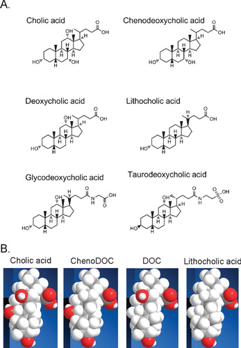
Figure 1. Molecular structures of the investigated bile acids. (A) Formulas of the 6 bile acids. The first row shows the primary bile acids cholic acid and chenodeoxycholic acid, the second row the secondary acids deoxycholic acid and lithocholic acid, and the third row the conjugated forms of deoxycholic acid, glycodeoxycholic acid and taurodeoxycholic acid. (B) Molecular representations of the unconjugated bile acids investigated. Space filling models of the indicated bile acids are shown to highlight the differences in position of the hydroxyl groups in the bile acids. All bile acids have been oriented such that the hydrophilic face is shown. The carboxyl group is in the upper right corner, and the 3-OH group common to all bile acids is at the bottom. Cholic acid has 2 additional hydroxyl groups at the C7 and C12 positions; DOC and chenoDOC have 1 additional hydroxyl group at the C12 and C7 positions, respectively. Carbon atoms are shown in grey, hydrogen atoms in white and oxygen atoms in red.
The high concentrations of bile in the duodenum and proximal jejunum are in part responsible for the low abundance of endogenous and pathogenic microorganisms in these more proximal regions of the intestine. The effect on cellular membranes is the main antibacterial mechanism of bile acids. Bile acids are able to solubilize membrane lipids and proteins (Vyvoda et al. Citation1977, Begley et al. Citation2005), and increase membrane permeability (Vyvoda et al. Citation1977, Noh and Gilliland Citation1993). Gram-negative organisms are naturally more resistant to the deleterious effects of bile acids than Gram-positive bacteria, in part due to the presence of lipopolysaccharides and the low permeability of the outer membrane (Nikaido Citation1996, Citation2003). But, even with the natural protection afforded by the outer membrane, V. cholerae grows poorly in the presence of bile acids (Provenzano et al. Citation2000, 2001). Bile and/or bile acids have a plethora of effects on bacterial cells. For example, in V. cholerae, they have been shown to increase motility (Gupta and Chowdhury Citation1997), to affect virulence gene expression (Gupta and Chowdhury Citation1997, Schuhmacher and Klose Citation1999), to promote biofilm formation (Hung et al. Citation2006), to induce the expression of genes encoding proteins involved in efflux of bile acids (Bina and Mekalanos Citation2001, Chatterjee et al. Citation2004, Cerda-Maira et al. Citation2008), and to induce the expression of the ompU porin gene (Provenzano et al. Citation2000) and the repression of the ompT porin gene (Bina et al. Citation2006). In most cases, these effects of bile are mimicked by individual or mixture of bile acids, but not always (for example, bile acids induce cholera toxin production (Hung and Mekalanos Citation2005), but bile decreases expression of the cholera toxin gene ctxA (Gupta and Chowdhury Citation1997, Schuhmacher and Klose Citation1999), indicating a complex interplay between bile and V. cholerae. Therefore, bile and its components may serve as an important environmental cue to signal entry of the bacteria into a host.
The general diffusion porins OmpU and OmpT of V. cholerae function as channels for the diffusion of small hydrophilic solutes across the outer membrane (Chakrabarti et al. Citation1996). Although their atomic structures are unknown, their reported trimeric assembly and high beta-strand content make them akin to the Escherichia coli porins with which they share some sequence homology (Chakrabarti et al. Citation1996, Simonet et al. Citation2003, Pagel et al. Citation2007). Therefore, they are believed to form trimeric β-barrels in the outer membrane, with each subunit containing a water-filled pore. The expression of the porins is differentially regulated by the trans-membrane protein ToxR, which plays an important role in the coordinated expression of virulence factors in response to environmental conditions, including the intestinal environment (Lee et al. Citation1999, Reidl and Klose Citation2002). Thus, it is believed that ompU expression is activated while ompT transcription is repressed when the bacteria are in the host. The two porins appear to confer differing physiological properties to the bacterial cells. Merely substituting OmpT for OmpU in the outer membrane leads to a number of attenuated pathogenic properties, such as decreased virulence factor production and intestinal colonization (Provenzano and Klose Citation2000). In addition, cells expressing ompT in place of ompU grow more poorly in the presence of the bile component deoxycholic acid (Provenzano et al. Citation2000, 2001). Thus it appears that OmpU may play a protective role for the bacteria. This protective phenotype is attributed to the permeation characteristics of OmpU and OmpT, in such that OmpT allows better permeation of bile acids than OmpU (Duret and Delcour Citation2006).
In fact, OmpT and OmpU have many diverging functional properties. Electrophysiological studies have reported differences in spontaneous activity, conductance, selectivity, voltage sensitivity, pH sensitivity and effective radius (Simonet et al. Citation2003, Duret et al. Citation2007, Duret and Delcour Citation2010). Importantly, OmpT is reversibly blocked by deoxycholic acid, presumably as it transits through the pore. OmpU, on the other hand, remained unblocked by deoxycholic acid, at least in the concentration range investigated (up to 250 μM) (Duret and Delcour Citation2006). These results indicate a preferential uptake of deoxycholic acid by OmpT. It is conceivable that deoxycholic acid plays the role of a signaling molecule, leading to the cell's adaptation to a bile containing environment. Since V. cholerae inhabits a section of the small intestine where various bile acids are present, the purpose of the work presented here is to test the effects of other bile acids on the OmpT porin using patch clamp electrophysiology.
Materials and methods
Chemicals, media and buffer composition
The porin-deficient Vibrio cholerae strain KKV884 (Provenzano et al. Citation2001) was used for expression of the ompT gene cloned into a pBAD30 plasmid. Cells were grown in Luria-Bertani broth (1% tryptone, 1% NaCl and 0.5% yeast extract) with appropriate antibiotics (ampicillin 0.1 mg/ml, streptomycin 0.1 mg/ml) and 0.01% L-arabinose. Tryptone and yeast extract were from Difco laboratories. N-Octyl-oligo-oxyethylene (Octyl-POE) was purchased from Axxora. Other chemicals were from Sigma. For electrophysiology, the following buffers were used: buffer A (150 mM KCl, 10 μM CaCl2, 0.1 mM K-EDTA, 5 mM HEPES pH 7.2) and buffer B (=buffer A + 20 mM MgCl2). Bile acid solutions were freshly made daily from Na+ salts, except for lithocholic acid that was purchased as the acid. A 1% (w:v) stock solution was made in buffer A and the pH adjusted to 7.2 using NaOH. The stock solution was diluted in buffer A to the appropriate concentration and the pH again checked. For lithocholic acid, which has a solubility limit of 2.3 mM (∼0.09%) (Oelberg et al. Citation1984), the solutions were made directly at the desired concentrations (<1 mM). The solutions were stable for several hours (after which they tend to take on a gel-like consistency), while each individual experiment lasted for about 30 min.
Protein purification
The purification of OmpT was essentially carried out according to a previously published protocol (Simonet et al. Citation2003). The protein was extracted through consecutive incubations with 1% and 3% octyl-POE at 4°C. The sample with the highest amount of protein was then applied to an anion exchange column (Resource Q, GE Healthcare). The proteins were eluted with an increasing salt gradient, 5–35%, of 1 M NaCl, 10 mM NaPi, 1% octyl POE, pH 7.6 in 40 min. Fractions were collected every 1.5 ml. Typically OmpT eluted from the column between 15% and 20% of buffer. Those fractions with the most protein and least contamination were pooled and loaded onto a size-exclusion column (Superdex 75 HR 10/30 Pharmacia). The column was eluted at a rate of 1.5 ml per min with 1% octyl-POE, 50 mM NaCl, 10 mM Na phosphate buffer, pH 7.6, for 340 ml of total elution. Fractions were collected every 2.5 ml, beginning after the first 40 ml of elution buffer had passed through the column. The purity of the samples was assessed by SDS-PAGE with protein visualization by Coomassie-stain. The trimeric composition of the protein was also verified by SDS-PAGE of non heat denatured samples. Pure protein was kept at −80°C in 1% octyl-POE, 10 mM Na phosphate buffer, pH 7.6, and 50 mM NaCl, prior to use in electrophysiology. Protein concentration was determined with the bicinchoninic assay (Pierce).
Reconstitution into liposomes and patch clamp electrophysiology
Pure protein was reconstituted into soybean phospholipids (L-α-Phosphatidylcholine Type II from Sigma Aldrich) at protein-to-lipid ratios of 1:5,000 to 1:7,000 (w/w), as described (Delcour et al. Citation1989, Simonet et al. Citation2003). Patch clamp experiments were performed on unilamellar blisters emerging from liposomes in the presence of 20 mM MgCl2 (buffer B) (Delcour et al. Citation1989). Patch pipettes were filled with buffer A and had a resistance of ∼10 MΩ. The pipette was brought into contact with the blister generating seals of 0.5–1 GΩ. Patches were excised by brief exposure to air. If the patch showed the activity of a single trimer, the buffer B in the bath was exchanged with Buffer A, so that the inside of the pipette and the bath had the same solution condition. For investigation of the effect of bile acids, 30 ml of the bile acid solution in buffer A at the appropriate concentration was perfused into the bath. An Axopatch 1D amplifier (Axon Instrument) was used to monitor currents under voltage-clamp conditions. The current was filtered at 1 KHz, digitized at 100 μs sampling intervals (ITC-18, Instrutech), and stored on a PC computer using the Acquire software (Bruxton).
Data analysis
Open probability (Po) analysis was carried out using a program specifically developed in the laboratory and written by Arnaud Baslé using Microsoft Visual Studio C.net. The open probability Po was calculated as the ratio of the observed integrated current obtained over a 1-min long recording to the total current expected for the same duration if the current value remained at the fully open level. The dose dependence curve in the presence of DOC was fitted to the Hill equation below using SigmaPlot (Marquardt-Levenberg algorithm):
where Po (BA) is the open probability of OmpT in the presence of the different bile acids at the different concentrations and Po (CON) is the open probability of OmpT in buffer without bile acids; n is the Hill coefficient and IC50 is the concentration of bile acid at which the Po is reduced by 50%, relative to the control. The Hill coefficient was found to be 2.5 as reported previously (Duret and Delcour Citation2006).
Results
The effect of various bile acids on the activity of the OmpT porin was investigated on patches containing a single trimer. Recordings from such patches were first obtained in control conditions, in the absence of any bile acid, and then after bath perfusions of a given bile acid at various concentrations on the same patch. Note that the solutions were buffered at pH 7.2, and actually contained a mixture of protonated and unprotonated bile acid. In control conditions, the OmpT porin is mostly in the open state, but displays frequent spontaneous closures and re-openings of single monomers (). This level of gating activity is higher than that of OmpU and the E. coli porins (Simonet et al. Citation2003, Baslé et al. Citation2004). Still, in the absence of bile acids, the current levels that are most often visited are those corresponding to 2 and 3 open monomers (O2 and O3 in ). The other traces of illustrate the behavior of OmpT in the presence of the highest concentration tested for a given bile acid. All the tested bile acids were highly soluble in the patch clamp buffer (pH 7.2), except for lithocholic acid whose solubility limit is 2.3 mM at pH 7.0 (Oelberg et al. Citation1984). All the tested concentrations are below the critical micellar concentration (CMC) of the bile acids in salt solutions (2–11 mM) (Roda et al. Citation1983, Carey Citation1984). Nevertheless, membrane disruptions or solubility issues limited our ability to test the effects of bile acids at concentrations greater than those indicated in .
Figure 2. The OmpT porin is blocked by some of the investigated bile acids. A 20-s representative recording of a single OmpT trimer activity is shown in the absence (Control) or the presence of the indicated bath-applied bile acids at the highest concentration tested. These concentrations were: 0.008% (0.2 mM) DOC, 0.03% (0.76 mM) chenoDOC, 0.04% (0.85 mM) glycoDOC, 0.01% (0.19 mM) tauroDOC, 0.06% (1.47 mM) cholic acid, and 0.02% (0.53 mM) lithocholic acid. The current levels corresponding to three, two and one open monomer(s) are labeled O3, O2 and O1, respectively; the current level for the closed channel is labeled C. The pipette voltage is +50 mV.
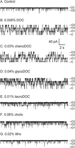
At 0.008% deoxycholic acid (DOC) and 0.03% chenodeoxycholic acid (chenoDOC), the channel undergoes much more frequent transitions between open and closed states, and is found in the fully closed state ∼50% of the time (, ). Increased gating activity is also observed at 0.04% glycodeoxycholic acid (glycoDOC) (), but the fully closed state is not observed as frequently as with DOC and chenoDOC. In the presence of even the highest concentrations tested of the other three bile acids, taurodeoxycholic acid, cholic acid and lithocholic acid, the traces are similar to the one in control conditions (, , ). The pattern of activity observed in the presence of DOC, chenoDOC and glycoDOC is reminiscent of the time-resolved blocking events reported for the E. coli porin OmpF in the presence of ampicillin (Nestorovich et al. Citation2002) or the maltoporin LamB in the presence of maltodextrins (Kullman et al. Citation2002). In our previous work analyzing the effect of DOC on OmpT and OmpU (Duret and Delcour Citation2006), we have argued that the observed transitions represent the transient block of each monomer as DOC penetrates inside the pore. Similarly, chenoDOC and glycoDOC also block OmpT, albeit at higher concentrations.
In order to quantify the effect and obtain a dose-dependence, we have plotted the ratio of the channel open probability in the presence of bile acids to the open probability in control conditions. The results are presented in in two separate panels on the same scale for the sake of clarity. Panel A shows the dose dependence of the most effective bile acids, and panel B of the least effective ones. To be accurate in the relative comparison between bile acids, the dose dependence needs to be shown in mM rather than % because the molecular mass of each bile acid is slightly different from the others. Except for the case of DOC, it was not possible to reliably fit the data obtained with the other bile acids, because we did not have enough points of low Po. Therefore, for the other effective bile acids (chenoDOC and glycoDOC), the lines in were drawn according to Equation (1) in Materials and methods using the same value for the Hill coefficient as for DOC (2.5, as previously published (Duret and Delcour Citation2006)), and adjusting the value of the IC50 to obtain the best fit by eye (values given in the Figure legend). shows the results obtained with the non-effective bile acids in the investigated concentration range, where the Po ratio hovers around 1.0 (no fit was performed and the data points are connected by lines). The data clearly indicates that the efficacy of bile acids towards block of OmpT depends on the nature of the bile acid, in the following sequence: DOC > chenoDOC > glycoDOC > cholic acid. We had difficulties maintaining patches with concentrations of tauroDOC higher than 0.2 mM (0.01%), and unfortunately we have too few points with tauroDOC to clearly ascertain whether its efficacy is equal or worse than glycoDOC. Similarly, because of the limit of solubility, we were not able to extend the concentration range for lithocholic acid beyond ∼0.5 mM (0.02%) for accurate measurements. Nevertheless, despite the limited range of concentrations tested, it is clear that both tauroDOC and lithocholic acid are far less effective than DOC and chenoDOC.
Figure 3. The bile acid mediated OmpT block is dose-dependent. The open probability of a single trimer was measured in the presence of various bath-applied concentrations of a given bile acid [Po(BA)] and in the absence of bile acids [Po(CON)] on the same patch, at +50 mV (pipette voltage). The graph plots the average (± SEM) of the ratio of such open probabilities (n > = 3) against bile acid concentration (in mM). In a few conditions, symbols do not have error bars because they represent only two experiments. The black line is a fit of the data in DOC to equation 1 (Materials and methods). The other lines of panel A are trend lines fitted through the points by eye (see text). The IC50s were found to be: 0.178 mM (0.007%) for DOC, 0.638 mM (0.025%) for chenoDOC and 1.282 mM (0.06%) for glycoDOC. In panel B, the individual data points are joined by lines to help visualization. The black horizontal line marks a Po ratio of 1.0.
![Figure 3. The bile acid mediated OmpT block is dose-dependent. The open probability of a single trimer was measured in the presence of various bath-applied concentrations of a given bile acid [Po(BA)] and in the absence of bile acids [Po(CON)] on the same patch, at +50 mV (pipette voltage). The graph plots the average (± SEM) of the ratio of such open probabilities (n > = 3) against bile acid concentration (in mM). In a few conditions, symbols do not have error bars because they represent only two experiments. The black line is a fit of the data in DOC to equation 1 (Materials and methods). The other lines of panel A are trend lines fitted through the points by eye (see text). The IC50s were found to be: 0.178 mM (0.007%) for DOC, 0.638 mM (0.025%) for chenoDOC and 1.282 mM (0.06%) for glycoDOC. In panel B, the individual data points are joined by lines to help visualization. The black horizontal line marks a Po ratio of 1.0.](/cms/asset/218c2b35-d2d8-4c06-9951-fc3b1e34bd43/imbc_a_519727_f0003_b.jpg)
The on- and off-rates of binding of a blocker to a channel can be obtained from the analysis of the open and closed (i.e., blocked) dwell times. However, this requires that the blocking events be clearly distinguished from intrinsic closures, or that the level of spontaneous closures in the absence of blocker be very low. This is not the case for OmpT, which displays a fairly active spontaneous closing activity. Thus, in the presence of bile acids, the open and closed dwell times represent a mixture of the spontaneous closing kinetics and the kinetics of block. We have attempted to sort this out by looking at dwell time distributions, but the results are inconclusive. In addition, there is some variability in the kinetics of the spontaneous closures, which further compounds the difficulty of analysis. Therefore, we have restricted our analysis to that of the relative decrease in open probability, as done in our earlier study (Duret and Delcour Citation2006).
An interesting aspect of the dose-dependent block by penetrating bile acids is the voltage-dependence of the effect. We previously reported that the DOC-mediated block of OmpT was favored at positive pipette voltages, regardless of the side of addition (pipette vs. bath) or even in the presence of DOC on both sides (Duret and Delcour Citation2006). If the block was due to the electrophoretic attraction of the negatively charged species into the pore lumen upon positive pipette voltage, one would expect the voltage-dependences to be mirror images when DOC is bath-applied versus pipette-applied. In addition, the pH dependence of the DOC effect hints that the protonated uncharged DOC is the active species (Duret and Delcour Citation2006). All together, these observations suggest that the binding site for DOC in the OmpT pore is intrinsically voltage-dependent. We have confirmed this observation here, as can been seen for DOC and the other two most potent bile acids, chenoDOC and glycoDOC (). Thus it appears that the effective bile acids share the same binding site inside the porin.
Figure 4. The bile acid mediated OmpT block is voltage-dependent. The open probability of a single trimer was measured in the presence of a given bath-applied bile acid [Po(BA)] and in the absence of the bile acids [Po(CON)] on the same patch. The graph plots the ratio of such open probabilities at various voltages. The concentrations of bile acids were as follows: 0.006% (0.15 mM) DOC, 0.03% (0.76 mM) chenoDOC, 0.03% (0.64 mM) glycoDOC, 0.01% (0.19 mM) tauroDOC, 0.06% (1.47 mM) cholic acid, 0.02% (0.53 mM) lithocholic acid. A voltage-dependence is found for the effective bile acids (DOC, chenoDOC and glycoDOC), but not the ineffective ones (cholic, litho and tauroDOC), as highlighted by the solid horizontal line (Po ratio of 1).
![Figure 4. The bile acid mediated OmpT block is voltage-dependent. The open probability of a single trimer was measured in the presence of a given bath-applied bile acid [Po(BA)] and in the absence of the bile acids [Po(CON)] on the same patch. The graph plots the ratio of such open probabilities at various voltages. The concentrations of bile acids were as follows: 0.006% (0.15 mM) DOC, 0.03% (0.76 mM) chenoDOC, 0.03% (0.64 mM) glycoDOC, 0.01% (0.19 mM) tauroDOC, 0.06% (1.47 mM) cholic acid, 0.02% (0.53 mM) lithocholic acid. A voltage-dependence is found for the effective bile acids (DOC, chenoDOC and glycoDOC), but not the ineffective ones (cholic, litho and tauroDOC), as highlighted by the solid horizontal line (Po ratio of 1).](/cms/asset/eab8eaee-3f7a-4d7a-84cc-d7c3eda4f795/imbc_a_519727_f0004_b.jpg)
Discussion
Using patch clamp electrophysiology, we report here the findings that OmpT, one of the general diffusion porins of the V. cholerae outer membrane, is differentially sensitive to channel block by six bile acids. The effectiveness of the tested bile acids as OmpT blockers is as follows: DOC > chenoDOC > glycoDOC > cholic acid. Lithoholic acid and tauroDOC are as ineffective as cholic acid in the concentration range tested. The extent of block by the bile acids is concentration- and voltage-dependent. The effective concentrations for the most potent bile acids are below the CMC (Roda et al. Citation1983), and thus the observed modulation of the channel can be attributed to the effect of monomers rather than micelles. The concentration of bile salts in bile is about 40 mM (Begley et al. Citation2005), but is lowered in the jejunum and ileum, where V. cholerae colonizes, due to reabsorption. The total postprandial concentration of unconjugated bile acids has been estimated between 0.25 and 1 mM in the ileum (Northfield and McColl Citation1973), and DOC and chenoDOC represents ∼25% and 35%, respectively, of the total bile acids (Carey and Duane Citation1994). Thus, the effective concentrations of DOC and chenoDOC on OmpT (∼ 200 μM) are within the physiological range in the region of the intestine colonized by V. cholerae.
The change in OmpT channel kinetics in the presence of DOC, chenoDOC and glycoDOC is similar to the pattern of time-resolved blocking events also observed when the E. coli porin OmpF is blocked by ampicillin (Nestorovich et al. Citation2002) or the maltoporin LamB by maltodextrins (Kullman et al. Citation2002). Ampicillin and maltodextrins are known to use OmpF and LamB, respectively, to cross the outer membrane into the cell (Nikaido Citation2003). Bezrukov and colleagues have argued that the observed blocking events in the presence of ampicillin or maltodextrins represent the transient binding and unbinding of the molecules inside their respective pores, and estimated, on the basis of kinetic parameters, that the unblocking events represents an actual translocation event 20–50% of the time (Kullman et al. Citation2002, Nestorovich et al. Citation2002). We have made a similar argument for DOC and OmpT since the molecular weight of DOC is smaller than the size exclusion limit for OmpT (Chakrabarti et al. Citation1996), DOC is known to reach the inner membrane in vivo (Hung and Mekalanos Citation2005, Cerda-Maira et al. Citation2008), and DOC can access its binding site inside OmpT from both sides of the channel (Duret and Delcour Citation2006). Therefore, the simplest explanation for the frequent and prolonged monomer closures observed here with DOC and chenoDOC, and to a lesser extent glycoDOC, is that the closures represent transient blockages of the open pores as the bile acids travel through the pore. As discussed previously (Duret and Delcour Citation2006), membrane-mediated effects of the bile acids cannot be completely dismissed, but the changes in activity are more likely to be due to a direct effect of the bile acid on OmpT because: (1) They are fully reversible, (2) they are voltage-dependent, and (3) they have the hallmark kinetic features of open channel block (Hille Citation1992).
Why then does OmpT show differing sensitivities to each bile acid? In the case of β-lactam antibiotics, Bezrukov and colleagues have demonstrated that time–resolved blocking events were observed with ampicillin and amoxicillin, two zwitterionic penicillins that make the most favorable interactions with the constriction zone of the OmpF pore. On the other hand, the dianionic carbenicillin does not fit tightly within the constriction zone, and also does not induce the blocking pattern observed with the other two antibiotics (Danelon et al. Citation2006). By analogy, we would like to propose that the differential effect of the bile acids on OmpT also reflect the strength of the interactions with the OmpT constriction zone. As seen in , the unconjugated bile acids investigated here have differing states of hydroxylation, though for all, the substituents are on the α face of the molecules making it hydrophilic, while the β face is substituent free, making it hydrophobic (Hofmann and Hagey Citation2008). Cholic acid has a total of 3 hydroxyl groups, chenoDOC and DOC both have 2 (though the locations of the hydroxyl groups vary), and lithocholic acid only has one hydroxyl group. Interestingly, the two bile acids most effective in blocking OmpT, chenoDOC and DOC, are dihydroxy. ChenoDOC is hydroxylated at C3 and C7, while DOC is hydroxylated at C3 and C12. Since all natural bile acids have a C3 hydroxyl group that originates from the cholesterol moiety, it appears that it is the number and location of other hydroxyl groups that may dictate the interactions for optimal binding.
Another parameter to be considered among bile acids is their state of ionization. All unconjugated bile acids have a pKa of ∼5.0, but conjugation decreases the pKa sharply to ∼2.6 for the glycine conjugates, and close to 0 for the taurine conjugates (Carey Citation1984, Hofmann and Roda Citation1984). In addition, we have previously suggested that it is the protonated neutral form of DOC that blocks OmpT, and not the charged species (Duret and Delcour Citation2006), implying that the carboxyl group may also participate in the binding site. Because of the difference in pKas between unconjugated and conjugated bile acids, one might have expected a stronger effect of the unconjugated bile acids, since a larger proportion of them would be protonated at neutral pH. However the conjugated forms of DOC – at least the glycine conjugate – are more potent than cholic acid and lithocholic acid. GlycoDOC is only different from DOC at the carboxyl moiety, while cholic acid and lithocholic acid share the carboxyl group with DOC, but differ in their state of hydroxylation. Therefore, we propose that the carboxyl group has a role in binding, since its protonation may be important (Duret and Delcour Citation2006) and the conjugated forms of DOC have a decreased potency relative to DOC, but the influence of the carboxyl group is likely to be of lesser importance than that of the hydroxyl groups. The decreased potency of glycoDOC (and tauroDOC) relative to DOC may be due to steric effects and/or the absence of specific interactions involving the protonated carboxyl group. For OmpF and ampicillin, it has been shown that multiple constriction zone residues participate in specific ionic, hydrogen bond and hydrophobic interactions with specific regions of the antibiotic molecule (Nestorovich et al. Citation2002, Danelon et al. Citation2006). We anticipate that multiple interactions are also required for the favorable binding of DOC or chenoDOC inside the OmpT pore. Because the number and position of the hydroxyl groups are of prime importance, it is likely that specific hydrogen bonds are formed between the bile acid molecule and the channel wall. It is unfortunate that the crystal structure of OmpT is not available to model the interactions, as has been done for OmpF and the β-lactam antibiotics.
Bezrukov and colleagues have argued that the presence of a binding site for ampicillin inside OmpF results in the preferred permeation of this antibiotic through the porin (Danelon et al. Citation2006). The temporary block of OmpF by ampicillin is indicative of an appreciable residency time in the pore, and this may be construed as impairment to permeation. However, one has to consider that the presence of the high affinity binding site will favor uptake of this molecule, and exert an effect that will be especially important at low concentrations. Thus, it is not surprising to find a strong correlation between the existence of a binding site in the pore and the efficacy of translocation of β-lactam antibiotics (Danelon et al. Citation2006). β-lactam antibiotics with low diffusion rates do not bind in the constriction zone of OmpF and do not cause time-resolved channel blocking events. By analogy, we infer that DOC and chenoDOC, which induce blocking events at low concentrations, would be taken up more favorably by V. cholerae cells than the other bile acids, including the conjugated forms of DOC. Interestingly, the recently discovered bile response genes breAB and breR were also found to be preferentially regulated by DOC and chenoDOC, relative to other bile acids, in a manner that mirrors their potency on OmpT, and the bile acids have been proposed to bind directly to the regulatory protein BreR (Cerda-Maira et al. Citation2008).We anticipate that, during the transit of the ingested V. cholerae cells through the small intestine, DOC and chenoDOC may play a role of signaling to the bacteria that: (1) The bacteria are inside a host, and (2) they are in a region that is favourable to bacterial growth, since deconjugation occurs only due to the activities of the endogenous flora. DOC may be particularly suitable as a signal, since the dehydroxylation of cholic acid into DOC is itself a microorganism-mediated event as well, and the other secondary bile acid, lithocholic acid, is relatively scarce (Carey and Duane Citation1994). Interestingly, the E. coli porins OmpF and OmpC are not affected by DOC (Delcour, unpublished).Thus, the preferential binding of DOC to OmpT (the porin that is present in the ingested bacteria as they first enter the host) over all other bile acids, and its resulting favored uptake may relate to the particular role of DOC as a signaling molecule in V. cholerae.
Acknowledgements
We thank Guillaume Duret for critically reading the manuscript. The work was supported by the Welch Foundation grant E-1597.
Declaration of interest: The authors report no conflicts of interest. The authors alone are responsible for the content and writing of the paper.
References
- Baslé A, Iyer R, Delcour AH. 2004. Subconductance states in OmpF gating. Biochim Biophys Acta 1664:100–107.
- Begley M, Gahan CG, Hill C. 2005. The interaction between bacteria and bile. FEMS Microbiol Rev 29:625–651.
- Bina JE, Mekalanos JJ. 2001. Vibrio cholerae tolC is required for bile resistance and colonization. Infect Immun 69:4681–4685.
- Bina JE, Provenzano D, Wang C, Bina XR, Mekalanos JJ. 2006. Characterization of the Vibrio cholerae vexAB and vexCD efflux systems. Arch Microbiol 186:171–181.
- Butler SM, Camilli A. 2005. Going against the grain: Chemotaxis and infection in Vibrio cholerae. Nat Rev Microbiol 3:611–620.
- Carey MC. 1984. Bile acids and bile salts: Ionization and solubility properties. Hepatology 4:66S–71S.
- Carey MC, Duane WC. 1994. Enterohepatic circulation. In: Arias IM, Boyer JL, Fausto N, Jakoby WB, Schachter DA, Shafritz DA, editors. The liver: Biology and pathobiology. 3d ed. New York: Raven Press Ltd. pp 719–767.
- Cerda-Maira FA, Ringelberg CS, Taylor RK. 2008. The bile response repressor BreR regulates expression of the Vibrio cholerae breAB efflux system operon. J Bacteriol 190:7441–7452.
- Chakrabarti SR, Chaudhuri K, Sen K, Das J. 1996. Porins of Vibrio cholerae: Purification and characterization of OmpU. J Bacteriol 178:524–530.
- Chatterjee A, Chaudhuri S, Saha G, Gupta S, Chowdhury R. 2004. Effect of bile on the cell surface permeability barrier and efflux system of Vibrio cholerae. J Bacteriol 186:6809–6814.
- Danelon C, Nestorovich EM, Winterhalter M, Ceccarelli M, Bezrukov SM. 2006. Interaction of zwitterionic penicillins with the OmpF channel facilitates their translocation. Biophys J 90:1617–1627.
- Delcour AH, Martinac B, Adler J, Kung C. 1989. Modified reconstitution method used in patch-clamp studies of Escherichia coli ion channels. Biophys J 56:631–636.
- Duret G, Delcour AH. 2006. Deoxycholic acid blocks Vibrio cholerae OmpT but not OmpU porin. J Biol Chem 281:19899–19905.
- Duret G, Delcour AH. 2010. Size and dynamics of the Vibrio cholerae porins OmpU and OmpT probed by polymer exclusion. Biophys J 98:1820–1829.
- Duret G, Simonet V, Delcour AH. 2007. Modulation of Vibrio cholerae porin function by acidic pH. Channels 1:70–79.
- Gupta S, Chowdhury R. 1997. Bile affects production of virulence factors and motility of Vibrio cholerae. Infect Immun 65:1131–1134.
- Hille B. 1992. Ionic channels of excitable membranes. Sunderland, MA: Sinauer Associates.
- Hofmann AF. 1994. Bile acids. In: Arias IM, Boyer JL, Fausto N, Jakoby WB, Schachter DA, Shafritz DA, editors. The liver: Biology and pathobiology. 3rd ed. New York: Raven Press Ltd. pp 677–718.
- Hofmann AF, Hagey LR. 2008. Bile acids: Chemistry, pathochemistry, biology, pathobiology, and therapeutics. Cell Mol Life Sci 65:2461–2483.
- Hofmann AF, Roda A. 1984. Physicochemical properties of bile acids and their relationship to biological properties: An overview of the problem. J Lipid Res 25:1477–1489.
- Hung DT, Mekalanos JJ. 2005. Bile acids induce cholera toxin expression in Vibrio cholerae in a ToxT-independent manner. Proc Natl Acad Sci USA 102:3028–3033.
- Hung DT, Zhu J, Sturtevant D, Mekalanos JJ. 2006. Bile acids stimulate biofilm formation in Vibrio cholerae. Mol Microbiol 59:193–201.
- Kullman L, Winterhalter M, Bezrukov SM. 2002. Transport of maltodextrins through maltoporin: A single-channel study. Biophys J 82:803–812.
- Lee SH, Hava DL, Waldor MK, Camilli A. 1999. Regulation and temporal expression patterns of Vibrio cholerae virulence genes during infection. Cell 99:625–634.
- Nestorovich EM, Danelon C, Winterhalter M, Bezrukov SM. 2002. Designed to penetrate: Time-resolved interaction of single antibiotic molecules with bacterial pores. Proc Natl Acad Sci USA 99:9789–9794.
- Nikaido H. 1996. Outer membrane. In: Neidhart FC, editor. Escherichia coli and Salmonella. Cellular and molecular biology. Washington, DC: ASM Press. pp 29–47.
- Nikaido H. 2003. Molecular basis of bacterial outer membrane permeability revisited. Microbiol Mol Biol Rev 67:593–656.
- Noh DO, Gilliland SE. 1993. Influence of bile on cellular integrity and beta-galactosidase activity of Lactobacillus acidophilus. J Dairy Sci 76:1253–1259.
- Northfield TC, McColl I. 1973. Postprandial concentrations of free and conjugated bile acids down the length of the normal human small intestine. Gut 14:513–518.
- Oelberg DG, Chari MV, Little JM, Adcock EW, Lester R. 1984. Lithocholate glucuronide is a cholestatic agent. J Clin Invest 73:1507–1514.
- Pagel M, Simonet V, Li J, Lallemand M, Lauman B, Delcour AH. 2007. Phenotypic characterization of pore mutants of the Vibrio cholerae porin OmpU. J Bacteriol 189:8593–8600.
- Provenzano D, Klose KE. 2000. Altered expression of the ToxR-regulated porins OmpU and OmpT diminishes Vibrio cholerae bile resistance, virulence factor expression, and intestinal colonization. Proc Natl Acad Sci USA 97:10220–10224.
- Provenzano D, Lauriano CM, Klose KE. 2001. Characterization of the role of the ToxR-modulated outer membrane porins OmpU and OmpT in Vibrio cholerae virulence. J Bacteriol 183:3652–3662.
- Provenzano D, Schuhmacher DA, Barker JL, Klose KE. 2000. The virulence regulatory protein ToxR mediates enhanced bile resistance in Vibrio cholerae and other pathogenic Vibrio species. Infect Immun 68:1491–1497.
- Reidl J, Klose KE. 2002. Vibrio cholerae and cholera: Out of the water and into the host. FEMS Microbiol Rev 26:125–139.
- Ridlon JM, Kang DJ, Hylemon PB. 2006. Bile salt biotransformations by human intestinal bacteria. J Lipid Res 47:241–259.
- Roda A, Hofmann AF, Mysels KJ. 1983. The influence of bile salt structure on self-association in aqueous solutions. J Biol Chem 258:6362–6370.
- Schuhmacher DA, Klose KE. 1999. Environmental signals modulate ToxT-dependent virulence factor expression in Vibrio cholerae. J Bacteriol 181:1508–1514.
- Simonet VC, Basle A, Klose KE, Delcour AH. 2003. The Vibrio cholerae porins OmpU and OmpT have distinct channel properties. J Biol Chem 278:17539–17545.
- Vyvoda OS, Coleman R, Holdsworth G. 1977. Effects of different bile salts upon the composition and morphology of a liver plasma membrane preparation. Deoxycholate is more membrane damaging than cholate and its conjugates. Biochim Biophys Acta 465:68–76.
- Wilson M. 2005. Microbial inhabitants of humans. Their ecology and role in health and disease. Cambridge, UK: Cambridge University Press.