
Abstract
GO-COO-β-CD/CA inclusion (carboxylated graphene-β-cyclodextrin/chlorhexidine acetate) was fabricated with a graphene-based drug carrier. The reaction time and ratio of carrier to drug were optimized by X-ray diffraction spectra to ensure the complete wrapping of CA. Hemolysis test and recalcification test demonstrated that the inclusion possessed good blood compatibility due to the inherent biocompatibility of β-CD molecules in the carrier. The inclusion displayed excellent inhibition effect on both gram negative bacteria of Escherichia coli and gram positive bacteria of Staphylococcus Aureus, while showing no cytotoxicity. More importantly, the drug efficiency was greatly improved with CA dosage as less as one-third of the pure drug due to the synergistic effect of the drug and carrier. Dynamic simulation implies that the delivery profile of CA from the inclusion is in accordance with the first-order dynamic equation, i.e. ln(1-Mt/M) = −kt.
Introduction
Graphene, with an atomically thin and two-dimensional (2D) layer, displays advantageous electrical conductivity and novel physico-chemical properties (Novoselov et al., Citation2005; Katsnelson et al., Citation2007; Geim, Citation2009; Lu et al., Citation2012). As a result of their unique chemical and physical properties, with respect to structures that can be oriented and surfaces that can be modified, graphene and graphene-based materials, such as graphene oxide (GO) and carboxylated graphene (GO-COOH), have become promising candidates in biotechnological applications, and thus have attracted enormous interest in biological studies and biotechnology. Various graphene-based nanomaterials have been used to fabricate functionalized biosystems integrated with nucleic acids (NAs), peptides, proteins and even cells (Kuila et al., Citation2011; Lu et al., Citation2012; Pei et al., Citation2012). Due to its merit of large specific surface area, a promising application of GO is in the fabrication of drug carriers (Shen et al., 2012; Wu et al., 2012). Hence, another exciting area of graphene research is drug delivery in living cells and many achievements using graphene as an advanced transporter for drug delivery have been acquired (Dembereldorj et al., Citation2012; Chowdhury et al., Citation2013; Yang et al., Citation2013). For instance, Dai’s group prepared polyethylene glycol functionalized graphene for the first time and successfully loaded an anti-cancer drug of Camptothecin by π–π stacking on modified graphene (Liu et al., Citation2008).
Cyclodextrin and its derivatives have the functionality of enhancing the solubility and dissolving rate of drugs with poor water solubility. Furthermore, they can improve the stability and bioavailability while inhibiting the stimulation and side effects of the drugs (Trotta et al., Citation2012; Yhaya et al., Citation2012; Baek et al., Citation2013). These advantages render cyclodextrin family among good candidates in medicines and biomedical applications (Chaturvedi et al., Citation2011; Moya-Ortega et al., Citation2012; Yannakopoulou Citation2012).
Recently we found that the combination of GO-COOH with β-cyclodextrin (β-CD) turns out to be a novel nanohybrid drug carrier of GO-COO-β-CD with the performance of higher drug loading owing to the nano-size effect of GO and good biocompatibility of β-CD.
Chlorhexidine acetate (CA) is a kind of cationic surfactant and is widely used because it is an effective drug with broad anti-bacterial and sterilization spectrum, for both gram negative bacteria and positive bacteria (Acar et al., Citation2011;Agarwal et al., Citation2012). Some limitations need to be addressed including change of taste, increase of oral surface color and calcification, light stimulation and reversible peeling of children (Ferraz et al., Citation2001; Meng et al., Citation2009; Murayama et al., Citation2011). Furthermore, in practical use, CA is usually applied in the form of diluted solution, which is effective in quick and temporary treatments. However, there remains a great challenge for long-term and controlled release of CA. Capitalizing the β-CD cavities with hydrophobic inner walls and hydrophilic outer walls as well as the synergistic effect of GO-COOH on drugs (Zhang et al., Citation2010), CA can be included in GO-COO-β-CD to improve its curative effect, bioavailability and medication safety and is thus used as model drug in the present study. Herein, we report our current work on the preparation of GO-COO-β-CD/CA inclusion and investigation on its blood compatibility, drug efficiency, anti-bacterial performance and drug delivery properties.
Experimental
Materials
CA was supplied by Jintan Aide Medicine Manufacturer (Jintan, China). β-CD, absolute ethanol, beef extract, peptone, glucose and agar were purchased from Sinopharm Chemical Reagent Co., Ltd. (Beijing, China). Escherichia coli (ATCC25922) and Staphylococcus aureus (ATCC25923) were obtained from Jiangsu Provincial Center for Disease Prevention and Control (Nanjing, China). GO-COO-β-CD was prepared according to the following procedures. Small amounts of raw GO-COOH and β-CD were ground in an agate mortar with dropwise addition of ethanol (0.7900 g) during the first 10 min. The paste was transferred into a beaker and 15 ml of ethanol was added. After further reaction for 3 h under ultrasonication, the resulting powder was heated at 75 °C for 24 h using a vacuum oven. Finally, the dried product was gently ground into a fine homogeneous powder.
Human Embryonic Kidney 293 cells, also referred as HEK 293, and Dulbecco’s modified Eagle's medium (DMEM) were from Thermo Fisher HyClone (Logan, UT). 3-(4,5-Dimethylthiazol-2-yl)2,5-diphenyl tetrazolium bromide (MTT) was from Amresco (Solon, OH). All other chemicals were of analytical grade.
Preparation of GO-COO-β-CD/CA inclusions
A certain amount of CA was dissolved in 5 ml of ethanol and was put in an agate mortar together with the as-prepared GO-COO-β-CD (pH = 8–9). The mixture was gently ground for some minutes and freeze-dried for 24 h before grey fine powder was obtained, which is referred to GO-COO-β-CD/CA inclusion. For comparison, a physical mixture was prepared by mixing together GO-COO-β-CD and CA. The amount of CA is equivalent to that applied in the inclusion.
Characterization
X-ray diffraction (XRD) patterns were recorded in a range from 3 °C to 80 °C (2θ) at a rate of 2 °C/min on a diffractometer (Rigaku D/max, Tokyo, Japan) using CuKα radiation. The diffracted intensity was recorded automatically. The CA amount for XRD assay was 50 mg and kept equivalent for all XRD characterizations. Ultraviolet-Visible spectrum was recorded using Cary 50 UV spectroscopy (Varian) (Palo Alto, CA). Aqueous stability of the inclusion was investigated by measuring the particle size and polydispersity index (PDI) at different temperatures using dynamic light scattering (ZetasizerNano-ZS90, Malvern, UK).
Assays of antibacterial property of GO-COO-β-CD/CA inclusion
Preparation of culture medium
Liquid culture medium was prepared according to the following steps. Three grams of beef extract was dissolved in 800 ml double-distilled water (DDW) followed by the addition of 10 g peptone and 5 g NaCl. The mixture was heated to boiling and diluted to 1000 ml. The pH value was adjusted to 7.0–7.2 and the medium was sterilized under high pressure for 15 min before placing it in a refrigerator (4 °C).
Nutrient agar culture medium was prepared as follows: 3 g beef extract, 10 g peptone and 5 g NaCl were dissolved in 1000 ml DDW. Fifteen percent NaOH was added until the pH value of the mixture reached 7.0–7.2. Fourteen grams of agar was then added while heating and stirring before sterilization under high pressure.
Culture and activation of the bacteria
Ten milliliters of liquid medium was placed in a sterilized test tube before E. coli or S. aureus were transferred into it. The test tubes were placed in a shaker at 37 °C for 24 h.
Antibacterial property assays
GO-COO-β-CD, CA and GO-COO-β-CD/CA powder and the physical mixture of GO-COO-β-CD and CA were pressed into round discs with a diameter of 13 mm. The discs were sterilized under pressure of 0.1 MPa for 20 min. Agar medium was casted into a plate and the activated bacterium suspension of E. coli or S. aureus was pipetted into 150 ml agar medium until the bacteria were dispersed homogeneously. The discs were placed on an agar plate before it was put in an incubator of 37 °C for 24 h.
The determination of minimum inhibitory concentration (MIC) was performed according to Meng et al., (Citation2009). GO-COO-β-CD/CA of different concentrations was added into the culture. The concentrations of E. coli and S. aureus were 106 cfu/ml. The mixtures were placed in a shaker of 37 °C for 24 h. GO-COO-β-CD was also added into the culture as control. The MIC is defined as the concentration at which no bacteria appear.
Blood compatibility assays
Hemolysis test
Totally 0.01 g of GO-COO-β-CD/CA, GO-COO-β-CD and CA were dissolved in 10 ml of 37% NaCl to make a 1 mg/ml solution. The solution was diluted to different concentrations. The diluted whole blood sample was mixed with different solutions and the mixtures were gently stirred until they were homogeneous. The mixtures were placed in an incubator of 37 °C for 60 min, followed by centrifugation for 10 min (1500 r/min). The upper clear solution was removed to a 96-well plate and UV absorbance measurement at 545 nm was performed. The measurements were repeated three times for each sample to obtain the standard deviation. Hemolysis rate was calculated according to the following equation:
where Dt is the absorbance of the sample, Dnc absorbance of negative control, i.e. 10 ml 0.9 % NaCl, and Dpc absorbance of positive control, i.e. 10 ml DDW.
Recalcification profile test
The whole blood sample was centrifuged for 10 min (3000 r/min) to obtain platelet poor plasma (PPP). CaCl2 and 0.1 ml upper clear solution were then added into PPP in a 96-well plate. The dynamic relationship was obtained with the optical density (OD) at a wavelength of 405 nm by BioTek synergy2 enzyme-labeled meter (Winooski, VT). 0.025 M CaCl2 in 0.1 ml PPP and pure 0.1 ml PPP were used as controls. All experiments were repeated three times.
Cytotoxicity assays
HEK293 cells were maintained in DMEM with 10% fetal bovine serum (FBS). The cells were rinsed briefly with sterilized phosphate buffer solution (PBS) and trypsinized before re-suspending in FBS-free DMEM with a concentration of 1×10⁵ cells/ml. The cells in 96-well plates at a density of 104 cells/well in DMEM were placed in an incubator with 5% CO2 at 37 °C. The cell differentiation and proliferation was observed with a converted optical microscope. Cytotoxicity of different samples was assessed in vitro using MTT assay. GeneO-COO-β-CD, CA and GeneO-COO-β-CD/CA were diluted with DMEM to various drug doses (0.2, 2 and 20 µg/ml) and the cells were placed in 100 µl of the above solutions, respectively. After 24 h of incubation, the media was removed and subsequently replaced with PBS containing 5 mg/ml MTT followed by incubation for another 4 h. After incubation, the medium was removed and the cells were lysed by adding dimethylsulfoxide (DMSO). The mixture was shaken for 10 min before absorbance and measurements at 490 nm were performed.
where RGR is the relative growth rate of cells.
Drug delivery profile
Release studies in vitro were carried out according to Chinese Pharmacopoeia, Ed 2010. Three dialysis bags were equilibrated with the dissolution medium of HCl (pH = 1.0) and PBS (pH = 7.0) for a few hours. Hundred milligrams of GO-COO-β-CD/CA was dispersed in HCl or PBS in the dialysis bags. The bags were dipped into receptor compartment containing 200 ml dissolution medium at 37 °C ± 0.5 °C and shaken at a rate of 100 rpm. Five milliliters of each solution was taken at 1, 120, 200 h and other time intervals, and another 5 mlof fresh solution was supplemented thereafter. UV characterization was performed on the taken solutions.
Results and discussion
In basic pH domain (pH = 8–9), some CA molecules may be adsorbed on the carboxylated surface, while others may enter the cavities of CD. Thus, there are two kinds of combination of GO-COO-β-CD with CA. In the formation process of the inclusion, parts of GO-COO-β-CD molecules are chemically bonded with carboxyl groups on the surface of GO-COOH slice layer, and some parts are padded between GO-COOH layers. The adsorbed CA is covered by β-CD assemblies, thus GO-COO-β-CD/CA inclusion formed.
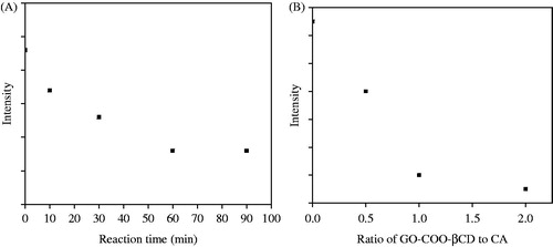
XRD was applied to verify the formation of GO-COO-β-CD/CA inclusion. The characteristic peaks of CA are positioned at 2θ = 17.78°, 20.22°, 22.82° and 23.16°. We found that the peak intensity at 17.78° gradually decreased with reaction time and almost disappeared at 60 min (). This can be explained by the fact that CA molecules entered the cavities of β-CD or adsorbed GO-COOH, but covered with β-CD assemblies to form a GO-COO-β-CD/CA inclusion. The optimum carrier/drug ratio was determined by changing the GO-COO-β-CD to CA ratio from 1:2 to 2:1. It was found that the characteristic peaks of CA disappeared at the ratio of 2:1, indicating a complete inclusion of CA with GO-COO-β-CD (). Thus, we chose 60 min as reaction time and 2:1 as ratio of GO-COO-β-CD to CA.
Figure 1. (A) Relationship between intensity at 2θ = 17.78° from XRD of CA and reaction time. (B) Ratio of GO-COO-β-CD to CA.
Stability of the inclusion in aqueous solution plays a crucial role in real applications. Variation of particle size and PDI of GO-COO-β-CD/CA with temperatures are shown in and also listed in for clear comparison. It is seen that the particle size slightly increases with temperature increase from 25 to 45 °C. The slight increase in size may be caused by the weakening of the hydrogen bonds between CD molecules at higher temperatures. Meanwhile, PDI falls in the range of 0.1–0.2, demonstrating the good dispersibility of the inclusion in aqueous solution at different temperatures.
Figure 2. Variation of particle size and PDI of GO-COO-β-CD/CA with temperatures. Inset shows the digital pictures of GO-COO-β-CD/CA aqueous solutions.
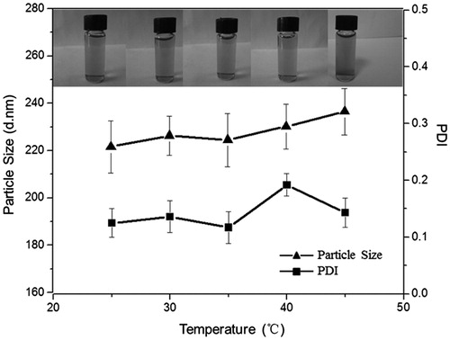
Table 1. Particle size and PDI of GO-COO-β-CD/CA at different temperatures.
displays the inhibition zone test of different samples with respect to E. coli and S. aureus. It is clearly seen that GO-COO-β-CD nearly has no inhibition effect on the bacteria ( and ) while GO-COO-β-CD/CA inclusion greatly inhibits the growth of the bacteria ( and ). An obvious inhibition zone was observed with the largest diameter of 11.5 mm as listed in . It is also observed that the inhibition zone of the inclusion is larger than that of pure CA ( and ). More importantly, the drug dosage in the inclusion is only one-third of pure CA. A reasonable explanation for the phenomena is that the solubility of CA in the inclusion is increased 6.56 times higher than that of pure CA, leading to much better inhibition effect. Moreover, the antibacterial property of the inclusion to both bacteria is improved compared with that of the physical mixture of the two components ( and ). In detail, the inhibition zones of the inclusion are 1.36 and 1.47 times as large as those of the physical mixtures to E. coli and S. aureus, respectively. The phenomena further demonstrate the synergistic effect of the drug and carrier. Good drug carriers can simultaneously improve the antibacterial performance of the drug (Liu et al., Citation2008) and GO-COO-β-CD is a promising candidate for use as a drug carrier.
Figure 3. (A) Optical images of inhibition zone for GO-COO-β-CD (a), CA (b), GO-COO-β-CD/CA (c) and physical mixture of GO-COO-β-CD and CA (d) against E. coli. (B) Optical images of inhibition zone for GO-COO-β-CD (a), CA (b), GO-COO-β-CD/CA (c) and physical mixture of GO-COO-β-CD and CA (d) against S. aureus.
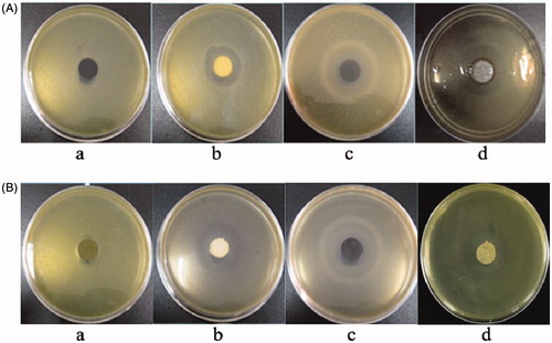
Table 2. Diameter of inhibition zone of GO-COO-β-CD, CA, GO-COO-β-CD/CA and physical mixture of GO-COO-β-CD and CA.
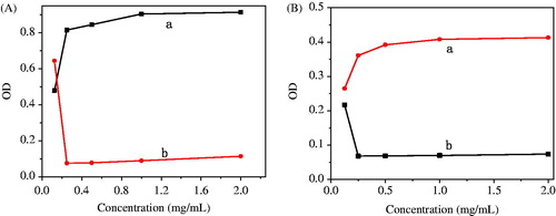
GO-COO-β-CD and GO-COO-β-CD/CA with different concentrations were added into the same amount of E. coli. From we see that the addition of GO-COO-β-CD clearly increased OD. However, with the addition of GO-COO-β-CD/CA, OD initially had a sharp decrease with the suspension changing from opaque to transparent due to the antibacterial performance of the inclusion. As we know, OD value is determined by the transparency of the solution/suspension. The subsequent increase of OD was simply caused by the increase of the concentration of GO-COO-β-CD/CA. Thus, the MIC of GO-COO-β-CD/CA inclusion is 0.25 mg/ml for E. coli. In comparison with the gram negative bacteria of E. coli, same investigation was performed on gram positive bacteria of S. aureus and a conclusion was drawn that the MIC for S. aureus is 0.25 mg/ml.
Figure 4. (A) MIC of GO-COO-β-CD (a) and GO-COO-β-CD/CA (b) at different apparent concentrations for E. coli. (B) MIC of GO-COO-β-CD (a) and GO-COO-β-CD/CA and (b) at different apparent concentrations for S. aureus..
Inhibition curve of GO-COO-β-CD/CA was recorded during 24 h and is shown in . In , trace a tells us the natural growth process of E. coli, i.e. the well with bacteria grow with time and reach a peak at 504 min, after which their growth gradually ceases with part of the bacteria dying. It is apparently seen that the addition of GO-COO-β-CD/CA (curve b to curve f) greatly changes the growth trend of E. coli. The decrease of OD demonstrates the inhibition effect of the inclusion and the bacterium growth is inhibited at the time of 200 min. The calculated inhibition rates based on are summarized in . Inhibition percentage varies with the concentration of GO-COO-β-CD/CA. When the concentration of the inclusion is 0.25 mg/ml, the inhibition rates are 88.35% and 88.96% for E. coli and S. aureus, respectively.
Figure 5. (A) Inhibition curve for E. coli at apparent concentrations of 0 (a), 2 (b), 1 (c), 0.5 (d), 0.25 (e) and 0.125 mg/ml (f). (B) Inhibition curve for S. aureus at apparent concentrations of 0 (a), 2 (b), 1 (c), 0.5 (d), 0.25 (e) and 0.125 mg/ml (f).
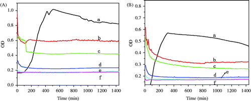
Table 3. Antimicrobial percentage of GO-COO-β-CD/CA for E. coli and S. aureus..
The hemolysis test is to determine the solubility of the blood cells in contact with foreign materials and has been a powerful in vitro test to evaluate the hemolysis property of biomedical materials and products. When blood cells are damaged, hemoglobin is subsequently delivered from the cells. Hemolysis property is thus known by measuring the absorbance of visible light and the results are listed in . It is known that for the materials having potential applications as drug or implanted devices, a hemolysis rate of lower than 5% is required. CA exhibits hemolysis effect when its concentration is relatively high, i.e. from 10 to 50 µg/ml. GO-COO-β-CD/CA, however, has no hemolysis effect in a wide range of concentration, from 0.1 to 50 µg/ml. This is due to the blood compatibility of the drug carrier of GO-COO-β-CD, hemolysis rate of which is lower than 5%. As we previously reported, the drug carrier of GO-COO-β-CD possesses a core-shell structure, in which β-CD molecules aggregate onto the surface of GO-COOH sheet and form biocompatible supramolecular vesicles. In the inclusion, CA is loaded in the cavity of the shells and its cytotoxicity is screened by the β-CD shells, thus its hemolysis effect is greatly depressed.
Table 4. Results of hemolysis test of GO-COO-β-CD, CA and GO-COO-β-CD/CA.
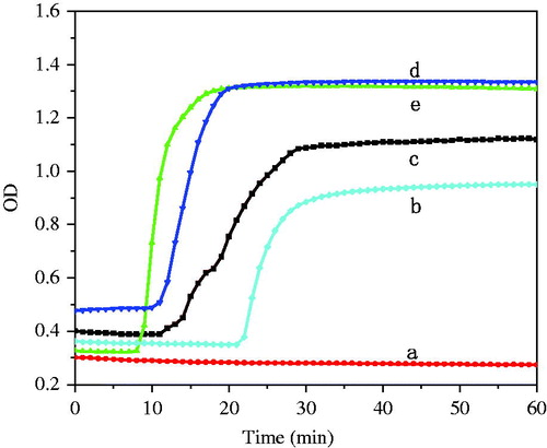
Recalcification is a powerful method to evaluate the function of endogenous blood coagulation system. When Ca2+ is added in PPP, fibrinogen is transferred to fibrin. Fibrin in turn crosslinks to form insoluble substance, thrombus. Recalcification time can be prolonged in the presence of anti-thrombin genic materials. A longer recalcification time means a better blood compatibility of the materials. From we see that the recalcification time of CA (d) is nearly as long as that of PPP+Ca2+ (e), indicating a poor blood compatibility of CA. However, the recalcification time of GO-COO-β-CD/CA (b) and GO-COO-β-CD (c) prolong ca 11 min. It is predictable because the drug carrier of GO-COO-β-CD has anions of OH- in the outer shell and –COO− in the core. The negative charges coordinate with antithrombogenicity factor and hamper the formation of the insoluble fibrin network, thus improve the blood compatibility of the inclusion.
Figure 6. Recalcification time of PPP (a), GO-COO-β-CD/CA (b), GO-COO-β-CD (c), CA (d) and PPP+Ca2+ (e).
Cytotoxicity is another important factor that determines the safety of biomedical materials. MTT test was employed to determine the cytotoxicity of different samples in our study. Optical microscopic images in reveal that HEK293 cells on negative control (a) and in the presence of GO-COO-β-CD, CA and GO-COO-β-CD/CA inclusion (b to d) exhibit normal shuttle shape and good attachment to the substrates. These results demonstrate that GO-COO-β-CD, CA and GO-COO-β-CD/CA have no cytotoxicity, promoting the growth of HEK293 cells. Experimental results are also listed in . Toxicity grade is classified according to USP (Trotta et al., Citation2012).
Figure 7. Morphology of HEK 293 cells after culture for 48 h on negative control (a), GO-COO-β-CD (b), CA (c) and GO-COO-β-CD/CA (d).
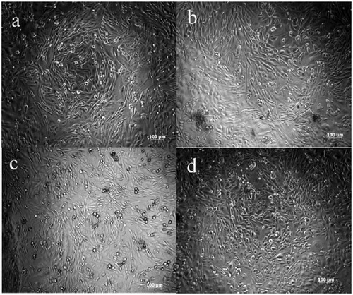
Table 5. Cytotoxicity of GO-COO-β-CD, CA and GO-COO-β-CD/CA.
We conclude that due to its significantly higher RGR than that of CA, the inclusion effectively reduced the cytotoxicity of the drug and thus improved the drug’s biological compatibility.
Based on the above discussion about the way of drug loading, we presume that the mode of CA release from GO-COO-β-CD largely depends on the type of interaction between the two components, i.e. adsorption and in the cavities. On one hand, CA can be released from the cavities of the self-assembled β-CD. On the other hand, CA adsorbed on the surface of GO-COOH can be released through the passageway between β-CD molecules. Results of UV characterization tell us that the CA delivery amount from GO-COO-β-CD/CA inclusion is in accordance with the following equation ():
Figure 8. Release profile of GO-COO-β-CD/CA in HCl (pH = 1.0) (A) and PBS (pH = 7.0) (B).
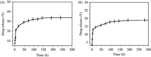
where Er is the cumulative delivery of CA, Ve the volume of the supplemented fresh medium, which was 5 ml in our experiments, V0 the volume of the starting solution i.e. 200 ml in our experiments, Ci the concentration of CA in the solution at i medium exchange, Cn the concentration of CA in the final solution, mdrug the mass of the starting GO-COO-β-CD/CA and D the drug load of GO-COO-β-CD. The cumulative release amount of GO-COO-β-CD/CA inclusion is 34.74% and 18.98% in HCl and PBS, respectively, indicating that GO-COO-β-CD/CA has good delivery profile under acidic condition. Er of CA is 17.42% at ca 5th hour, which ensures quick curative effect. Moreover, CA continues releasing until ca. 150 h, ensuring long-term drug effect. No obvious abrupt drug release was observed in the delivery process in both media. All the results demonstrate that GO-COO-β-CD is an ideal drug carrier with desired drug release profile.
The first- and second-order dynamic equations were applied to simulate the experimental data. The dynamic simulation implies that CA delivery from the inclusion is in accordance with the first-order dynamic equation, i.e. ln(1 − Mt/M)= −kt, where t is the time, Mt the cumulative release amount at t, and M the total release amount at ∞, with R2 of 0.9653 and 0.9813 for delivery in HCl and PBS, respectively.
Conclusion
We designed a new drug carrier of GO-COO-β-CD and fabricated a GO-COO-β-CD/CA inclusion. The paper systematically studied the bacterium inhibition performance, blood compatibility of the inclusion and release profile of the drug. Results demonstrate that the inclusion has good biocompatibility, no cytotoxicity and largely improved drug efficiency due to the synergistic effect of the drug and carrier. In particular, the antibacterial property of the inclusion is greatly enhanced with only one-third drug dosage in comparison with that of pure CA. Dynamic simulation of the experimental data implies that CA delivery from the inclusion is in accordance with the first order dynamic equation. Thus, GO-COO-β-CD/CA can be used as a promising form of medication in vivo applications. With the success of the above studies, it is likely that graphene-based nanocarriers will find widespread applications in biomedicine in the future.
Declaration of interest
The project was funded by Natural Science Foundation of Jiangsu Province, China (Grant No. BK2012845), Specialized Research Fund for the Doctoral Program of Higher Education of China (20123219110010), Prospective Joint Research Project of Jiangsu Province (BY2011109) and Priority Academic Program Development of Jiangsu Higher Education Institutions (PAPD). All authors also thank the Foundation of Jiangsu Collaborative Innovation Center of Biomedical Functional Materials, Jiangsu Six Category Outstanding Talent (NY-031) and Jiangsu Agriculture Science and Technology Innovation Fund [SCX(13)3283].
References
- Acar A, Uygur F, Diktas H, et al. (2011). Comparison of silver-coated dressing (Acticoat (R)), chlorhexidine acetate 0.5% (Bactigrass (R)) and nystatin for topical antifungal effect in Candida albicans-contaminated, full-skin-thickness rat burn wounds. Burns 37:882–5
- Agarwal A, Nelson TB, Kierski PR, et al. (2012). Polymeric multilayers that localize the release of chlorhexidine from biologic wound dressings. Biomaterials 33:6783–92
- Baek JS, Cho CW. (2013). 2-Hydroxypropyl-beta-cyclodextrin-modified SLN of paclitaxel for overcoming p-glycoprotein function in multidrug-resistant breast cancer cells. J Pharm Pharmacol 65:72–8
- Chaturvedi K, Ganguly K, Kulkarni AR, et al. (2011). Cyclodextrin-based siRNA<AQ1/> delivery nanocarriers: a state-of-the-art review. Expert Opin Drug Del 28:1455–68
- Chowdhury SM, Lalwani G, Zhang KV, et al. (2013). Cell specific cytotoxicity and uptake of graphene nanoribbons. Biomaterials 34:283–93
- Dembereldorj U, Kim M, Kim S, et al. (2012). A spatiotemporal anticancer drug release platform of Pegylated graphene oxide triggered by glutathione in vitro and in vivo. J Mater Chem 22:23845–51
- Ferraz CCR, Gomes B, Zaia AA, et al. (2001). In vitro assessment of the antimicrobial action and the mechanical ability of chlorhexidine gel as an endodontic irrigant. J Endodont 27:452–5
- Geim AK. (2009). Graphene: status and prospects. Science 324:1530–34
- Katsnelson MI, Novoselov KS. (2007). Graphene: new bridge between condensed matter physics and quantum electrodynamics. Solid State Commun 143:3–13
- Kuila T, Bose S, Khanra P, et al. (2011). Recent advances in graphene-based biosensors. Biosens Bioelectron 26:4637–48
- Liu Z, Robinson JT, Sun X, et al. (2008). Pegylated nanographene oxide for delivery of water-insoluble cancer drugs. J Am Chem Soc 130:10876–7
- Lu F, Zhang SH, Gao HJ, et al. (2012). Protein-decorated reduced oxide graphene composite and its application to SERS. Acs Appl Mater Inter 4:3278–84
- Meng N, Zhou NL, Zhang SQ, et al. (2009). Controlled release and antibacterial activity chlorohexidine acetate (CA) intercalated in montmorillonite. Int J Pharmaceut 382:45–9
- Moya-Ortega MD, Alvarez-Lorenzo C, Concheiro A, et al. (2012). Cyclodextrin-based nanogels for pharmaceutical and biomedical applications. Int J Pharmaceut 428:152–63
- Murayama N, Terada Y, Okuaki M, et al. (2011). Dose assessment of 2% chlorohexidine acetate for canine superficial pyoderma. Vet Dermatol 22:449–53
- Novoselov KS, Geim AK, Morozov SV, et al. (2005). Two-dimensional gas of massless Dirac fermions in graphene. Nature 438:197–200
- Pei H, Li J, Lv M, et al. (2012). A graphene-based sensor array for high-precision and adaptive target identification with ensemble aptamers. J Am Chem Soc 134:13843–9
- Shen AJ, Li DL, Cai XJ, et al. (2012). Multifunctional nanocomposite based on graphene oxide for in vitro hepatocarcinoma diagnosis and treatment. J Biomed Mater Res A 100A:2499–506
- Trotta F, Zanetti M, Cavalli R. (2012). Cyclodextrin-based nanosponges as drug carriers. Beilstein J Org Chem 8:2091–9
- Wu J, Wang YS, Yang XY, et al. (2012). Graphene oxide used as a carrier for adriamycin can reverse drug resistance in breast cancer cells. Nanotechnology 23:355101 (1--9)
- Yang GH, Cao JT, Li LL, et al. (2013). Carboxymethyl chitosan-functionalized graphene for label-free electrochemical cytosensing. Carbon 51:124–33
- Yannakopoulou K. (2012). Cationic cyclodextrins: cell penetrating agents and other diverse applications. J Drug Deliv Sci Tech 22:243–9
- Yhaya F, Binauld S, Kim Y, et al. (2012). Shell cross-linking of cyclodextrin-based micelles via supramolecular chemistry for the delivery of drugs. Macromol Rapid Comm 33:1868–74
- Zhang LM, Xia JG, Zhao QH, et al. (2010). Functional graphene oxide as a nanocarrier for controlled loading and targeted delivery of mixed anticancer drugs. Small 6:537–44