
Abstract
The aim was to develop albumin anchored docetaxel lipid nanoemulsion (ALNE) for improving tumor targeted delivery. The O/W lipid nanoemulsion, LNEs were prepared by homogenization and ultrasonication processes. The size of globules and zeta potential were measured by Malvern Zetasizer. Albumin was coupled to stearylamine containing lipid nanoemulsion (SALNE) globules using water soluble EDC reaction. The drug content and entrapment efficiencies for the LNEs were determined by the high-performance liquid chromatography. The in vitro cytotoxic studies of the delivery systems were performed on MCF-7 and Hela cells. The IC 50 values of ALNE on both the cell lines were statistically significant. The in vivo antitumor activity was tested on solid tumors induced in C57BL/6 mice. This study revealed that the percentage tumor inhibition for the groups treated with DLNE, SALNE and ALNE when compared with untreated control was found to be 55.62 ± 5.41%, 54.27 ± 4.85% and 80.01 ± 2.74%, respectively. Furthermore, in vivo distribution studies were carried out in breast cancer MDA-MB231 xenografted Balb/c mice. The LNEs were loaded with fluorescent DiD oil and the distribution in different organs after 6 h was tracked using Caliper life sciences in vivo imaging system. The studies revealed that ALNE was superior in tumor targeting activity when compared with DLNE and SALNE by 3.04 and 2.26 folds, respectively. The average radiance values of ALNE on the tumor tissue were statistically significant when compared with DLNE, SALNE at p < 0.01. In addition, this strategy can become a platform technology for other lipophilic drugs to target tumors.
Introduction
Docetaxel is used in the treatment of breast and ovarian cancers and currently administered in the form of parenteral intravenous (iv) infusion. Docetaxel is available as an injectable dosage form containing tween 80 and ethanol. It may cause adverse effects such as severe hypersensitivity reactions, neutropenia, neurotoxicity, musculoskeletal toxicity and cumulative fluid retention. To overcome the excipient-related adverse effects various dosage forms have been investigated. Few of them are liposomes, nanoparticles, solid dispersion, prodrug, freeze dry powder and so on (Zhang et al., Citation2014).
The cytotoxic activity of this drug is not only specific against tumor tissues but also on healthy tissues. The best way of circumventing this problem is to deliver the drug in a manner that is preferentially localized at the desired (target) site of action, that is, at cancer tissue.
Albumin emerged as a versatile and potent carrier for drug targeting and for improving the pharmacokinetic profile (Kratz, Citation2008). Apart from this, the high abundance and small size (∼66 kDa) of albumin make it suitable for transporting many metabolic compounds and therapeutic drugs. Many drug formulations for tumor targeting were developed based on the knowledge that albumin gets accumulated in solid tumors. A successful formulation in the market is Abraxane®, an albumin-bound paclitaxel nanoparticle (Gradishar et al., Citation2005; Ibrahim et al., Citation2005; Green et al., Citation2006) and was developed by M/s Abraxis Bioscience (Los Angeles, CA) in which a lipophilic drug and human serum albumin were passed through a jet under high pressure to form nab-paclitaxel nanoparticles with a mean particle size of 130 nm. This paclitaxel nanoparticle is water-soluble and was first approved by the FDA in 2005 for the treatment of metastatic breast cancer. Phase II clinical trials on nab-docetaxel (ABI008), Albumin-doctaxel nanoparticles were conducted. It is prepared by the high pressure homogenization method. DOXO-EMCH (albumin-bound derivative of doxorubicin) was also developed (Kratz et al., Citation2007; Unger et al., Citation2007) for clinical trials against sarcoma and gastric cancer. Methotrexate–albumin conjugate (Stehle et al., Citation1997; Vis et al., Citation2002; Fiehn et al., Citation2004) was evaluated clinically. Anti-inflammatory drug, diclofenac was targeted to inflammatory site using albumin tagged lipid nanoemulsion (Prabhakar et al., Citation2012). Further, Albuferon, a fusion protein of albumin and interferon, was clinically assessed for the treatment of hepatitis C and could become an alternative to pegylated interferon (Chemmanur & Wu, Citation2006; Kratz, Citation2008). Additionally, many therapeutic peptides and proteins were physically or covalently linked with albumin to improve stability, nonimmunogenicity, toxicity and pharmacokinetic profile. The enhanced uptake of albumin-based drug delivery systems in tumors is mediated by hyper vasculature, enhanced permeability of vascular tissue and enhanced retention in tumor tissue, primarily due to lack of or the impaired lymphatic system (Sinha et al., Citation2006; Kratz, Citation2008). Non-targeted docetaxel lipid nanoemulsions were reported (Gao et al., Citation2008; Venkateshwarlu et al., Citation2010), which were easy to formulate and no albumin ligand attached lipid emulsions were reported. However, folate-decorated docetaxel-loaded human serum albumin nanoparticles were reported (Shougang et al., Citation2015) for effective targeted activity on human hepatoma cell lines and better in vivo antitumor activity. In addition, Pegylated docetaxel lipid nanoemulsions were reported (Muzammil & Kishan, Citation2013) and claimed to be biocompatible and very stable.
In this work, albumin-tagged docetaxel lipid nanoemulsion formulations were prepared, characterized and evaluated for cytotoxic activity on two different cell lines and tumor inhibition study in C57BL/6 mice. Further, the targeting potential of the delivery system in the tumor-induced Balb/C mice was assessed by using small animal in vivo imaging system (IVIS).
Materials and methods
Materials
Purified olive oil (Lipoid, Ludwigshafen, Germany), egg phosphatidyl choline (EPC-80) (Lipoid, Ludwigshafen, Germany), cholesterol and glycerol (Merck, Mumbai, India), α-tocopherol (Sigma, Mumbai, India), oleic acid (S.D. Fine chemicals, Mumbai), docetaxel (A kind gift from M/s Dr Reddy’s Laboratories, Hyderabad, India), Bovine serum albumin (Merck Ltd., Mumbai, India), EDC (1-Ethyl-3-(3-dimethylamino) propyl carbodiimide) (Sigma, Mumbai, India), Coomassie Brilliant Blue G (Sigma, Mumbai, India), TRIS (Promega corporation, Mumbai, India), EDTA (ethylene diamine tetraacetic acid disodium salt) (S.D. Fine Chemicals, Mumbai, India), Sephadex-G-75 (Pharmacia Fine Chemicals, Uppsala, Sweden), Sephadex mini column (Bio rad, Hercules, CA), sodium dihydrogen phosphate, disodium hydrogen phosphate (S.D. Fine Chemicals, Mumbai, India), centrisart tubes (Sartorius, Goettingen, Germany), dialysis membrane (Himedia, Mumbai, India), chloroform, acetonitrile (Merck, Mumbai, India). HeLa – Human cervical carcinoma cell line (NCCS, Pune, India), MCF-7 – Human breast carcinoma cell line (NCCS, Pune, India), B16F10 Murine Malanoma cells (NIPER, Hyderabad, India), RPMI-1640 medium (Himedia, Mumbai, India), dimethylsulphoxide (DMSO) (Merck India Ltd, Mumbai, India), SDS lysis buffer (Himedia, Mumbai, India), trypsin (Himedia, Mumbai, India), MTT (3-(4, 5-dimethylthiazol-2yl)-2,5-diphenyltetrazolium bromide) (Himedia, Mumbai, India) and DiD oil (1,1′-dioctadecyl-3,3,3′,3′-tetramethyl indocarbocyanine perchlorate) (Sigma, Mumbai, India). All other chemicals used were of reagent grade.
Methods
Preparation of lipid nanoemulsions containing docetaxel
The lipid nanoemulsions (LNEs) were prepared by hot homogenization followed by the ultrasonication process (Varshika et al., Citation2009). Required quantities of ingredients were weighed (). The oil soluble substances were added to the oily phase and the water soluble substances to the aqueous phase. Then both the phases were heated to 70 °C. The aqueous phase was added to oily phase slowly drop wise with stirring and then homogenized for about 3 min at 15 000 rpm. The coarse emulsion was then sonicated for about 20 min at 50% amplitude with 12T probe of sonicator (Vibracell, Sonics material Inc., Newtown, CT) and characterized for size, polydispersity and zeta potential.
Table 1. Composition of different docetaxel LNEs.
Attachment of albumin to surface of LNE globules
The stearylamine lipid nanoemulsion (SALNE) was chosen for coupling with albumin. Two milliliters of SALNE was suspended in phosphate-buffered saline (pH 7.4) containing albumin (10 mg) and EDC (28.5 mg) and then incubated for 2 h at room temperature (Vandana et al., Citation2005). The unbound albumin was removed using a Sephadex G-75 minicolumn.
Preparation of sephadex minicolumn
One gram of Sephadex G-75 was soaked in 25 ml of Tris EDTA buffer pH 7.5 (Tris10 mM and EDTA 1 mM) for about 24 h. Gel was filled carefully without any air bubbles into empty plastic column (Bio-Rad, Hercules, CA) having reservoir of 10 ml. The cap was closed and centrifuged at 4000 rpm for 10 min for uniform setting of gel. The remaining amount of gel was added and the procedure was repeated to get a uniform column. The plastic cap at the other end was tipped off, and then the column material was equilibrated with phosphate-buffered saline pH 7.4 and used to entrap the unbound albumin.
Removal of unbound albumin by gel filtration
The emulsion was loaded onto the column of sephadex G-75. The sephadex G-75 had capacity to entrap the molecules having molecular size range of 3000–80 000 D (Porath & Flodin, Citation1959).
The unbound albumin got entrapped in the core of the gel pores and allowed the emulsion to pass through the voids. The emulsion was collected and used for determining the amount of albumin bound to the emulsion droplets.
Quantification of albumin
The amount of albumin coupled at the surface of the lipid emulsions was determined by the modified Bradford dye assay using Coomassie Blue G dye (Georgiou et al., Citation2008). The amount of albumin bound to emulsion globules was found by using the calibration curve obtained with standard concentrations of albumin. The albumin coupling efficiency was expressed as molecules of albumin per oil globule of the emulsion.
Coomassie Brilliant Blue G-250 – TCA reagent preparation
Coomassie stock reagent was prepared by dissolving 60 mg Coomassie Brilliant Blue (CBB) in 100 ml of 1 N HCl (designated as CBB–HCl stock) by stirring. The CBB–HCl was stirred for 40 min, filtered through Whatman filter paper and stored at 4 °C while protected from light. Freshly, the CBB–TCA reagent (20 ml) was prepared by adding 20 ml CBB–HCl stock to the solution containing 1% absolute ethanol, 2% TCA (0.4 g TCA) and adjusting the pH of the resulting mixture to 0.4 by the addition of approximately 1.67 g solid Na3PO4 12H2O with continuous stirring. The reagent was cleared from the blue particulate matter formed by centrifugation at 5000g for 5 min at room temperature.
Estimation of albumin bound to globules
The standard assay (performed in a final volume of 1 ml) consisted of mixing 0.05 ml albumin solution with 0.95 ml of hydrophobic CBB–TCA reagent. After 5–10 min incubation at room temperature, the absorbance of the mixture was measured at λmax 610 nm against an appropriate reagent blank. For making a linear standard curve, BSA standard solutions of 10–60 μg/ml were used.
Distribution studies of fluorescent LNEs in tumor-induced mice by using the IVIS
Female Balb/c mice (6–8 weeks) were subcutaneously xenografted by MDA-MB 231 cells and tumors were developed in three weeks (Taitz et al., Citation1995; Taheri et al., Citation2012; Devika et al., Citation2013). After tumor growth, mice (n = 3 for each group) were anaesthetized with 2% isoflurane and were injected into the tail vein with fluorescent tagged LNEs at a dose of 10 mg/kg (). The animals were imaged after 6 h using Caliper life sciences IVIS, Massachusetts, USA. After whole animal imaging, the mice were sacrificed and dissected for imaging major organs (liver, spleen, lungs, heart, kidneys and tumor tissue). Image display analysis was performed using living image software (version 4.3.1, Xenogen, Alameda, USA). Data were obtained from the fluorescent images by drawing the region of interest (ROI) on the subcutaneous tumors. Similar ROI was also positioned on the isolated major tissues/organs with the ROI tools available in the software. The number of photons emitted by the subject was measured in the form of average radiance (photons/sec/cm2/sr) (Goutayer et al., Citation2010; Hussaini et al., Citation2014). Statistical analysis was performed, using one-way analysis of variance (ANOVA) by the Newman–Keuls multiple comparison test by using (GraphPad software inc., Lajolla, CA) software. Statistical significance was assigned for values of p < 0.01.
In vivo antitumor effect of DOC LNEs
C57BL/6 mice (about 5–6 weeks old, 18–29 g) were subcutaneously injected at the lower right flank region with 0.1 ml of cell suspension containing 0.5 × 10⁶ B16F10 cancer cells. The mice (24 no.) whose tumor was palpable were randomly divided into four groups with each group containing six mice and the day was designated as “Day 1”. On Day 1 tumor size was measured using digital screw gauge (M & W Precision tools, Lancashire, UK) and the mice were administered by iv. route with DOC-LNEs (DOC-LNE, SALNE and albumin-LNE) (10 mg/kg). Tumor size was measured after seven days duration during the study. After this measurement one more dose was given. The tumor volume was calculated by the formula W × L2/2 (Liu et al., Citation2012), where W is the widest diameter and L is the longest diameter. Tumor volume was measured regularly for 29 days. In all, four times the injections were given, with a gap of seven days interval. The mice were sacrificed at day 29 and the tumors were stripped and weighed. For the obtained data statistical analysis was performed using one-way ANOVA by the Newman–Keuls multiple comparison test using GraphPad prism 5 software. Statistical significance was assigned for values of p < 0.01.
Characterization
Determination of globule size, polydispersity index and zeta potential (Zp)
The LNE was diluted (1:100) with double-distilled water and was taken in the cuvette. The cuvette was placed inside the sample holder of the instrument (Malvern Nano ZS90, Worcestershire, UK) for measurement of size and zeta potential ().
Table 2. Physical characters of the prepared DLNEs (n = 3).
In vitro drug release studies of docetaxel containing LNE formulations
The drug release was studied by the open tube method using cellulose membrane (DM 60 molecular weight cut-off 12 000–14 000, Himedia, Mumbai, India). Initially, the dialysis membrane was hydrated overnight in phosphate buffer pH 7.4 at room temperature. The emulsions/solution (1 ml) were placed in dialysis open tube and then suspended in 100 ml of 30%v/v ethanol phosphate buffer pH 7.4 mixture in 250 ml beakers as dialysis medium, which was stirred continuously on a magnetic stirrer (Remi equipments, Mumbai, India). At different time intervals, that is, 0.25, 0.5, 1, 2, 3, 4, 6, 8, 10 and 12 h during dialysis, 1 ml of samples were withdrawn from the beaker and replaced by equal volume of fresh medium for analyzing the drug content. The amount of drug in collected samples was calculated using the linear calibration curve plotted with known concentrations of drug by measuring the absorbance at λmax 227 nm on a UV–visible spectrophotometer (SL 159, ELICO, Hyderabad, India). The release pattern of the drug from different formulations was calculated by plotting cumulative percentage drug released versus time.
Determination of total drug content and entrapment efficiency
High-performance liquid chromatography (HPLC) system consisting of a LC-20AD solvent delivery system containing double reciprocating plunger pump (Shimadzu, Kyoto, Japan), a SPD-20A UV–visible variable wavelength detector with deuterium lamp (Shimadzu), and a 250 × 4.6 mm, 5 μm, C-18 reverse phase analytical column (Lichrospher®, Merck, Mumbai, India) was used to determine total drug content and entrapment efficiency (EE) of the formulations. The mobile phase consisted of 65 parts of acetonitrile and 35 parts of double distilled water. The flow rate was 1 ml/min and the detection was performed at λmax of 227 nm (Ciccolini et al., Citation2001).
Drug content
Each docetaxel-loaded LNE (0.1 ml) was diluted to 1 ml and further diluted twice (1:10) with methanol, and 20 μl sample was injected to determine drug content by the reported HPLC method (Ciccolini et al., Citation2001).
Entrapment efficiency
EE was determined by measuring the concentration of free drug (unentrapped) in the aqueous medium (Varshika et al., Citation2009). The aqueous medium was separated by ultra-filtration using centrisart tubes (Sartorius, Goettingen, Germany), which consisted of filter membrane (M.Wt. cut-off 20 000 Da) at the base of the sample receiver chamber. About 2 ml of the formulation was placed in the donor chamber and centrifuged at 6500 rpm for 15 min (C24 centrifuge, Remi Instruments, Mumbai, India). The LNEs along with encapsulated drug remained in the donor chamber and aqueous phase moved into the receiver chamber through filter membrane.
The amount of docetaxel in the aqueous phase was estimated by the HPLC method and the EE was calculated.
In vitro cytotoxic evaluation of LNEs by the MTT assay method
The adherent cells (Hela and MCF-7) were trypsinized with 0.3 ml of 0.25% trypsin-EDTA and incubated for 2–5 min. Then flask was gently “tapped” for dislodging the cells. Cells were washed and resuspended in fresh medium. Cell suspension was mixed thoroughly by pipetting several times to get a uniform single cell suspension (van de Loosdrecht et al., Citation1994). The cell count was made by using a cell counter (Moxiflow V2.0, ORFLO, Ketchum, USA) and was found to be 78 000 Hela cells per ml and 75 000 MCF-7 cells per ml. Different dilutions of drug solutions (DSs) and formulations were made in medium (RPMI-1640) with final DMSO concentration in the well to be less than 1%. About 100 μl of cell suspension was transferred aseptically to each well of a 96-well plate and to it 100 μl of 1% medium/DS (n = 6) in medium was added along with control. The plate was then incubated at 37 °C for 48 h in CO2 incubator (MRC, Hasselroth, Germany). After incubation for 48 h, 20 μl of MTT was added to each well. The plate was incubated for 2 more hours. Subsequently, 80 μl of lysis buffer was added to each well, the plate was wrapped in aluminum foil to prevent the oxidation of the dye and the plate was placed on a shaker for overnight. The absorbances were recorded on the ELISA reader (Biotek, Winooski, USA) at 562 nm wavelength. The absorbance of the test was compared with that of DMSO control to get the percentage inhibition.
Results and discussion
The LNEs were prepared by hot homogenization and ultrasonication processes. The initial typical formula was selected based on previous literature on parenteral lipid emulsions (Gao et al., Citation2008; Venkateshwarlu et al., Citation2010). Control emulsion (docetaxel lipid nanoemulsion (DLNE)) consisted of 10% (w/v) olive oil as the oil core, 1.2% (w/v) EPC-80 as a phospholipid emulsifier, oleic acid (0.3%) as a negative charge inducer, α-tocopherol (0.25%) as antioxidant and glycerol (2.25%) was included to maintain the isotonicity of the formulation for iv administration (). For preparation of albumin anchored docetaxel lipid nanoemulsion (ALNE), the stearyl amine lipid nanoemulsion was used. The purpose of using stearyl amine in the formulation was to attach albumin ligand to the emulsion globules. Stearylamine contained a long C-18 fatty chain which would hold the albumin molecule at the surface of globule after attachment.
Albumin was coupled to SALNE globules by using the water soluble EDC method (Vandana et al., Citation2005). The stoichiometry was calculated based on the assumption that each amino group of SA would react with albumin molecule. The amount of albumin bound to emulsion globules was calculated (). The average amount of albumin bound was 62.7 ± 0.6 %w/w of total amount of albumin used in reaction. The estimated average number of albumin molecules attached to the surface of each emulsion globule was 1093.43 ± 107.37. However, in the case of samples used in cytotoxic and animal study, the estimated number of albumin molecules bound to each globule was found to be 1125 and 1132, respectively.
Table 3. Estimation of amount of albumin bound to each globule of docetaxel LNE.
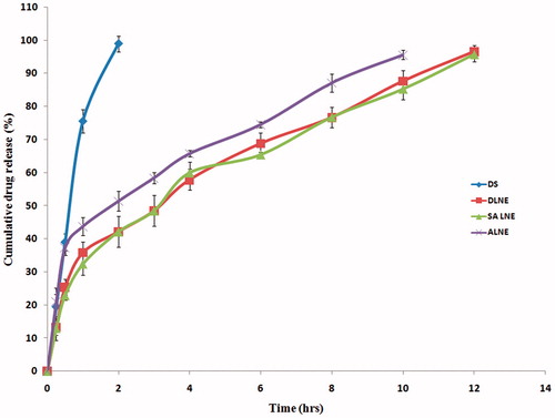
The size of albumin LNE globules increased when compared with control emulsion DLNE (). The sizes of albumin attached globules were reported to be increased when compared with stearyl amine-loaded LNE (Prabhakar et al., Citation2012). The in vitro drug release from DLNE and SALNE after 12 h was found to be 96.56 ± 1.79 and 95.64 ± 2.15, respectively. The in vitro release of the drug from albumin-coupled LNE after 10 h was found to be 95.52 ± 1.4 and from DS after 2 h was found to be 98.9 ± 2.3 (). The in vitro release of the drug from albumin-coupled LNEs was faster than the control emulsion, that is, DLNE. This could be due to change in surface characters, that is, increased hydrophilicity (Prabhakar et al., Citation2012).
Figure 1. In vitro drug release profiles of DS, DLNE, SALNE and ALNE formulations (n = 3).
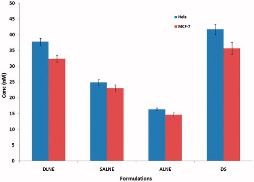
The in vitro cytotoxic studies were performed on two cell lines (MCF-7 and Hela). The IC 50 values on MCF-7 cell lines of DLNE, SALNE, ALNE and DS were found to be 32.31 ± 1.2, 22.97 ± 1.1, 14.52 ± 0.65 and 35.68 ± 1.9 nM, respectively (). The IC 50 values on Hela cell lines of DLNE, SALNE, ALNE and DS were found to be 37.79 ± 1.12, 24.84 ± 0.98, 16.33 ± 0.43 and 41.71 ± 1.67 nM, respectively (). The IC 50 values of MCF7 cell lines were less than that of the Hela cell lines, and this difference might be due to docetaxel being more cytotoxic toward the breast cancer cells (MCF7). When compared with DS, ALNE was found to have 2.45 and 2.56 folds more cytotoxic activity on MCF7 and Hela cell lines. Similarly, when compared with DLNE, ALNE was found to have 2.21 and 2.31 folds cytotoxic activity on MCF7 and Hela cell lines, respectively. The results of one-way ANOVA by the Newman–Keuls multiple comparison test when applied, it was found that ALNE was statistically significant with DS, DLNE, at p < 0.05. Previous studies Priyanka et al. Citation(2014) reported that methotrexate coupled with BSA were higher cytotoxic on MCF-7 breast cancer cells when compared with an equivalent dose of free methotrexate. The enhanced activity is attributed to the preferential uptake of Au-BSA-MTX particles by MCF-7 cells due to the presence of BSA.
Figure 2. In vitro cytotoxic study-a comparison of IC50 values of DLNE on Hela and MCF-7 cell lines (Mean ± SD, n = 6). (DLNE: Docetaxel lipid nanoemulsion, SALNE: stearyl amine containing lipid nanoemulsion, ALNE: albumin containing lipid nanoemulsion, DS: drug solution).
The in vivo imaging studies were performed on the tumor-induced female Balb/c mice. The fluorescence intensity (radiance) was measured after 6 h of iv administration of the fluorescent dye-loaded LNEs. The photons emitted from the tumor site of the balb/c mice after the administration of DLNE, SALNE and ALNE were found to be 2.6e+03 ± 300, 2.88e+03 ± 220.55, 7.38e+03 ± 534.84 (p/s/cm2/sr)/(μW/cm2), respectively (). After that animals were sacrificed and their major tissues such as liver, spleen, lungs, heart, tumor and kidneys were isolated and the average radiance was measured by the IVIS (). There is a decrease in average radiance value at the tumor site of the mice when compared with that of isolated tumor mass. It could be due to the role of skin acting as a barrier and reducing the fluorescence in the whole animal. Organs with the brightest yellow color represented the highest accumulation of the fluorescent oil globule at the site. Subsequently, ROI on individual organs and average radiance values are shown in . During this study, fluorescent intensity was found to be more with the RES organs than with the non RES organs. It might be due to the presence of macrophages and rich blood supply to the RES organs. The sequence of organ fluorescence intensity is Liver > Spleen > Lungs > Kidney> Tumor > Heart. Similar type of tissue distribution was reported in the previous literature (Hollis et al., Citation2013; Liu et al., Citation2014). When ALNE was administered, the average radiance emitted by tumor was 3.04 times more than that of DLNE formulation. Similarly, when compared with SALNE, it was found to be 2.26 times. The results of one-way ANOVA by the Newman–Keuls multiple comparison test when applied, it was found that ALNE was statistically significant when compared with DLNE and SALNE at p < 0.01.
Figure 3. Fluorescence emitted from the mice after 6 h of administration of different LNEs. (Note: Encircled areas depict the tumors. The increased fluorescence intensity is observed when compared with control animal (before treatment), after the administration of DiD oil-loaded DLNE, SALNE and ALNE.
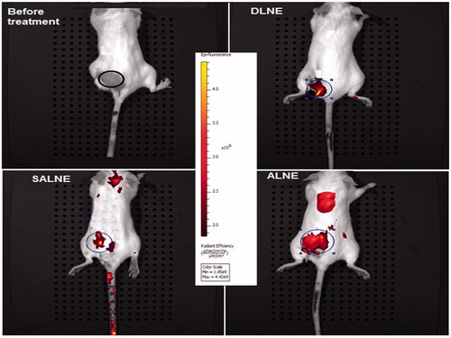
Figure 4. A comparison: fluorescent images of the major organs/tissues after 6 h of administration of different LNEs in tumor-induced mice.
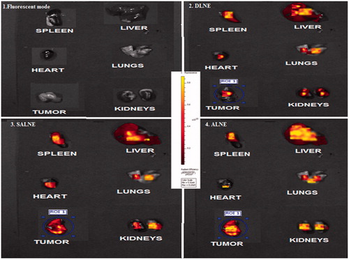
Table 4. Average radiance values of the major organs/tissues after 6 h of administration of different docetaxel LNEs (n = 3) containing fluorescent DiD oil.
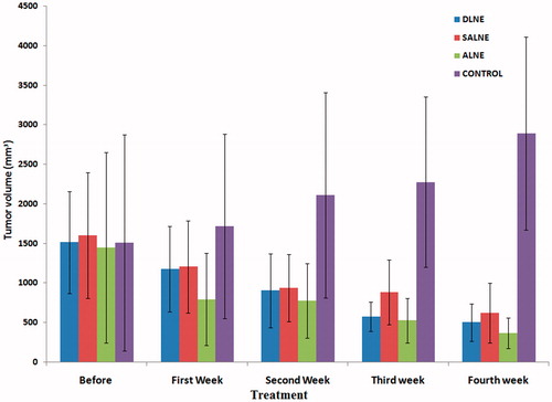
The antitumor effect of the delivery system was noticed from the tumor regression study in mice treated with DLNE, SALNE and ALNEs (). The initial average tumor volumes in the DLNE, SALNE and ALNE groups were found to be 1512.17 ± 647.59, 1601.37 ± 798.46 and 1444.61 ± 1206.68 mm3, respectively. The tumor volumes were significantly reduced after four dose treatments with DLNE, SALNE and ALNE and were found to be 500.64 ± 235.75, 620.85 ± 377.89 and 364.49 ± 195.26 mm3, respectively. While the untreated control group showed enormous increase in tumor volume, that is, 2892.76 ± 1219.77 mm3.
Figure 5. Progressive tumor volume reduction after weekly treatments with various LNEs (n = 6).
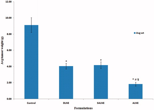
After four treatments the tumors were isolated by sacrificing the mice and weighed. The average tumor weights for the groups treated with DLNE, SALNE and ALNE were found to be 4.05 ± 0.46, 4.18 ± 0.41 and 1.83 ± 0.23 g, respectively (). The results of one-way ANOVA by the Newman–Keuls Multiple comparison test when applied, it was found that ALNE was statistically significant with DLNE and SALNE at p < 0.01. The percentage tumor inhibition for the groups treated with DLNE, SALNE and ALNE when compared with control (untreated group) was found to be 55.62 ± 5.41%, 54.27 ± 4.85% and 80.01 ± 2.74%, respectively. In this study, no significant difference was noticed between DLNE and SALNE. However, significant difference was noticed from these two when compared with ALNE. This clearly indicated the role of albumin in their improved antitumor effect of docetaxel. Folate-decorated docetaxel-loaded human serum albumin nanoparticles had shown effective antitumor activity (Shougang et al., Citation2015). The enhanced uptake of albumin-based drug delivery systems in solid tumors is mediated by the pathophysiology of tumor tissue, characterized by neoangiogenesis, hypervasculature, a defective vascular architecture and an impaired lymphatic drainage. Accumulation of nabpaclitaxel was also found due to transcytosis initiated by binding of albumin to a cell surface, 60 kDa glycoprotein (gp60) receptor (albondin) as well as due to binding of albumin to secreted protein acid and rich in cysteine. Albumin binds to the gp60 receptor, which in turn results in binding of gp60 with an intracellular protein (caveolin-1) and subsequent invagination of the cell membrane to form transcytotic vesicles, that is, caveolae (John et al., Citation2003; Desai et al., Citation2006). Similar potent antitumor activity was reported Kim et al. Citation(2011) by using albumin-bound curcumin nanoparticles (CCM-HSA-NPs), and this was claimed due to the enhanced water solubility, increased accumulation in tumors and ability to traverse vascular endothelial cell. Furthermore, studies were reported (Shen et al., Citation2011) claiming that folic acid-conjugated albumin nanospheres had improved the targeting on HeLa cell lines. Nab-paclitaxel, an albumin bound delivery system showed superior antitumor efficacy over paclitaxel in a number of human tumor xenograft models (Desai et al., Citation2006). Taken together, our studies with ALNEs were found to be superior in delivery of docetaxel to tumors as discussed above and caused significant reduction in size of solid tumors. Thus, this strategy of ligation of albumin to delivery vehicle is a potential approach for targeting of docetaxel to tumors.
Figure 6. Bar diagram showing a comparative reduction in final tumor weights after four treatments with various LNEs (n = 6). Note: *Statistically significant when compared with control at p < 0.01. #Statistically significant when compared with DLNE at p < 0.01. $Statistically significant when compared with SALNE at p < 0.01.
Conclusion
Albumin-coupled DLNE was prepared and characterized. The albumin-coupled lipid nanoemulsion was found to have nano range globule size, EE and retarded release for about 10 h. The in vitro cytotoxic studies clearly revealed the significant effect on two cancer cell lines. Furthermore, imaging studies in tumor-induced balb/c mice were conducted and the results revealed that ALNE had the significant targeting efficiency by 3.04 folds when compared with control LNE. The pharmacodynamic studies also revealed the antitumor activity of the ALNE based on the tumor volume regression when compared with control LNE. Thus, albumin-coupled DLNE could become a potential targeted drug delivery system for cancer. Furthermore, this strategy can be extended as platform technology for targeted delivery of other lipophilic anti-cancer drugs.
Acknowledgements
Gift samples of docetaxel were provided from M/s Dr Reddy’s Laboratories, Hyderabad, India and Egg Lecithin from M/s Lipoid, Ludwigshafen, Germany. Dr Y Narsimha Reddy, UCPSc, Kakatiya University, Warangal, Telangana, India for the help in cytotoxic studies. Mrs Ramadevi from NIN, Tarnaka, Hyderabad, India for the help in the iv administration of fluorescent LNEs during the in vivo imaging studies. Mr T. Dinesh and Dr G. Chandraiah, NIPER, Hyderabad, India for their help in animal studies. Mr Gulzaar Hasan from CCMB, Tarnaka, Hyderabad, India for his help in imaging study.
Declaration of interest
First author would like to thank UGC New Delhi for BSR Fellowship and financial support from UGC Major Research Project (F. No. 36-133/2008 (SR) Dated 26-03-2009). The authors would like to declare that they have no conflicts of interest.
References
- AT Chemmanur, GY Wu. (2006). Drug evaluation: albuferon-alpha A — an antiviral interferon-alpha/albumin fusion protein. Curr Opin Investig Drugs 7:750–8
- J Ciccolini, J Catalin, MF Blachon, et al. (2001). Rapid high-performance liquid chromatographic determination of docetaxel (Taxotere) in plasma using liquid-liquid extraction. J Chromatogr B Biomed Sci Appl 759:299–306
- N Desai, V Trieu, Z Yao, et al. (2006). Increased antitumor activity, intratumor paclitaxel concentrations, and endothelial cell transport of cremophor-free, albumin-bound paclitaxel, ABI-007, compared with cremophor-based paclitaxel. Clin Cancer Res 12:1317–24
- C Devika Nandan, P Reshmi, U Saji, et al. (2013). Therapeutic properties of boswellic acid nanoparticles in prostate tumor–bearing BALB/c mice model. J Nanopharm Drug Deliv 1:30–7
- C Fiehn, U Muller-Ladner, S Gay, et al. (2004). Albumin-coupled methotrexate (MTX-HSA) is a new antiarthritic drug which acts synergistically to MTX. Rheumatology 43:1097–105
- K Gao, J Sun, K Liu, et al. (2008). Preparation & characterization of a submicron lipid emulsion of docetaxel: submicron lipid emulsion of docetaxel. Drug Dev Ind Pharm 34:1227–37
- CD Georgiou, K Grintzalis, G Zervoudakis, et al. (2008). Mechanism of Coomassie brilliant blue G-250 binding to proteins: a hydrophobic assay for nanogram quantities of proteins. Anal Bioanal Chem 391:391–403
- M Goutayer, S Dufort, V Josserand, et al. (2010). Tumor targeting of functionalized lipid nanoparticles: assessment by in vivo fluorescence imaging. Eur J Pharm Biopharm 75:137–47
- WJ Gradishar, S Tjulandin, N Davidson, et al. (2005). Phase III trial of nanoparticle albumin-bound paclitaxel compared with polyethylated castor oil-based paclitaxel in women with breast cancer. J Clin Oncol 23:7794–803
- MR Green, GM Manikhas, S Orlov, et al. (2006). Abraxane®, a novel Cremophor®-free, albumin-bound particle form of paclitaxel for the treatment of advanced non-small-cell lung cancer. Ann Oncol 17:1263–8
- CP Hollis, HL Weiss, M Leggas, et al. (2013). Biodistribution and bioimaging studies of hybrid paclitaxel nanocrystals: lessons learned of the EPR effect and image-guided drug delivery. J Control Release 172:12–21
- SSQ Hussaini, H Tanvirul, A Amer, L Xinli. (2014). Hyaluronan polymer length, grafting density, and surface poly(ethylene glycol) coating influence in vivo circulation and tumor targeting of hyaluronan-grafted liposomes. ACS Nano 8:5423–40
- NK Ibrahim, B Samuels, R Page, et al. (2005). Multicenter phase II trial of ABI-007, an albumin-bound paclitaxel, in women with metastatic breast cancer. J Clin Oncol 23:6019–26
- TA John, SM Vogel, C Tiruppathi, et al. (2003). Quantitative analysis of albumin uptake and transport in the rat microvessel endothelial monolayer. Am J Physiol Lung Cell Mol Physiol 284:L187–96
- TH Kim, HH Jiang, YS Youn, et al. (2011). Preparation and characterization of water-soluble albumin-bound curcumin nanoparticles with improved antitumor activity. Int J Pharm 403:285–91
- F Kratz, G Ehling, HM Kauffmann, et al. (2007). Acute and repeat-dose toxicity studies of the (6-maleimidocaproyl) hydrazone derivative of doxorubicin (DOXOEMCH), an albumin-binding prodrug of the anticancer agent doxorubicin. Hum Exp Toxicol 26:19–35
- F Kratz. (2008). Albumin as a drug carrier: design of prodrugs, drug conjugates and nanoparticles. J Control Release 132:171–83
- Q Liu, R Li, Z Zhu, et al. (2012). Enhanced antitumor efficacy, biodistribution and penetration of docetaxel-loaded biodegradable nanoparticles. Int J Pharm 430:350–8
- Y Liu, J Sun, H Lian, et al. (2014). Folate and CD44 receptors dual-targeting hydrophobized hyaluronic acid paclitaxel-loaded polymeric micelles for overcoming multidrug resistance and improving tumor distribution. J Pharm Sci 103:1538–47
- S Muzammil Afzal, V Kishan. (2013). Preparation, characterization and in vitro evaluation of stealth docetaxel lipid nanoemulsions for efficient cytotoxicity. IJDD 5:188–95
- J Porath, P Flodin. (1959). Gel filtration: a method for desalting and group separation. Nature 183:1657–9
- K Prabhakar, S Muzammil Afzal, G Surendar, et al. (2012). Albumin coupled lipid nanoemulsions of diclofenac for targeted delivery to inflammation. Nanomed: Nanotechnol Biol Med 8:1162–71
- M Priyanka, T Amruta, S Anjali, et al. (2014). In situ synthesized BSA capped gold nanoparticles: effective carrier of anticancer drug methotrexate to MCF-7 breast cancer cells. Mater Sci Eng: C 34:158–67
- Z Shen, Y Li, K Kohama, et al. (2011). Improved drug targeting of cancer cells by utilizing actively targetable folic acid-conjugated albumin nanospheres. Pharmacol Res 63:51–8
- J Shougang, G Xianfeng, Z Xiuhua, et al. (2015). Preparation, characterization, and antitumor activities of folate-decorated docetaxel-loaded human serum albumin nanoparticles. Drug Delivery 22:206–13
- R Sinha, GJ Kim, S Nie, et al. (2006). Nanotechnology in cancer therapeutics: bioconjugated nanoparticles for drug delivery. Mol Cancer Ther 5:1909–17
- G Stehle, A Wunder, H Sinn, et al. (1997). Pharmacokinetics of methotrexate-albumin conjugates in tumor bearing rats. Anticancer Drugs 8:835–44
- A Taheri, R Dinarvand, F Ahadi, et al. (2012). The in vivo antitumor activity of LHRH targeted methotrexate–human serum albumin nanoparticles in 4T1 tumor-bearing Balb/c mice. Int J Pharm 431:183–9
- A Taitz, G Petruzzelli, AS Pak, et al. (1995). Immune parameters of mice bearing human head and neck cancer. Cancer Immunol Immunother 40:283–91
- C Unger, B Häring, M Medinger, et al. (2007). Phase I and pharmacokinetic study of the (6-maleimidocaproyl) hydrazone derivative of doxorubicin. Clin Cancer Res 13:4858–66
- AA van de Loosdrecht, RH Beelen, GJ Ossenkoppele, et al. (1994). A tetrazolium-based colorimetric MTT assay to quantitate human monocyte mediated cytotoxicity against leukemic cells from cell lines and patients with acute myeloid leukemia. J Immunol Methods 174:311–20
- S Vandana, DV Kohli, SK Jain. (2005). Transferrin coupled liposomes as drug delivery carriers for brain targeting of 5-florouracil. J Drug Target 13:245–50
- E Varshika, K Prabhakar, V Kishan. (2009). Preparation, characterization and in vivo pharmacodynamic evaluation of parenteral diclofenac submicron lipid emulsions. PDA J Pharm Sci Technol 63:380–9
- I Venkateshwarlu, K Prabhakar, A Mubarak, et al. (2010). Development and in vitro cytotoxic evaluation of parenteral docetaxel lipid nanoemulsions for application in cancer treatment. PDA J Pharm Sci Technol 64:233–41
- AN Vis, A van der Gaast, BW van Rhijn, et al. (2002). A phase II trial of methotrexate-human serum albumin (MTX-HSA) in patients with metastatic renal cell carcinoma who progressed under immunotherapy. Cancer Chemother Pharmacol 49:342–5
- H Zhang, J Dou, Y Zhai, et al. (2014). Advances in the formulations of noninjection administration of docetaxel. J Drug Target 22:87–94