Abstract
Bone marrow mesenchymal stem cells (BMMSCs) are multipotent stem cells that can differentiate into different blastoderm cells in vitro. In this study, BMMSCs in rabbit bone marrow were isolated by density gradient centrifuge separation, purified and expanded in vitro. BMP-2 and FGF 2 were used for differentiation into osteoblasts, and the results demonstrated that bone morphogenetic proteins (BMPs) could affect the differential direction of the BMMSCs. PCR assays indicated that BMP signals pathway played important roles in osteoblasts differentiation of BMMSCs, and the members included BMPRI, Smad 1, Smad 5, Smad 8, Runx 2, collage type I and osteopontin. This study provides a theoretical basis and experimental evidence for the therapeutic application of BMMSCs to the treatment of bone injury.
Keywords::
Introduction
Mesenchymal stem cells (MSCs) are promising candidates for cellular therapies and tissue engineering strategies. They are a population of adult stem cells and derive their name from an intrinsic ability to differentiate into multiple cell lineages of mesodermal origin, such as bone. MSCs were initially identified in postnatal bone marrow and later in peripheral blood, periosteum, muscle, adipose tissue, and connective tissue (Noren Hooten and Evans 2011, Pittenger et al. 1999, Zuk et al. 2001). Bone marrow mesenchymal stem cells (BMMSCs) are easy to isolate, culture, and manipulate in vitro and have great plasticity; for these reasons they have become an important tool in cell replacement therapy and are considered as candidates for different clinical applications (Bongso and Richards 2004). Osteoblasts are cells which originate from bone marrow and contribute to the production of new bone. These cells build up the matrix of bone structure and play a role in the mineralization of bone matrix. Therefore, this study aims to establish a system for differentiation of osteblastes from BMMSCs and explain the mechanism of such differentiation, which can be used for repairing bone injury.
Materials and methods
Isolation and culture of BMMSCs
MSCs were collected from the bone marrow of six one-week-old rabbits, supplied by the Laboratory Animal Center of Huazhong University of Science and Technology, Wuhan, RP China. The animals were sacrificed with a lethal dose of sodium pentobarbital. All animal experiments were performed in accordance with the guidelines established by the Institutional Animal Care and Use Committee at UMDNJ-RWJMS. First, bone marrow samples were drained from rabbit tibia, pelleted by centrifugation, resuspended with phosphate buffer solution (PBS) and fractionated on density gradient (Percoll, 1.077 g/ml, Amersharm Biosciences, Sweden) for 30 min at 400 g. The interface was collected and seeded at 1 × 10⁴ cells/well in six-well culture plates with 1-ml medium containing L-DMEM (Gibco, USA) + 10% (v:v) fetal bovine serum (FBS) (Gibco, USA) + 10 U/ml penicillin + 10 ug/ml streptomycin. Cultures were maintained at 37.5°C in a humidified atmosphere containing 95% air and 5% CO2. The culture medium was changed every three days and the non-adherent cells were removed after three days. BMMSC morphology was observed by phase-contrast microscopy and photographed (Olympus, Japan). When the cultures became 80–90% confluence, the cells were detached with 0.25% trypsin and 0.02% EDTA for 3 min at room temperature, and reseeded at 1 × 10⁴ cells/well in six-well culture plates with 1-ml L-DMEM for serial passaging.
For growth kinetics assay, the cells were harvested and plated in 24-well microplates at 1 × 10⁴ cells/well. After being cultured for 7 days, the cells from 3 wells were counted randomly each day. Growth curves were plotted according to the mean values, and the population doubling time (PDT) was calculated correspondingly.
Antigen characteristics analysis of BMMSCs.
Surface antigens of BMMSCs at different passages were detected by immunofluorescence staining and RT-PCR. BMMSCs were fixed in 4% paraformaldehyde for 20 min, then blocked for 15 min with methanol containing 0.1% Triton X-100 and 0.3% H2O2 to eliminate endogenetic peroxides, and then incubated in goat serum working solution for 30 min to block nonspecific binding. The BMMSCs were then incubated with primary antibodies at room temperature for 1 hr. The primary antibodies included CD29, CD 31, CD34, CD44, CD71, ICAM-1, SSAE-4, CD90 and CD105 (all from Abcam, US). The MSCs were incubated with secondary antibodies conjugated with FITC (goat anti-mouse IgG, Boster, China). For negative control, PBS was used to replace primary antibodies. BMMSCs were mounted in vector shield containing 4’,6-diamidino-2-phenylindole nuclear stain (Sigma, US). The cells were examined using phase contrast fluorescence microscope (Olympus, Japan).
PCR
RNA was extracted from BMMSCs using Trizol reagent (Invitrogen, USA). Total RNA was reverse transcribed, followed by 35 PCR cycles using RNA PCR kit ver 3.0 (TARAKA, China). Information of gene specific primer pairs was listed in . PCR was performed in 50 μl of mixture containing 10 μl of 5× PCR Buffer (TARAKA, China), 28.5 μl of ddH2O, 0.25 μl of Ex-Taq (TARAKA, China), 0.5 μl of forward and reverse primers, and 1.5 μl of template cDNA. The cycling conditions consisted of one initial 2-min cycle at 94°C, followed by 30 30-s cycles at 94°C (denaturation), one 30-sec cycle at 50–60°C (annealing), and one 2-min cycle at 72°C (extension). PCR products were detected by 2.5% agarose gel electrophoresis. Real-time PCR was carried out using a DyNAmo SYBR Green qPCR kit (Finnzymes, Espoo, Finland). The PCR mixture, which contained 20 pmol of forward and reverse primers and 2 μl of cDNA, was subjected to amplification with a DNA Engine Opticon 1 (MJ Research, San Francisco, CA, USA). The cycles were set at 95°C for 10 min for preheating, followed by 40 cycles at 94°C for 15 sec, at 55°C for 30 sec and at 72°C for 30 sec. The amplicons were detected directly by measuring the increase in fluorescence caused by the binding of the SYBR Green I dye to gene-specific and amplified double-strand DNA using a DNA Engine Opticon 1. Following the completion of the PCR reaction, the temperature was raised from the annealing temperature to 95°C for melting curve analysis. The expression level was calculated by the 2- Ct method and compared with the relative expression.
Table I. Primer sequences used in RT-PCR and qPCR assay.
Osteogenic differentiation
The BMMSCs were plated to 24-well plates for osteogenic differentiation. When cell confluence reached 80% the medium was removed. The cells were washed with PBS 3 times. Then, DMEM medium containing 0.5 nmol/L FGF 2 and 100 μg/L BMP 2 (Peprotech, US) was added to the induced group (group A) (Rickard et al. 1994, Maegawa et al. 2007, Varkey et al. 2006). The co-culture group was set up by adding about 2 g of bone particles of rabbit tibia to the transwell (Corning, US) and was cultured with conventional complete L-DMEM medium (group B). Meanwhile, group C (group A + group B) was organized. The control group was cultured with conventional complete L-DMEM medium, and both the induced group and the control medium were collected every 3 days to detect alkaline phosphatase (ALP) concentration using alkaline phosphatase assay kit (Abcam, US).
Two weeks later, the cells were examined for the formation of calcium node using Alizarin Red staining, and the expressions of osteoblasts specific genes and BMP signals pathway members via RT-PCR and real time PCR assay.
Statistical analysis
Statistical analyses of the data were performed with a one-way ANOVA followed by the Tukey-Kramer honestly significant difference (HSD) test for the three sets of results. A P-value of less than 0.05 was considered significant. Statistical analyses were done with a JMP® Statistical Discovery Software (SAS Institute, Cary, NC).
Results
Morphological observation of BMMSCs
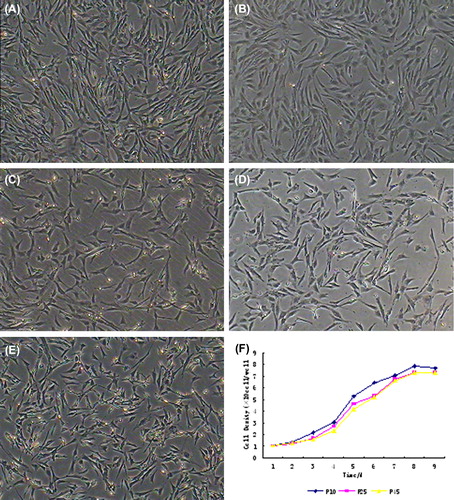
BMMSCs were large, lucent and strongly refractive. Their nuclei were oval and mixed with some blood cells. The non-adherent cells were removed on the third day, and MSCs were fusiform and showed cell-like clone with slower growth. Four days later, MSCs proliferated rapidly. We observed a delayed outgrowth of exponentially growing cell populations with spindle-shaped morphology, indicative of MSCs (Colter et al. 2001) (). The cells doubled every 30.92 hr and were maintained in culture for more than 100 days with no sign of senescence or differentiation. The growth curves of BMMSCs at different passages were S-shaped (). Two or three days later, MSCs entered the exponential growth phase, and seven to nine days later, entered platform growth phase.
Figure 1. Morphology and growth curves of BMMSCs (100×) at (A) P5, (B) P15, (C) P25, (D) P35, (E) P45, and at (F) passages 10, 25 and 45 for comparison. The cells expanded easily and exhibited fibroblast-like morphology. The growth curves of BMMSCs at different passages were S-shaped, and the PDTs of BMMSCs were 30.21, 31.78, 35.19 and 35.87 hr respectively.
Surface antigen characteristics of BMMSCs
Immunoflourescence staining results showed that BMMSCs at different passages expressed antigens CD44, CD29, ICAM-1, CD71, CD73, CD90, CD105 and SSEA-4, but did not express antigens CD31 or CD34. CD31 antigen is a special marker of endothelial cells, and CD34 antigen is a hematopietic marker. There was no significant difference in the positive rates at different passages (P > 0.05) (). RT-PCR assays consistently showed that BMMSCs at different passages were CD29, ICAM-1, SSEA-4, CD44, CD71 and CD73 positive, CD31 and CD34 negative ()
Figure 2. Surface antigen characteristics of BMMSCs at different passages. Immunoflourescence staining results showed that BMMSCs at different passages expressed antigens CD44, CD29, ICAM-1, CD71, CD73, CD90, CD105 and SSEA-4, but not antigens CD31 and CD34.
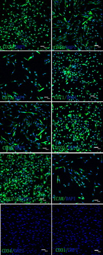
Figure 3. Detection of surface markers of BMMSCs via RT-PCR. RT-PCR assays showed that BMMSCs at different passages were positive for CD29, ICAM-1, SSEA-4, CD44, CD71 and negative for CD73, CD31 and CD34.
Osteogenic differentiation of BMMSCs
After the induction with osteoblast inducers, MSCs had apparent changes in appearance. From the 3rd day after the induction, BMMSCs in group C changed from fusiform to three-dimension, becoming larger and changing into polygon. As time went by, triangle or polygonal cells increased, and then grew into multilayers, and many crystal particles could be observed. The cell morphology of group A changed on the 5th day, and group B on the 7th day. Alizarin red staining of BMMSCs was positive on day 20 after induction. The positive region was brightly red, showing clear calcium nodules (), while the control group was negative. The ALP concentration assay of medium showed that ALP concentration increased with induced time extension (). RT-PCR and real-time PCR indicated that after incubation with BMP-2 and FGF 2, the specific genes, including BMPRI, Smad 1, Smad 5, Smad 8, Runx 2, collage type I and osteopontin were detected, and gene expression level showed a time-lapse increase ().
Figure 4. Osteogenic differentiation of BMMSCs. A: The control group. B: After incubation in osteogenic medium for 17 days, the cells metamorphosed from fusiform to tridimensional shapes and the nodules increased in number and size with prolonged inducing time. C: About 20 days later, the nodules were obviously observed following Alizarin Red staining. Cells cultured in complete medium were not influenced in morphology or stained by Alizarin Red (200×).
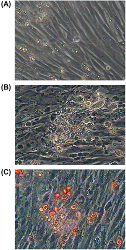
Figure 5. Detection of ALP concentration in inducer medium. The ALP concentration assay of medium showed that ALP concentration increased with induced time extension. Group C are significantly different (P < 0.05) than other group.

Figure 6. PCR analyses of BMP signals pathway members. A BMP regulation of osteoblast differentiation, ║ cell membrane, ║ karyotheca,Id inhibitor of DNA binding, dominant negative helix-loop-helix protein. B to H qPCR analyses of BMP signals pathway members. I RT-PCR analyses of BMP signals pathway members.
Discussion
Cell therapy has emerged as a strategy for the treatment of many human diseases, especially bone injury. At present, tissue stem cells were tentatively expanded and orientationally induced in vitro to some needed cells, which are then implanted into patients to repair damage, to replace regressive tissues and to improve the function of hereditarily defective tissues. It was reported that BMMSCs could be used to repair and reconstruct some tissues such as bone cartilage, lung, brain and liver (Pereira et al. 1998, Zhao et al. 2005, Liang et al. 2011, Thanabalasundaram et al. 2012). At present, the number of MSCs is not enough for tissue engineering. Accordingly, it is necessary to expand and purify MSCs in vitro. In vivo, the functions related to the developing and obtaining of tissue stem cells are related to gene-encoded transcription factors and extracellular signals (Majumdar et al. 1998). However, the differentiation mechanism of tissue stem cells in vitro is not clear yet. In this study, we performed the differentiation of BMMSCs into osteoblasts in vitro and detected related genes. The results demonstrated that BMPs could affect the differential direction of BMMSCs.
BMP signals are mediated by type I and type II BMP receptors and their downstream molecules Smads 1, 5 and 8. BMPs are involved in the control of bone formation. BMPs are known to play important roles in cartilage and bone formation (Urist 1997). BMP-2, a prototype of BMPs, exerts multiple effects on osteoblastic lineage cells (Yamaguchi et al. 2000, Emes et al. 2010). BMP-2 induces the differentiation of mesenchymal cells into osteoblasts, and promotes the maturation of osteoblasts by increasing the expression of Cbfa1/Runx2 and osteoblasts marker genes. Activation of type I BMP receptor phosphorylates Smad 1 and Smad 5 which can then associate with Smad 4 in a heteromeric complex. This complex is then translocated to the nucleus where it can activate gene transcription. Although the smad proteins are important molecules in the BMP pathway, other signaling pathways, such as extracellular signal-regulated kinase (ERK1/2), PKC and cAMP-dependent protein kinase A (PKA) may also play a role in BMP-2-induced osteoblast differentiation (Yamaguchi et al. 2000).
Conclussion
In this study, BMMSCs were isolated from rabbit tibia, and their morphology and antigen expression were investigated. We also induced BMMSCs to differentiate into osteoblasts, which proved that BMP signals pathway controls osteoblasts differentiation. The present study has important bearing on the potential application of MSCs as an adult stem cell source for regenerative therapies.
Declaration of interest
The authors report no conflicts of interest. The authors alone are responsible for the content and writing of the paper.
References
- Bongso A, Richards M. 2004. History and perspective of stem cell research. Best Pract Res Clin Obstet Gynaecol. 18:827–842.
- Colter DC, Sekiya I, Prockop DJ. 2001. Identification of a subpopulation of rapidly self-renewing and multipotential adult stem cells in colonies of human marrow stromal cells. Proc Natl Acad Sci USA. 98:7841–7845.
- Emes Y, Aybar B, Vural P, Saral NY, Atalay B, Kaya AS, et al. 2010. Effects of bone morphogenetic proteins on osteoblast cells: vascular endothelial growth factor, calcium, inorganic phosphate, and nitric oxide levels. Implant Dent. 19:419–427.
- Liang ZX, Sun JP, Wang P, Tian Q, Yang Z, Chen LA. 2011. Bone marrow-derived mesenchymal stem cells protect rats from endotoxin-induced acute lung injury. Chin Med J. 124:2715–2722.
- Maegawa N, Kawamura K, Hirose M, Yajima H, Takakura Y, Ohgushi H. 2007. Enhancement of osteoblastic differentiation of mesenchymal stromal cells cultured by selective combination of bone morphogenetic protein-2 (BMP-2) and fibroblast growth factor-2 (FGF-2). J Tissue Eng Regen Med. 1:306–313.
- Majumdar MK, Thiede MA, Mosca JD, Moorman M, Gerson SL. 1998. Phenotypic and functional comparison of cultures of marrow-derived mesenchymal stem cells (MSCs) and stromal cells. J Cell Physiol. 176:57–66.
- Noren Hooten N, Evans MK. 2011. The ultimate transformers: mesenchymal stem cells. Cell Cycle. 10:4189–4190.
- Pereira RF, O’Hara MD, Laptev AV, Halford KW, Pollard MD, Class R, et al. 1998. Marrow stromal cells as a source of progenitor cells for nonhematopoietic tissues in transgenic mice with a phenotype of osteogenesis imperfecta. Proc Natl Acad Sci USA. 95:1142–1147.
- Pittenger MF, Mackay AM, Beck SC, Jaiswal RK, Douglas R, Mosca JD, et al. 1999. Multilineage potential of adult human mesenchymal stem cells. Science. 284:143–147.
- Rickard DJ, Sullivan TA, Shenker BJ, Leboy PS, Kazhdan I. 1994. Induction of rapid osteoblast differentiation in rat bone marrow stromal cell cultures by dexamethasone and BMP-2. Dev Biol. 161:218–228.
- Thanabalasundaram G, Arumalla N, Tailor HD, Khan WS. 2012. Regulation of differentiation of mesenchymal stem cells into musculoskeletal cells. Curr Stem Cell Res Ther. 7:95–102.
- Urist MR. 1997. Bone morphogenetic protein: the molecularization of skeletal system development. J Bone Miner Res. 12:343–346.
- Varkey M, Kucharski C, Haque T, Sebald W, Uludag H. 2006. In vitro osteogenic response of rat bone marrow cells to bFGF and BMP-2 treatments. Clin Orthop Relat Res. 443:113–123.
- Yamaguchi A, Komori T, Suda T. 2000. Regulation of osteoblast differentiation mediated by bone morphogenetic proteins, hedgehogs, and Cbfa1. Endocr Rev. 21:393–411.
- Zhao DC, Lei JX, Chen R, Yu WH, Zhang XM, Li SN, Xiang P. 2005. Bone marrow-derived mesenchymal stem cells protect against experimental liver fibrosis in rats. World J Gastroenterol. 11:3431–3440.
- Zuk PA, Zhu M, Mizuno H, Huang J, Futrell JW, Katz AJ, et al. 2001. Multilineage cells from human adipose tissue: implications for cell-based therapies. Tissue Eng. 7:211–228.