Abstract
Objective: Surface matching is a relatively new method of spatial registration in neuronavigation. Compared to the traditional point matching method, surface matching does not use fiducial markers that must be fixed to the surface of the head before image scanning, and therefore does not require an image acquisition specifically dedicated for navigation purposes. However, surface matching is not widely used clinically, mainly because there is still insufficient knowledge about its application accuracy. This study aimed to explore the properties of the Target Registration Error (TRE) of surface matching in neuronavigation.
Materials and Methods: The surface matching process was simulated in the image space of a neuronavigation system so that the TRE could be calculated at any point in that space. For each registration, two point clouds were generated to represent the surface extracted from preoperative images (PCimage) and the surface obtained intraoperatively by laser scanning (PClaser). The properties of the TRE were studied by performing multiple registrations with PClaser point clouds at different positions and generated by adding different types of error.
Results: For each registration, the TRE had a minimal value at a point in the image space, and the iso-valued surface of the TRE was approximately ellipsoid with smaller TRE on the inner surfaces. The position of the point with minimal TRE and the shape of the iso-valued surface were highly random across different registrations, and the surface registration error between the two point clouds was irrelevant to the TRE at a specific point. The overall TRE tended to increase with the increase in errors in PClaser, and a larger PClaser made it less sensitive to these errors. With the introduction of errors in PClaser, the points with minimal TRE tended to be concentrated in the anterior and inferior part of the head.
Conclusion: The results indicate that the alignment between the two surfaces could not provide reliable information about the registration accuracy at an arbitrary target point. However, according to the spatial distribution of the target registration error of a single registration, enough application accuracy could be guaranteed by proper visual verification after registration. In addition, surface matching tends to achieve high accuracy in the inferior and anterior part of the head, and a relatively large scanning area is preferable.
Introduction
Navigation systems have become routine equipment in neurosurgery, and can help neurosurgeons reduce the operating time and improve the quality of the surgery Citation[1]. The working principle of neuronavigation is as follows: An image space is constructed in the system using the images of a patient and a transformation between the image space and the patient space is calculated by spatial registration. Then, the position of real surgical tools in the patient space is transformed into the image space, and virtual surgical tools are generated and overlaid on the images accordingly. The relative positions of the virtual tools and images can provide guidance for the surgical operation on the real patient. If there is no error in the entire process, the positional relationship between the virtual tools and the images should be identical to that between the real tools and the real patient.
Spatial registration is one of the most important steps in the clinical application of neuronavigation, and to a large extent determines the navigation accuracy. The methods of spatial registration used in neuronavigation can be broadly classified into two groups: point matching methods and surface matching methods Citation[2]. In point matching, the surgeon selects a group of corresponding points in each of the two spaces and closed-form solutions are used to calculate a transformation between the two spaces by matching the corresponding points Citation[3]. In surface matching, the surgeon first extracts a point cloud representing the face of the patient in the image space, then a second point cloud representing the patient's face in the patient space is obtained by laser scanning during the surgery. Iterative methods can then be used to calculate a transformation between the two spaces by matching these two point clouds Citation[4].
Point matching is simple and stable, and has been widely used in neuronavigation in a clinical setting. Its major disadvantage is that it usually requires an image scan specifically dedicated for navigation since fiducial markers must be fixed to the patient's head prior to scanning. These fiducial markers then serve as the corresponding points in the registration. The use of fiducial markers and the dedicated image acquisition increase the cost and time of preparation. In contrast, surface matching imposes no special requirements on the images used for navigation. Usually, the original diagnostic images are available and can be used for surface matching, so no dedicated image acquisition is necessary.
Though surface matching offers the advantages mentioned above, it is still not widely used clinically. A major reason for this is that the Target Registration Error (TRE) in surface matching is still not well understood. After two spaces are registered with one another, any point in one space can be transformed into the other space, and the TRE of this point is the distance between the transformed position and its real corresponding point in that space. Some published studies have compared the TRE between point matching and surface matching, but the results were not consistent. Usually, the image space and patient space were registered by these two approaches then the TRE was measured at a small number of anatomical or artificial landmarks for each method. In some studies the TRE calculated for these two registration methods was similar Citation[5–7], while in others the TRE for point matching was smaller than that for surface matching Citation[8–10]. According to the research on TRE distribution in point matching Citation[11], Citation[12], the TRE varies at different points in the same registration. Similarly, the TRE of different points must also differ in surface matching. Therefore, the TRE values in these studies were easily biased by the selection of points to be measured. In addition, the measured TRE may also have been influenced by the accuracy of the position tracking device of the navigation system, the accuracy of the measuring tools, human error, the resolution of the images used in navigation, and so on. All these factors, individually or in accumulation, may account for the inconsistency across these various studies.
There have also been some investigations of the distribution of the TRE for the surface matching method in neuronavigation. Raabe et al. Citation[13] and Krishnan et al. Citation[14] measured the TRE at a series of anatomical landmarks in real neuronavigation and found that the TRE was relatively smaller at the front of the head and larger at the occipital and temporal regions. However, they could not give the detailed distribution of TRE for the whole head because of the limitation on the number and position of anatomical landmarks available during a real neuronavigation procedure. In particular, most of the landmarks were on the skull and there were none available on the brain, so it was not possible to obtain data for the brain. Schamir et al. Citation[15] proposed a method to calculate TRE at different points according to the theory of Fitzpatrick and West Citation[11]. They gave the distribution of TRE for the whole head, and concluded that it was minimal at a point at the center of the face and increased according to the distance from that point. However, the method proposed by Fitzpatrick and West Citation[11] is used to calculate the TRE in point matching and requires definite correspondence between fiducial points, which don’t exist in the surface matching method. Therefore, it is still not clear if it is also applicable to calculating the TRE in surface matching.
In this study we simulated surface matching in the image space which enabled us to calculate the TRE at every point in the head. We have analyzed the distribution properties of the TRE in the surface matching method and offer some practical suggestions on the clinical application of surface matching in neuronavigation.
Materials and methods
We chose the images of a patient who underwent neuronavigation for our experiment. First, we extracted a point cloud PCimage to represent the head surface in the image space. Then we selected a subset of PCimage, transformed it by a transformation with random parameters Ttransform, and added random errors to each point to obtain a new point cloud. This new point cloud was denoted as PClaser, which represented the head surface in the patient space that is obtained by laser scanning on the real patient. Finally, we registered the two point clouds using the Iterative Closest Point (ICP) algorithm and calculated a transformation Tregistration from the patient space to the image space. For each point in one space, we could calculate the corresponding point in the other space by Ttransform or , since the patient space represented by PClaser was generated from the image space. Therefore, we could calculate the TRE at every point in the image space. At the same time, the TRE values calculated were not influenced by the navigation system or human errors, and they reflected solely the properties of surface matching.
Two types of laser scanning devices were used in previous studies of surface matching in neuronavigation. One type was a line scanning device, such as the z-touch (BrainLAB, Feldkirchen, Germany) Citation[9], Citation[14] or Fazer (Medtronic, Inc.) Citation[10], which was usually included as part of a commercial neuronavigation system. The other type was a surface scanning device that scanned a 3D surface as a whole instead of several lines, such as the VI 900 (Minolta, Osaka, Japan) Citation[16] or faceSCAN II (Breuckmann, Meersburg, Germany) Citation[15]. Surface scanning devices are very expensive and are not an integral part of commercial neuronavigation systems; if neurosurgeons wish to use a surface scanning device, they have to purchase it separately and integrate it into the neuronavigation system themselves. It is therefore difficult for surface scanning devices to be widely used in clinical neuronavigation. In this study, we only simulate the type of surface matching that uses line scanning devices. The experiments were done using a neuronavigation system developed in our laboratory.
Generation of the point cloud PCimage and PClaser
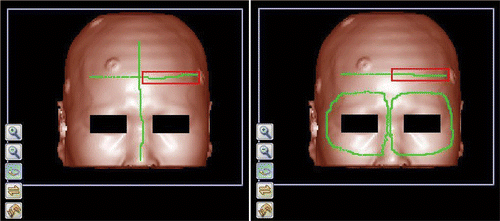
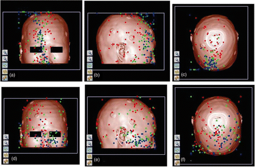
The CT images were formatted into 3D isotropic volume data with voxel size of 1.28*1.28*1.28 mm. We segmented out the head from the background by thresholding and extracted the voxels on the surface to form the point cloud PCimage. We then selected some voxels from PCimage on several lines and generated PClaser by transforming them using a transformation with random parameters and adding random errors to each point. PClaser point clouds were generated in two different areas and denoted as PClaser1 and PClaser2. The three-dimensional visualization of the CT volume data and the original position of PClaser1 and PClaser2 in the image space are shown in . PClaser1 consisted of 487 points and PClaser2 of 1216 points.
Figure 1. Three-dimensional visualization of the CT volume data and illustration of the original position of PClaser in the image space. The green lines in the left and right panels are the original positions of PClaser1 and PClaser2, respectively. In each case, the portion of the green line falling within the red rectangle is the area where anisotropic errors were added.
After selecting PClaser in its original position, we first transformed it into another space that was used to represent the patient space by a random rigid transformation Ttransform. The six parameters of Ttransform were random values between −2 and 2. The unit of translation parameters was the voxel and the unit of rotation parameters was the angle. In the clinical application of surface matching in neuronavigation, the patient space and image space are coarsely aligned by matching some anatomical landmarks prior to laser scanning and surface matching, and the residual errors between the two spaces are not very large. Therefore, the range of parameters used in this study was sufficient to simulate the scenario found in the clinical environment.
Next, for the point cloud transformed into the patient space, we added some random errors to obtain the final point cloud that mimicked the surface points obtained by laser scanning in the patient space. In a real neuronavigation application, many factors can result in differences between the point cloud obtained by laser scanning and that extracted from preoperative image data, such as the physiological shift of facial skin, inclusion of foreign objects in the laser scanning, and so on. Marmulla et al. Citation[17] studied the differences caused by physiological shift of facial skin in 20 patients and found that the mean difference for each patient ranged from 0.6 mm to 2.3 mm. To study the influence of different types of error on the laser scanning, we added the following three types of error:
Isotropic random error (IE). For each point in the point cloud, a random value between −2 and 2 was added to each of its three coordinates.
Small anisotropic random error (SAE). After adding isotropic random error, we added anisotropic errors to a part of the point cloud. For each point in this part, a random value between 0 and 4 was added to each of its three coordinates.
Large anisotropic random error (LAE). After adding isotropic random error, anisotropic error was added to the same part of the point cloud where small anisotropic random error was added, and the random value added changed to between 2 and 6.
Surface matching registration algorithm
The ICP algorithm proposed by Besl and McKay Citation[4] is widely used to register two point clouds. A major disadvantage of the ICP algorithm is that it is easy to get stuck at a local minimum. To deal with this problem, we performed multiple registrations with a small disturbance of the position of PClaser, and took the one with the minimal Surface Registration Error (SRE) as the registration result between the two point clouds. The SRE is the average distance between each of the points in PClaser and its nearest point in PCimage after registration. The initial positions of PClaser were obtained by transforming it from its original position by different transformations Tsearch. The translation parameters of Tsearch were −0.667 or 0.667 voxels, and the rotation parameters of Tsearch were −0.667 or 0.667 angles. There were 64 Tsearch transformations in total, so 64 registrations were performed for each pair of PCimage and PClaser point clouds.
After registration, a transformation Tregistration from the patient space to the image space was calculated. For any point Pi in the image space, we calculated its corresponding point Pp in the patient space by multiplying it with Ttransform. After registration, Pp was transformed back into the image space by multiplying it with Tregistration, and this point was denoted as Pi′. The TRE at point Pi was the distance between Pi and Pi′.
Experiments on the properties of TRE
We performed the following four experiments using the above-mentioned data and algorithm:
We generated one PClaser1 and one PClaser2 by adding isotropic random errors and registered each of them to PCimage. We calculated the TRE at each voxel of the volume data and displayed the TRE at three orthogonal sections to study the property of the spatial distribution of TRE in a single registration.
As shown in , we generated 50 PClaser point clouds for each combination of scanning area and error type and registered them with PCimage. After each registration, we calculated the TRE at selected sampling points and studied the property of the statistical distribution of the TRE at these points. As illustrated in , the sampling points are on three line segments that are parallel to the coordinate axes and pass through a point at the center of the volume data, and they are denoted as X′, Y′ and Z′, respectively. The sampling points were taken from left to right on X′, from superior to inferior on Y′, and from anterior to posterior on Z′. The sampling points were distributed evenly on the line segments with a distance of five voxels between neighboring points. There were 25, 26 and 29 sampling points on X′, Y′ and Z′, respectively.
We calculated the SRE of each registration in experiment 2 and studied the property of the statistical distribution of the SRE in each group.
We recorded the position of the point with minimum TRE in each registration in experiment 2 and studied their spatial distribution in the image space.
Figure 2. The position of the TRE sampling points. The red, green and blue lines are the X′, Y′ and Z′ sampling line segments, respectively. They intersect at a point at the center of the head, and are parallel to the X, Y and Z coordinate axes of the volume data.
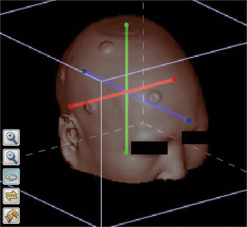
Table I. Generation method and number of point clouds in the patient space.
Results
illustrates the distribution of TRE on three orthogonal sections for a PClaser1 and a PClaser2. The TRE has minimal value at a point in the image space, and this point is denoted as the center point. Iso-valued surfaces of TRE are approximately ellipsoidal surfaces around this center point. The value of the inner iso-valued surfaces is smaller, and that of the outer ones is larger. illustrates the statistical distribution of the TRE on the sampling points with different scanning areas and error types. shows the statistical distribution of the SRE, and illustrates the spatial distribution of the center points.
Figure 3. Spatial distribution of TRE in surface matching. The left set of images represent a case of registration with scanning area PClaser1. The right set of images represent a case of registration with scanning area PClaser2. In both cases, the upper left, upper right and lower left panels correspond to the coronal, sagittal and axial sections that pass through the center point, which is indicated by a small red cross. The lower right panel in each set is the 3D visualization of the head segmented out from the background by thresholding. On the section images, the TRE at the points where the CT value is larger than the threshold is mapped into a gray value, with dark gray indicating a smaller TRE and light gray indicating a larger TRE. The TRE in the area within the innermost iso-valued surface is 0.5 voxels in the left image and 1.6 voxels in the right image, and the TRE increases by 0.1 voxels with each iso-valued surface moving outward.
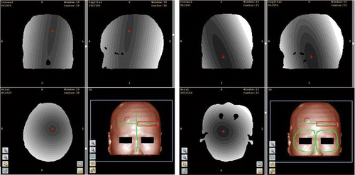
Figure 4. Statistical distribution of TRE on sampling axes with different scanning areas and error types. Small dots and triangles indicate the mean values of the TRE for PClaser1 and PClaser2, respectively, and the horizontal bars indicate the standard deviation. IE, SAE and LAE are the three types of errors: isotropic random error, small anisotropic random error, and large anisotropic random error. (a), (c) and (e) show the TRE distributions on the X′, Y′ and Z′ sampling axes when PClaser1 was used. (b), (d) and (f) show the TRE distributions on the X′, Y′ and Z′ sampling axes when PClaser2 was used.
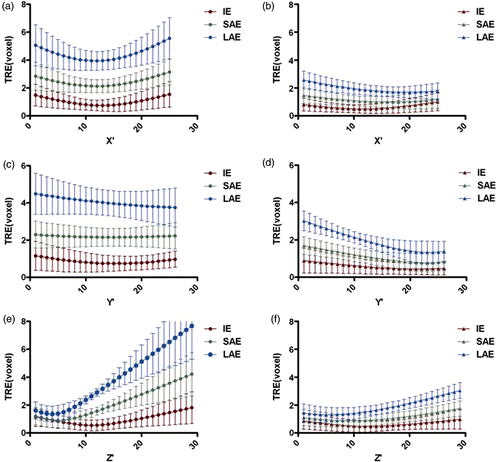
Figure 5. Statistical distribution of SRE for registrations with different scanning areas and error types. Small dots indicate the mean value of the SRE for registrations with PClaser1, and small triangles indicate the mean value of SRE for registrations with PClaser2. The horizontal bars indicate the standard deviation. IE, SAE and LAE are the three types of error: isotropic random error, small anisotropic random error, and large anisotropic random error.
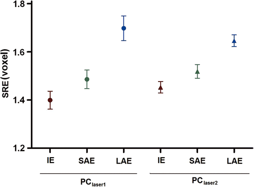
Figure 6. Spatial distribution of the center points for registrations with different scanning areas and error types. Fifty registrations were performed for each combination of scanning area and error type. The red, green and blue dots indicate the position of the center points for IE, SAE and LAE, respectively. (a), (b) and (c) are the distributions for the scanning area PClaser1, viewed from the front, right and top, respectively. (d), (e) and (f) are the distributions for the scanning area PClaser2, viewed from front, right and top, respectively.
Discussion
Spatial distribution of the TRE in a single registration
From we can see that the TRE at an arbitrary point depends on the position of the center point and the configuration of the iso-valued surfaces. Generally speaking, the center points are distributed sparsely over a wide range of areas, as indicated in . In addition, the configuration of the iso-valued surfaces differs for different registrations. Therefore, the spatial distribution of TRE for a single registration is highly random, and it is difficult to predict the TRE exactly at an arbitrary position. From and we can find some relativity between SRE and TRE. These two values increase at the same time along with the increase in anisotropic errors for the same scanning area. However, for different scanning areas, there is no similar relativity between SRE and TRE. If the same types of error are added, the mean value of the SRE for PClaser2 is larger than that for PClaser1 in , but the TRE for PClaser2 is smaller than that for PClaser1 in . This means that it is reasonable to optimize registration by minimizing SRE if the scanning area is not changed, but the difference in SRE values for registrations with different scanning areas does not provide comparison information regarding their TREs.
For a single registration by surface matching, the TRE at an arbitrary point is highly random, and the SRE cannot provide useful information about the TRE at a specific point. However, according to the spatial distribution of TRE illustrated in , if the TRE at the whole head surface is acceptable, the TRE at the inner regions of the head should also be acceptable. In practice, neurosurgeons can choose a series of points sparsely distributed on the head and verify the TRE at these points to guarantee the application accuracy of surface matching.
Statistical distribution of the TRE in different regions
From we can see the following:
For the same scanning area and the same sampling axis, the mean value of TRE increases with the introduction of anisotropic error and the increase in its value. This means that shape difference results in a large TRE in surface matching, and neurosurgeons should try to avoid this kind of difference in practice. For example, they should instruct the patient to relax during image scanning to avoid deforming the face by their expression. They should also be very careful when scanning the patient's head with the laser scanner to avoid including foreign objects such eye masks, intubation tubes, etc.
For each sampling axis, the mean value and standard deviation of TRE of PClaser2 is smaller than that of PClaser1 when the same type of error is used. This means that a larger scanning area tends to result in a smaller and more stable TRE. The TRE of PClaser1 increases rapidly with the introduction of anisotropic errors, so this is not a practical scanning area for real neuronavigation. In contrast, PClaser2 is a practical scanning area since the TRE is still not very large even when there are relative large anisotropic errors.
From , we can see that the TREs at the first 10 sampling points on the Z′ axes are small for all scanning areas and error types. This means that surface matching tends to achieve a small TRE on the front part of the head; however, the point with the smallest TRE is not in the center of the face but in the front part of the brain.
indicates that the TRE at the inferior part of head is smaller than that at the superior part when PClaser2 is used. According to , the mean value of the TRE is less than two voxels in the anterior and inferior half of the sampling axes, even when relatively large anisotropic errors are added. shows that the center points tend to be concentrated on the anterior and inferior part of the head with the introduction of anisotropic errors. This is also one of the reasons why the TRE at the anterior and inferior part of the head is relatively small.
Comparison to previous similar studies
The finding of this study that TRE is smaller in the anterior part of the head is consistent with the results of previous clinical studies Citation[13], Citation[14]. Though we were unable to provide a comparison between the accuracy of surface matching and that of point matching, we proved that surface matching could achieve high accuracy in the anterior and inferior part of the head if a proper scanning area was used.
The result for the spatial distribution of TRE in this study is different from that reported by Shamir et al. Citation[15]. Shamir et al. calculated TRE according to the formula proposed by Fitzpatrick and West Citation[11], which was used to calculate the expected TRE in point matching. According to this formula, the expectation of TRE at the center of the fiducial points is zero. Schamir et al. scanned part of the face in the patient space, and the center of this point cloud was at the center of the face, meaning that the point with minimal TRE was at the center of the face. In addition, they thought that the TRE of a point was proportional to its distance from this center point, but this is not consistent with the formula in the study by Fitzpatrick and West Citation[11]. The extension of this method into surface matching may therefore be unreliable.
Conclusion
From the experimental results presented, we can conclude the following:
Large scanning areas are better than small ones.
General speaking, TRE in the anterior part of the head is smaller than that in the posterior part, so surface matching is more suitable for operations performed in the anterior part of the head than in other regions.
Visual verification is obligatory to guarantee the application accuracy of surface matching in neuronavigation.
Declaration of interest: Dr. Song is supported by the National 863 Program of the Ministry of Science and Technology of China (Grant No. 2009AA02Z415) and the Shanghai Leading Academic Discipline Project of the Shanghai Municipal Education Commission (No. B112). Dr. Wang is supported by the Science & Technology Commission of Shanghai Municipality (Grant Nos. 08dz1900705, 09410702800 and 10dz2211800).
References
- Peters TM. Image-guidance for surgical procedures. Phys Med Biol 2006; 51(14)R505–R540
- Eggers G, Mühling J, Marmulla R. Image-to-patient registration techniques in head surgery. Int J Oral Maxillofac Surg 2006; 35(12)1081–1095
- Eggert DW, Lorusso A, Fischer RB. Estimating 3-D rigid body transformations: A comparison of four major algorithms. Mach Vis Appl 1997; 9(5–6)272–290
- Besl PJ, McKay ND. A method for registration of 3-D shapes. IEEE Trans Pattern Anal Mach Intell 1992; 14(2)239–256
- Cao A, Thompson RC, Dumpuri P, Dawant BM, Galloway RL, Ding S, Miga MI. Laser range scanning for image-guided neurosurgery: Investigation of image-to-physical space registrations. Med Phys 2008; 35(4)1593–1605
- Marmulla R, Mühling J, Wirtz CR, Hassfeld S. High-resolution laser surface scanning for patient registration in cranial computer-assisted surge. Minim Invasive Neurosurg 2004; 47(2)72–78
- Mascott CR, Sol JC, Bousquet P, Lagarrigue J, Lazorthes Y, Lauwers-Cances V. Quantification of true in vivo (application) accuracy in cranial image-guided surgery: Influence of mode of patient registration. Neurosurgery 2006; 59(1)146–155
- Hoffmann J, Westendorff C, Leitner C, Bartz D, Reinert S. Validation of 3D-laser surface registration for image-guided cranio-maxillofacial surgery. J Craniomaxillofac Surg 2005; 33(1)13–18
- Schlaier J, Warnat J, Brawanski A. Registration accuracy and practicability of laser-directed surface matching. Comput Aided Surg 2002; 7: 284–290
- Schicho K, Figl M, Seemann R, Donat M, Pretterklieber ML, Birkfellner W, Reichwein A, Wanschitz F, Kainberger E, Bergmann H, et al. Comparison of laser surface scanning and fiducial marker-based registration in frameless stereotaxy – Technical note. J Neurosurg 2007; 106(4)704–709
- Fitzpatrick JM, West JB. The distribution of target registration error in rigid-body point-based registration. IEEE Trans Med Imag 2001; 20(9)917–927
- West JB, Fitzpatrick JM, Toms SA, Maurer CR, Maciunas RJ. Fiducial point placement and the accuracy of point-based, rigid body registration. Neurosurgery 2001; 48(4)810–816
- Raabe A, Krishnan R, Wolff R, Hermann E, Zimmermann M, Seifert V. Laser surface scanning for patient registration in intracranial image-guided surgery. Neurosurgery 2002; 50(4)797–801
- Krishnan R, Raabe A, Seifert V. Accuracy and practicability of laser surface scanning for registration in image guided neurosurgery. In: Buzug TM, Lueth TC, editors. Perspective in image-guided surgery: Perspective in Image-Guided Surgery: Proceedings of the Scientific Workshop on Medical Robotics, Navigation and Visualization, Remagen, Germany March 2004. pp 31–36.
- Shamir RR, Freiman M, Joskowicz L, Spektor S, Shoshan Y. Surface-based facial scan registration in neuronavigation procedures: A clinical study. J Neurosurg 2009; 111(6)1201–1206
- Marmulla R, Mühling J, Eggers G. Image-to-patient registration by natural anatomical surfaces of the head. Central European Journal of Medicine 2007; 2(1)89–102
- Marmulla R, Muehling J, Lueth T, Hassfeld S. Physiological shift of facial skin and its influence on the change in precision of computer-assisted surgery. Br J Oral Maxillofac Surg 2006; 44(4)273–278