Abstract
The aim of this study was to compare the accuracy of preoperative templating in total knee arthroplasty (TKA) using conventional two-dimensional (2D) and computed tomography (CT)-based three-dimensional (3D) procedures, and to confirm the necessity of 3D evaluation for preoperative planning. One hundred consecutive primary TKAs were analyzed. Preoperative templating was performed for each TKA using both conventional 2D radiographs and a CT-based 3D image model created using KneeCAS software. Accuracies with regard to the predicted and actual implant sizes were determined for each procedure. The 3D procedure was found to be more accurate (59%) than the 2D procedure (56%) in predicting implant size, but the difference was not statistically significant (p = 0.67). Computer-assisted surgery systems are often used for preoperative planning in TKA. However, our results do not support the superiority of 3D preoperative templating over 2D conventional evaluation in predicting implant size. Thus, 3D templating may not be necessary for preoperatively predicting implant size in TKA, and can only be used as an approximate guide.
Introduction
Preoperative planning is an important part of the total knee arthroplasty (TKA) procedure. The functional results of TKA depend on patient factors and complications, but are also related to the position and size of the prosthesis. Both oversizing and undersizing of the prosthesis components can cause pain and/or functional impairment. Therefore, accurate preoperative templating to predict the implant size and position is important for obtaining a successful outcome in TKA. Accordingly, the use of a templating system has been recommended in joint arthroplasty, and such a system is now routinely used with most prosthesis designs Citation[1–3].
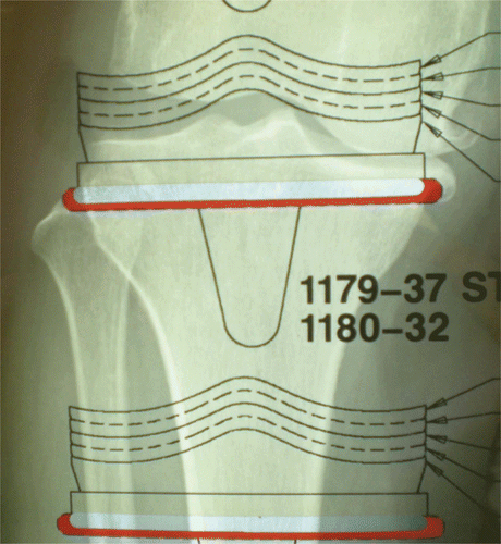
Standard preoperative templating for TKA is performed by placing acetate overlays of the knee implants on conventional radiographs of the knee. However, previous research suggests that preoperative analog templating in TKA yields inaccurate results concerning the implant size selection Citation[4–7]. On the other hand, several studies have shown that computer-assisted navigation is more accurate than conventional instrumentation, and CT scans are useful for preoperative planning Citation[8–10].
The aim of this study was to compare the accuracy of preoperative templating with regard to implant size selection in TKA for conventional two-dimensional (2D) and computed tomography (CT)-based 3D procedures in order to confirm the efficacy of using 3D evaluations for preoperative planning.
Patients and methods
This study analyzed 100 primary TKA procedures performed during the period between December 2005 and May 2009 at a single institution (Ishii Orthopaedic and Rehabilitation Clinic) under the direction of a single senior surgeon (Y. I.). The mean age of the patients was 73.3 years (range: 33 to 90 years). There were 16 male patients and 72 female patients, with twelve bilateral cases. Patients with a history of previous surgery of the involved knee were excluded. A prospective study was undertaken. All patients received LCS knee implants (Depuy Orthopaedics, Warsaw, IN). In all knees, the femoral components were fixed without cement and the tibial components were fixed with cement. The LCS component was available in 6 sizes.
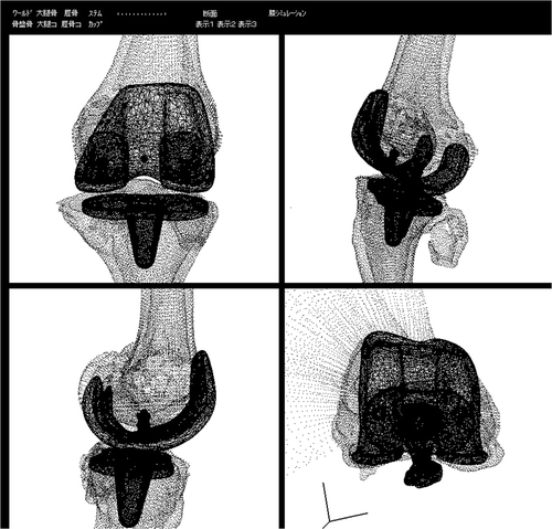
Preoperative templating was performed for each TKA using conventional 2D radiographs (both anteroposterior and lateral views), which were analyzed by a single senior surgeon (). Preoperative CT scans of the knee were performed and a 3D digital model of the knee was reconstructed from the CT data using KneeCAS (LEXI, Inc., Tokyo, Japan) as described in a previous report Citation[11], Citation[12]. KneeCAS is software for preoperative TKA planning, postoperative evaluation, and surgical support. In this study, the software was used only for predicting implant size. The manufacturer provided us with computer aided design (CAD) models of different sizes for the implant. By superimposing the CAD model of the implant on the CT-based 3D digital model of the knee, a radiology technologist predicted the implant size without any knowledge of the 2D procedure (). The 3D implant model was selected taking into consideration the individual anatomy of the patient so as to exclude overhang or notching. The size of the implant that had been inserted during surgery was determined from the surgical notes, and this was used as the gold standard.
Figure 1. Conventional 2D templating: anteroposterior view of the knee demonstrating templating of the tibia.

Figure 2. CT-based 3D image model (superimposing the CAD model of the implant) created using KneeCAS (LEXI, Inc., Tokyo, Japan).

The accuracy and reliability were assessed for all measurements of the two different templating procedures (i.e., the 2D and CT-based 3D procedures). The Chi-square test for independence for paired observations was used to analyze the accuracy. The Wilcoxon signed-rank test was used for the differences between the 2D procedure and the CT-based 3D procedure with regard to the mean absolute differences between the planned and actual implant size. In all tests, p < 0.05 was considered significant. The weighted kappa test was used to analyze the reliability. The weighted kappa test takes a value between 0 and 1; if all responses are in agreement, kappa is 1. If there is no more agreement than that which would be expected by chance, kappa is 0. A more detailed presentation of the kappa statistics can be found in .
Table I. Kappa coefficients and levels of agreement.
This study was approved by our institutional review board, and all patients provided informed consent.
Results
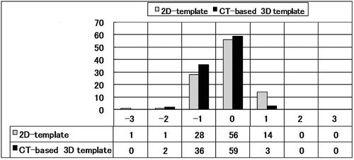
Since the templated and implanted sizes coincided closely for both the femoral and tibial components in all 100 cases, the evaluations of both components were thus performed simultaneously. The results are shown in and . Only 56% (56/100) of the 2D procedures were found to be an exact match. This figure increased to 98% (98/100) for templates that were within one size above or below that actually used, while 2% (2/100) were two sizes or more adrift. In comparison, 59% (59/100) of the CT-based 3D procedures were an exact match, 98% (98/100) were within one size, and 2% (2/100) were two sizes or more adrift. The CT-based 3D procedure was thus slightly more accurate than the 2D procedure. However, the difference was not statistically significant (p = 0.67).
Figure 3. Error from implanted size. There was a tendency to underestimate the size in the CT-based 3D procedure.

Table II. Error from implanted size. The accuracy increased to 98% for templated sizes within one size above or below that actually used for both the 2D procedure and the CT-based 3D procedure.
The CT-based 3D procedure was on average slightly more accurate than the 2D procedure with a mean absolute error of 0.43 versus 0.47. However, again the difference was not statistically significant (p = 0.59).
Further analysis of the data shows that templating using the 2D procedure was one size below in 28% (28/100) of the procedures and one size above in 14% (14/100), while templating using the CT-based 3D procedure was one size below in 36% (36/100) of the procedures and one size above in 3% (3/100). There was a tendency to underestimate the size in the CT-based 3D procedure, and that tendency was statistically significant (p = 0.033).
The weighted kappa coefficients for the 2D procedure and CT-based 3D procedure are shown in . The weighted kappa coefficient of the 2D procedure was 0.49 (indicating a moderate agreement), while that of the CT-based 3D procedure was also 0.49 (again indicating a moderate agreement). The results of the weighted kappa coefficients were not statistically significant (p = 0.65).
Table III. Weighted kappa coefficients for the 2D procedure and the CT-based 3D procedure.
Discussion
A templating system is recommended for joint arthroplasty, and is routinely used with most prosthesis designs. There are many advantages to preoperatively templating the size of the prosthesis in arthroplasty. Doing so involves selecting the correct implant size, determining the alignment, position and orientation of the prosthesis, and minimizing the surgical exposure. Theoretically, this should allow for a reduction in surgical time and reduce the incidence of complications.
Preoperative templating in TKA is routinely performed by the surgeon prior to surgery. Standard preoperative templating for TKA is performed by placing acetate overlays of the knee implants on conventional radiographs of the knee. A more accurate prediction of the component size can not only influence the clinical results, but also reduce the number of surgical instruments that are prepared for an operation, shorten the operation time, and possibly reduce the risk of infection.
However, previous research suggests that preoperative analog templating with regard to implant size selection in TKA may provide inaccurate results. Heal and Blewitt Citation[6] found preoperative radiologic templating in TKA to be accurate in just 57% of cases, and thus questioned its benefit in preoperative management. Arora et al. Citation[4] reported that templating in TKA was only accurate for both tibial and femoral components in 53.2% of cases, and concluded that preoperative templating is neither accurate nor reproducible. Aslam et al. Citation[5] reported that the exact size of the prosthesis was correctly predicted for 49% of femoral and 67% of tibial components, and that the statistical agreement between the templated size and the actual implant size was only fair to moderate using the weighted kappa test; they determined that acetate templating for TKAs was prone to error and could only be used as an approximate guide. The present study found that the 2D procedure showed similar results, with an accuracy of 56% and moderate statistical agreement using the weighted kappa test.
Computer navigation systems are gaining in popularity and several studies have suggested that there is improved alignment when such systems are used Citation[8], Citation[13]. However, van der Linden-van der Zwaag et al. Citation[14] reported there was a risk of oversizing the femoral component of TKA when using computer assisted orthopaedic surgery. It is therefore still unclear whether these emerging technologies offer a real cost-benefit or result in an improved outcome. Accordingly, the present study evaluated whether preoperative CT-based 3D templating, which is now one of the functions of computer-assisted surgery systems, improves the accuracy of the component size prediction in comparison to 2D templating. Several reports have previously compared the accuracy of preoperative templating for TKA using conventional analog 2D and digital 2D procedures. The et al. Citation[15] reported that planning of component sizes for TKA is an accurate procedure when performed digitally, while Specht et al. Citation[16] reported the digital 2D technique to be more accurate than acetate templating for tibial component size. There are also several reports comparing the accuracy of preoperative templating in total hip arthroplasty with a conventional 2D and CT-based 3D procedure Citation[17], Citation[18]. Sugano et al. Citation[17] reported that in cases without large anteversion and/or external rotation contracture, the simple X-ray template procedure might be sufficient for THA planning. However, there have so far been no reports which compare conventional 2D and CT-based 3D procedures.
This study is the first to investigate and compare the results of the 2D procedure and CT-based 3D procedure in TKA. The accuracy of the 2D procedure was 56% in the current series, and that of the CT-based 3D procedure was 59%. The weighted kappa test showed agreement between the templated size and the actual implant size to be moderate in both procedures. The use of different magnifications according to the preoperative flexion contracture of each patient in the 2D procedure might be the main reason for the “moderate” results obtained in this study. Observer error in interpreting the radiographs, the rotation of the radiographs, and magnification errors caused by a fixed flexion deformity have all been identified as possible reasons for the inaccuracy of the templates in TKA procedures Citation[6]. The observer in the 3D procedure, who had no previous experience of performing TKA surgery, might thus have paid special attention to the oversizing of implants, because the femoral components were fixed without cement (which is a requirement of the press-fit procedure) and the tibial components need to avoid soft tissue friction. Therefore, there was a tendency to underestimate the size in the CT-based 3D procedure, and that tendency was found to be statistically significant in this study. Conversely, the accuracy increased to 98% for templated sizes within one size above or below that used in both the 2D procedure and the CT-based 3D procedure.
These results indicate that neither the 2D procedure nor the CT-based 3D procedure for TKAs is reliable and accurate, and that they can only be used as an approximate guide. However, predicting implant sizes to within one size provides a tool for stock control, thereby allowing the number of surgical instruments that must be prepared for an operation to be reduced, and the surgical time and risk of infection can also be lowered as a result. We recognize that further study may be needed to address the specific questions of reproducibility and interobserver reliability of the 3D procedure among different experienced surgeons in order to fully demonstrate the advantages of the 3D procedure. We also recognize that our study only concerns the accuracy of implant size selection in preoperative templating; we do not refer to the overall planning of the TKA procedure (component placement, alignment, etc.).
Computer-assisted surgery systems are often used for preoperative planning in TKA. However, the present results do not support the superiority of 3D preoperative templating over 2D conventional evaluation in predicting implant size. Therefore, 3D templating may not be necessary for preoperatively predicting the implant size for TKA, and can only be used as an approximate guide.
Declaration of interest: No benefits in any form have been received or will be received from a commercial party related directly or indirectly to the subject of this article.
References
- Eggli S, Pisan M, Müller ME. The value of preoperative planning for total hip arthroplasty. J Bone Joint Surg Br 1998; 80(3)382–390
- Müller ME. Lessons of 30 years of total hip arthroplasty. Clin Orthop Relat Res 1992; 274: 12–21
- Clarke IC, Gruen T, Matos M, Amstutz HC. Improved methods for quantitative radiographic evaluation with particular reference to total-hip arthroplasty. Clin Orthop Relat Res 1976; 121: 83–91
- Arora J, Sharma S, Blyth M. The role of pre-operative templating in primary total knee replacement. Knee Surg Sports Traumatol Arthrosc 2005; 13(3)187–189
- Aslam N, Lo S, Nagarajah K, Pasapula C, Akmal M. Reliability of preoperative templating in total knee arthroplasty. Acta Orthop Belg 2004; 70(6)560–564
- Heal J, Blewitt N. Kinemax total knee arthroplasty: Trial by template. J Arthroplasty 2002; 17(1)90–94
- Howcroft DW, Fehily MJ, Peck C, Fox A, Dillon B, Johnson DS. The role of preoperative templating in total knee arthroplasty: Comparison of three prostheses. Knee 2006; 13(6)427–429
- Chauhan SK, Scott RG, Breidahl W, Beaver RJ. Computer-assisted knee arthroplasty versus a conventional jig-based technique. A randomised, prospective trial. J Bone Joint Surg Br 2004; 86(3)372–377
- Jazrawi LM, Birdzell L, Kummer FJ, Di Cesare PE. The accuracy of computed tomography for determining femoral and tibial total knee arthroplasty component rotation. J Arthroplasty 2000; 15(6)761–766
- Müller W, Bockholt U, Voss G, Lahmer A, Börner M. Planning system for computer assisted total knee replacement. Stud Health Technol Inform 2000; 70: 214–219
- Sato T, Koga Y, Omori G. Three-dimensional lower extremity alignment assessment system. Application to evaluation of component positioning after total knee arthroplasty. J Arthroplasty 2004; 19: 620–628
- Sato T, Koga Y, Sobue T, Omori G, Tanabe Y, Sakamoto M. Quantitative 3-dimensional analysis of preoperative and postoperative joint lines in total knee arthroplasty: A new concept for evaluation of component alignment. J Arthroplasty 2007; 22: 560–568
- Chauhan SK, Clark GW, Lloyd S, Scott RG, Breidahl W, Sikorski JM. Computer-assisted total knee replacement. A controlled cadaver study using a multi-parameter quantitative CT assessment of alignment (the Perth CT Protocol). J Bone Joint Surg Br 2004; 86(6)818–823
- van der Linden-van der Zwaag HM, Wolterbeek R, Nelissen RG. Computer assisted orthopedic surgery; its influence on prosthesis size in total knee replacement. Knee 2008; 15(4)281–285
- The B, Diercks RL, van Ooijen PM, van Horn J. Comparison of analog and digital preoperative planning in total hip and knee arthroplasties. A prospective study of 173 hips and 65 total knees. Acta Orthop 2005; 76(1)78–84
- Specht LM, Levitz S, Iorio R, Healy WL, Tilzey JF. A comparison of acetate and digital templating for total knee arthroplasty. Clin Orthop Relat Res 2007; 464: 179–183
- Sugano N, Ohzono K, Nishii T, Haraguchi K, Sakai T, Ochi T. Computed-tomography-based computer preoperative planning for total hip arthroplasty. Comput Aided Surg 1998; 3(6)320–324
- Viceconti M, Chiarini A, Testi D, Taddei F, Bordini B, Traina F, Toni A. New aspects and approaches in pre-operative planning of hip reconstruction: A computer simulation. Langenbecks Arch Surg 2004; 389(5)400–404
