Abstract
The accuracy of a commercial imageless navigation system for hip resurfacing and its reproducibility among different surgeons and for varying femoral anatomy was tested by comparing conventional and navigated implantation of the femoral component on different sawbones in a hip simulator. The position of the component was measured on postoperative radiographs. Variance for varus/valgus alignment and anteversion was higher for conventional implantation. Among the three surgeons, operation time, chosen implant size and anteversion were significantly different for conventional implantation but not for the navigated method. Using navigation, no difference was found for normal and abnormal anatomy. Values obtained with the navigation system were consistent with those measured on radiographs. Navigation appeared to be accurate and helped to reduce outliers. This was true for the three different surgeons and in varying anatomical situations.
Introduction
Hip resurfacing arthroplasty is regarded as a demanding operation that should be performed only by experienced surgeons. Although advances in surgical techniques and improvements in design have led to good short- and mid-term results, many concerns still remain. In particular, malpositioning of the femoral component may lead to serious complications, such as fracture of the femoral neck or impingement with painful reduction of range of motion. The incidence of femoral neck fracture is reported to be between 1.5% and 7.2% Citation[1], Citation[2] and is attributed to patient-related factors such as bone density and weight, as well as to operation-related factors, e.g., notching of the femoral neck or varus positioning of the component Citation[2–5]. Valgus positioning, on the other hand, may cause femoral neck notching or impairment of vascularity of the femoral head Citation[5], Citation[6]. In addition to choosing the appropriate implant size, the surgeon must position the femoral component correctly with regard to the offset between the femoral head and neck; otherwise, the risk of impingement increases and range of motion decreases Citation[7], Citation[8].
In total hip arthroplasty, computer navigation has been shown to improve the placement of the acetabular component Citation[9–12], whereas in hip resurfacing it should help to improve accuracy in positioning of the femoral component. Some studies have shown improvement in varus/valgus alignment of the femoral component with the use of computer navigation Citation[13–16]. Nevertheless, there are further requirements which must be met with the use of a navigation system, such as reproducibility among different surgeons and guidance in patients with abnormal anatomy.
The aim of our study was to answer the following three questions:
Is it possible to position the femoral component more accurately with regard to varus/valgus alignment and ante-/retroversion, as well as offset, compared to conventional implantation?
Is it possible to reproduce results with different surgeons?
Are any differences observed with regard to normal and abnormal anatomy?
Materials and methods
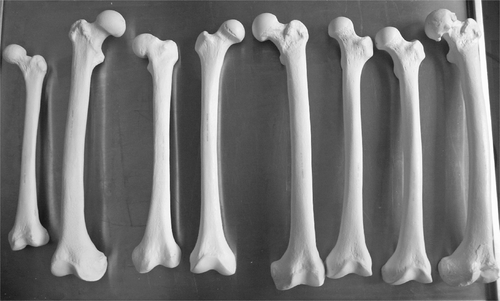
Six sets of eight different femurs (Sawbones®, Sawbones Europe AB, Malmö, Sweden) were used. In each set, six of the femurs were left-sided and two were right-sided. Five of the femurs were of normal anatomy but differed in size or shape, whereas the other three femurs represented abnormal anatomies, with one dysplastic arthritic femoral head, one dysplastic hip with 155° valgus, and one hip with pistol grip deformity after slipped epiphysis ().
Figure 1. The set of 8 different femurs, including 5 femurs with normal anatomy and 3 with abnormal anatomy. The abnormal femurs comprise one with a pistol grip deformity after slipped epiphysis (3rd from left), one with valgus deformity (6th from left), and one with a severely arthritic head (8th from left).
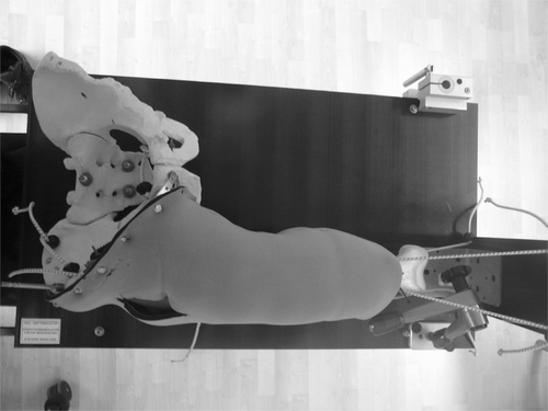
Three experienced orthopedic surgeons performed conventional and navigated implantations of the femoral component for hip resurfacing (MITCH®, Stryker, Kalamazoo, MI) on the femurs. Each surgeon operated on two sets of the eight different femurs in random order. Thus, a total of 48 operations were performed, 16 by each surgeon. All implantations were performed in a hip simulator which was developed in an earlier study Citation[17]. This consisted of a pelvis and femur surrounded by foam and neoprene resembling the muscles which normally surround the hip. It was fixed on a board by means of adjustable arms, which allowed the hip joint to be held in different positions (see ). The operations were performed via a direct anterior approach.
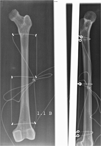
For computer navigation we used the software module for hip resurfacing of the Stryker iNfinitus® imageless navigation system (Stryker, Kalamazoo, MI). For in vitro application, the femur was fixed in the hip simulator, a pelvic and a femoral tracker were inserted, and landmarks were registered. The femur was dislocated and the head and neck registered. The computer provided information concerning the planned neck angle, anteversion and size of the component. The guide wire was inserted under navigation control, after which followed reaming and component implantation. Subsequently, all femurs were fixed on a board and radiographs were taken with standardized anterio-posterior (a.p.) and lateral views (see ).
The caput-collum-diaphyseal angle (CCD), anteversion (AV), and superior (sup.), inferior (inf.), anterior (ant.) and posterior (post.) offset were measured by four different investigators. The CCD was determined by measuring the angle between the shaft axis and the pin canal of the femoral component on the a.p. view, and the AV as the angle between the central axis of the femoral neck and the pin canal on the lateral view. Superior and inferior offsets were defined as the distance between a tangent along the reamed border of the head and a tangent along the femoral neck on the a.p. view, and anterior and posterior offsets as the distance between the tangent along the reamed border of the head and the femoral neck on the lateral view. Inter-observer reliability was tested for measured CCD, AV and offset values.
For statistical comparison among the different methods and surgeons the Mann-Whitney test and the Kruskal-Wallis test for non-parametric samples were used. The level of significance was p < 0.05.
Results
The measured CCD, AV and offset values showed no significant differences among the four investigators, thus these measurements could be defined as reliable.
In the comparison between navigated and conventional implantation, there was a significantly longer operation time for the navigated procedures. The insertion of trackers and registration of landmarks to enable the software to calculate the specific anatomy was the main contributor to the longer operation time.
No significant difference was found for the size of the chosen femoral component. While the target CCD angle was usually 135°, there were no significant differences for the CCD, and none for the AV angle and offset values between navigated and conventional implantation. For the results of navigated and conventional implantation, including p-values, see .
Table I. Mean results, standard deviation, range and p-values for comparison of conventional and navigated method. Significant differences are in highlighted in bold.
Looking at variances, there was a higher variance for the CCD and AV angle in conventional implantation compared to that in the navigated procedure, as shown in and .
Figure 4. CCD angle in conventional and navigated implantation for all surgeons. The box is defined by the upper and lower quartiles and the whiskers contain 95% of the observations. The dark line in the box is the median; data represented by black dots are outliers.
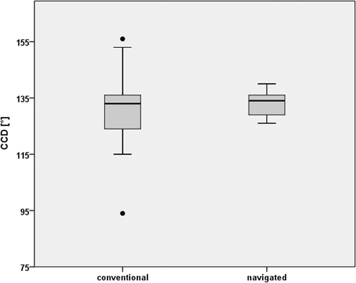
Figure 5. AV angle in conventional and navigated implantation for all surgeons. The box is defined by the upper and lower quartiles and the whiskers contain 95% of the observations. The dark line in the box is the median; data represented by black dots are outliers.
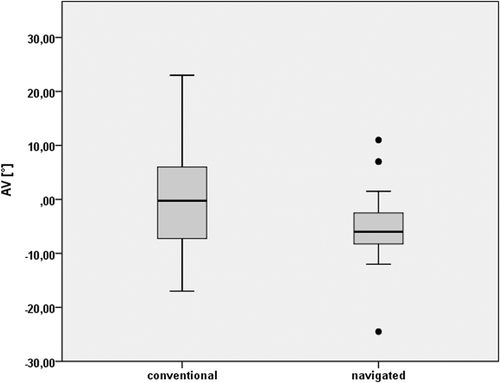
With the conventional method, the three surgeons showed significant differences with regard to operation time, chosen size of the femoral component, AV angle and anterior offset. With navigation these four parameters were comparable (see for results). However, the CCD and AV box plots showed narrower variances for all surgeons for the CCD and AV in navigation ( and ).
Figure 6. CCD angle for the three different surgeons with conventional and navigated implantation. The box is defined by the upper and lower quartiles and the whiskers contain 95% of the observations. The dark line in the box is the median.
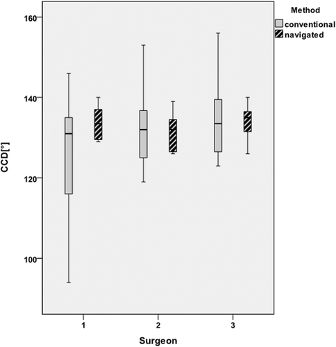
Figure 7. AV angle for the three different surgeons with conventional and navigated implantation. The box is defined by the upper and lower quartiles and the whiskers contain 95% of the observations. The dark line in the box is the median; data represented by black dots are outliers.
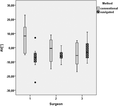
Table II. Mean results, standard deviation, range and p-values for comparison among the three different surgeons for both methods. Significant differences are highlighted in bold.
Comparing the implantations in the three femurs with abnormal anatomy to those in femurs with normal anatomy, no significant differences were found when using the conventional or navigated method, except for the AV angle in femurs with normal anatomy. The results are presented in .
Table III. Mean results, standard deviation, range and p-values for comparison between abnormal and abnormal anatomy for both methods. Significant differences are highlighted in bold.
No significant difference was found between the CCD and AV angle on the navigation screen (134° [SD 1.9°, range 129° to 137°] for the CCD; −1.5° [SD 8.7°, range −20° to 8°] for the AV angle) and the measured angle on the radiographs (133° [SD 4.5°, range 126° to 140°] for the CCD; −4.9° [SD 7.2, range −2.4° to 11°] for the AV angle). This was also true for all three surgeons. The planned CCD angle was 135° (SD 0.4°, range 135° to 137°) and the planned AV angle was 1.9° (SD 9.1°, range −23° to 9°). Comparing the planned angle and the measured angle on the radiographs revealed a significant difference for the CCD angle (p = 0.028) but not for the AV angle (p = 0.215).
Discussion
Hip resurfacing is a demanding operation, especially with regard to the positioning of the femoral component. The exact size and alignment of the component must be chosen precisely in order to avoid notching or impingement. Above all, malpositioning with regard to varus/valgus alignment can lead to serious complications, such as femoral neck fracture. The requirements for a navigation system therefore include the ability to provide accurate placement of the femoral component as well as reliable measurements compared to measurements obtained from X-ray or CT images. Also, the technique should be easy to apply and reproducible for different surgeons.
In our study we used different sets of sawbones and compared conventional to navigated implantation of the femoral component among three different surgeons. Although we performed the implantation in a hip simulator, this does not, of course, mirror exactly the situation of operating on a real patient. The longer operation time for the navigated procedures might be reduced to some extent as it becomes more routine. However, there are system-inherent processes like landmark registration and insertion of trackers which will always require a certain amount of time.
Some studies have reported more accurate placement of the femoral component, mainly with respect to varus/valgus alignment, by using navigation in sawbones Citation[18], cadavers Citation[13–16], Citation[19] or patients Citation[20–24]. However, other investigators have found no improvement in positioning Citation[25], Citation[26]. It has also been shown that measurements provided by the navigation system are consistent with those obtained from postoperative radiographs Citation[19], Citation[20], Citation[25] or CT images Citation[27], Citation[28]. Our study did not show a significant difference in femoral component positioning between conventional and navigated implantation. It did, however, show an obviously narrower box plot for navigated implantation with respect to the CCD and AV angle. This result was consistent for all three surgeons and also for the abnormal femurs. Moreover, observed differences between the three surgeons during conventional implantation with regard to the size of component, AV angle and anterior offset diminished with the use of navigated implantation. Thus, we conclude that the navigation system can reduce outliers in positioning the femoral component, even with different surgeons and abnormal femurs.
Only a few authors have tested navigation for hip resurfacing with abnormal anatomy. In computer simulations a high degree of accuracy was achieved, even for abnormal anatomy Citation[29–31], and this could also be confirmed in patients Citation[23], although the number of patients with abnormal anatomy in the latter study was very low. On the other hand, it was also shown with computer models that the accuracy of navigation decreased in pistol grip deformity Citation[30]. Whether navigation can help specifically with abnormal femurs cannot be definitely answered from our results alone. The number of abnormal femurs is certainly too small to show any significant results, but we could observe that, as in the normal femurs, outliers were reduced with navigation, and this was also found for all participating surgeons.
In contrast to standard total hip replacement, the offset between the femoral head and neck in hip resurfacing is often preserved. However, patients with underlying abnormal offset as in pistol grip deformity are often candidates for hip resurfacing, and it is particularly important in these patients to identify the correct femoral neck axis in order to position the femoral component correctly with regard to posterior and anterior offset. If it is placed too far posterior, the already low anterior offset will be further reduced, and this can result in anterior impingement Citation[32]. Optimal placement and correct size of the component are important to reduce impingement and maximize range of motion Citation[7], Citation[8]. Offset parameters and the size of the component were significantly different among the three surgeons when using conventional implantation, but this difference diminished with the use of navigated implantation. Currently, navigation does not provide direct information for offset values, but the information regarding the CCD, AV and component size help to place the component in a correct position. Nevertheless, information concerning offset would probably further enhance the outcome for navigation.
Measurements were consistent between the navigation system and the postoperative measurements on radiographs. Since we also ruled out a bias in the measurements by comparing four different investigators, navigation appeared to give accurate information concerning measurements. We found a difference between the planned and postoperatively measured CCD angles, but this can be explained by the fact that sometimes the planned angle was adjusted intraoperatively. The accurate information provided by the navigation system is better reflected by comparing the definite angle on the navigation screen and the postoperatively measured angle.
Overall, our results are similar to those achieved in testing navigation in knee arthroplasty and in acetabular component placement in hip arthroplasty. Many investigators comparing conventional and navigated implantation in total knee replacement or acetabular component placement in total hip arthroplasty found no significant improvement by comparing means for different parameters, but could demonstrate a reduction in the number of outliers Citation[9–12], Citation[33], Citation[34]. This can, of course, significantly reduce complications, especially in hip resurfacing. In decreasing the variance of the CCD and AV angles of the femoral component, the risk of notching, impingement and femoral neck fracture can be minimized. In our study the navigation system proved to be accurate and the results were reproducible for different surgeons. These results have to be confirmed in future clinical studies.
Acknowledgements
We thank Prof. Dr. Robert L. Snipes for reviewing the manuscript as a native English speaker and Dennis Huber for statistical help.
Declaration of interest: The authors report no declarations of interest.
References
- Mont MA, Seyler TM, Ulrich SD, Beaulé PE, Boyd HS, Grecula MJ, Goldberg VM, Kennedy WR, Marker DR, Schmalzried TP, et al. Effect of changing indications and techniques on total hip resurfacing. Clin Orthop Relat Res 2007; 465: 63–70
- Shimmin AJ, Back D. Femoral neck fractures following Birmingham hip resurfacing: A national review of 50 cases. J Bone Joint Surg Br 2005; 87(4)463–464
- Beaulé PE, Lee JL, Le Duff MJ, Amstutz HC, Ebramzadeh E. Orientation of the femoral component in surface arthroplasty of the hip. A biomechanical and clinical analysis. J Bone Joint Surg Am 2004; 86-A(9)2015–2021
- Beaulé PE, Dorey FJ, Le Duff M, Gruen T, Amstutz HC. Risk factors affecting outcome of metal-on-metal surface arthroplasty of the hip. Clin Orthop Relat Res 2004; 418: 87–93
- Jolley MN, Salvati EA, Brown GC. Early results and complications of surface replacement of the hip. J Bone Joint Surg Am 1982; 64(3)366–377
- Beaulé PE, Campbell PA, Hoke R, Dorey F. Notching of the femoral neck during resurfacing arthroplasty of the hip: A vascular study. J Bone Joint Surg Br 2006; 88(1)35–39
- Beaulé PE, Harvey N, Zaragoza E, Le Duff MJ, Dorey FJ. The femoral head/neck offset and hip resurfacing. J Bone Joint Surg Br 2007; 89(1)9–15
- Beaulé PE, Antoniades J. Patient selection and surgical technique for surface arthroplasty of the hip. Orthop Clin North Am 2005; 36(2)177–185, viii-ix
- Ecker TM, Tannast M, Murphy SB. Computed tomography-based surgical navigation for hip arthroplasty. Clin Orthop Relat Res 2007; 465: 100–105
- Nogler M, Kessler O, Prassl A, Donnelly B, Streicher R, Sledge JB, Krismer M. Reduced variability of acetabular cup positioning with use of an imageless navigation system. Clin Orthop Relat Res 2004; 426: 159–163
- Dorr LD, Malik A, Wan Z, Long WT, Harris M. Precision and bias of imageless computer navigation and surgeon estimates for acetabular component position. Clin Orthop Relat Res 2007; 465: 92–99
- Gandhi R, Marchie A, Farrokhyar F, Mahomed N. Computer navigation in total hip replacement: A meta-analysis. Int Orthop 2009; 33(3)593–597
- Davis ET, Gallie P, Macgroarty K, Waddell JP, Schemitsch E. The accuracy of image-free computer navigation in the placement of the femoral component of the Birmingham Hip Resurfacing: A cadaver study. J Bone Joint Surg Br 2007; 89(4)557–560
- Hodgson A, Helmy N, Masri BA, Greidanus NV, Inkpen KB, Duncan CP, Garbuz DS, Anglin C. Comparative repeatability of guide-pin axis positioning in computer-assisted and manual femoral head resurfacing arthroplasty. Proc Inst Mech Eng H 2007; 221(7)713–724
- Hodgson AJ, Inkpen KB, Shekhman M, Anglin C, Tonetti J, Masri BA, Duncan CP, Garbuz DS, Greidanus NV. Computer-assisted femoral head resurfacing. Comput Aided Surg 2005; 10(5–6)337–343
- Belei P, Skwara A, De La Fuente M, Schkommodau E, Fuchs S, Wirtz DC, Kämper C, Radermacher K. Fluoroscopic navigation system for hip surface replacement. Comput Aided Surg 2007; 12(3)160–167
- Wihan S. [Development of a surgical simulator for hip resurfacing by comparing datas at cadavers] [Degree dissertation] [In German]. Medical University of Innsbruck, 2009.
- Cobb JP, Kannan V, Brust K, Thevendran G. Navigation reduces the learning curve in resurfacing total hip arthroplasty. Clin Orthop Relat Res 2007; 463: 90–97
- Schnurr C, Nessler J, Meyer C, Schild HH, Koebke J, König DP. How accurate is image-free computer navigation for hip resurfacing arthroplasty? An anatomical investigation. J Orthop Sci 2009; 14(5)497–504
- Ganapathi M, Vendittoli PA, Lavigne M, Gunther KP. Femoral component positioning in hip resurfacing with and without navigation. Clin Orthop Relat Res 2009; 467(5)1341–1347
- Olsen M, Davis ET, Waddell JP, Schemitsch EH. Imageless computer navigation for placement of the femoral component in resurfacing arthroplasty of the hip. J Bone Joint Surg Br 2009; 91(3)310–315
- Romanowski JR, Swank ML. Imageless navigation in hip resurfacing: Avoiding component malposition during the surgeon learning curve. J Bone Joint Surg Am 2008; 90(Suppl 3)65–70
- Olsen M, Schemitsch EH. Computer navigated hip resurfacing for patients with abnormal femoral anatomy. Bull NYU Hosp Jt Dis 2009; 67(2)159–163
- Resubal JR, Morgan DA. Computer-assisted vs. conventional mechanical jig technique in hip resurfacing arthroplasty. J Arthroplasty 2009; 24(3)341–350
- Kruger S, Zambelli PY, Leyvraz PF, Jolles BM. Computer-assisted placement technique in hip resurfacing arthroplasty: Improvement in accuracy?. Int Orthop 2009; 33(1)27–33
- Shields JS, Seyler TM, Maguire C, Jinnah RH. Computer-assisted navigation in hip resurfacing arthroplasty – a single-surgeon experience. Bull NYU Hosp Jt Dis 2009; 67(2)164–167
- Bailey C, Gul R, Falworth M, Zadow S, Oakeshott R. Component alignment in hip resurfacing using computer navigation. Clin Orthop Relat Res 2009; 467(4)917–922
- Barrett AR, Davies BL, Gomes MP, Harris SJ, Henckel J, Jakopec M, Kannan V, Rodriguez y Baena FM, Cobb JP. Computer-assisted hip resurfacing surgery using the acrobot navigation system. Proc Inst Mech Eng H 2007; 221(7)773–785
- Cobb JP, Kannan V, Dandachli W, Iranpour F, Brust KU, Hart AJ. Learning how to resurface cam-type femoral heads with acceptable accuracy and precision: The role of computed tomography-based navigation. J Bone Joint Surg Am 2008; 90(Suppl 3)57–64
- Pitto RP, Malak S, Anderson IA. Accuracy of computer-assisted navigation for femoral head resurfacing decreases in hips with abnormal anatomy. Clin Orthop Relat Res 2009; 467(9)2310–2317
- Ma B, Bergeron SG, Grant HJ, Rudan J, Antoniou J. Quantifying degree of difficulty in hip-resurfacing of pistol-grip deformity. Bull NYU Hosp Jt Dis 2009; 67(2)154–158
- Schmalzried TP, Silva M, de la Rosa MA, Choi ES, Fowble VA. Optimizing patient selection and outcomes with total hip resurfacing. Clin Orthop Relat Res 2005; 441: 200–204
- Nogler M, Mayr E, Krismer M, Thaler M. Reduced variability in cup positioning: The direct anterior surgical approach using navigation. Acta Orthop 2008; 79(6)789–793
- Haaker RG, Tiedjen K, Ottersbach A, Rubenthaler F, Stockheim M, Stiehl JB. Comparison of conventional versus computer-navigated acetabular component insertion. J Arthroplasty 2007; 22(2)151–159

