
Abstract
Osteoarthritis (OA) is the most common chronic disease of our joints, manifested by a dynamically increasing degeneration of hyaline articular cartilage (AC). While currently no therapy can reverse this process, the few available treatment options are hampered by the inability of early diagnosis. Loss of cartilage surface, or extracellular matrix (ECM), integrity is considered the earliest sign of OA. Despite the increasing number of imaging modalities surprisingly few imaging biomarkers exist. In this narrative review, recent developments in optical coherence tomography are critically evaluated for their potential to assess different aspects of AC quality as biomarkers of OA. Special attention is paid to imaging surface irregularities, ECM organization and the evaluation of posttraumatic injuries by light-based modalities.
Introduction
Osteoarthritis (OA) develops as a consequence of structurally and functionally compromised articular cartilage (AC). Such damage can result from a variety of causes; in this regard, loss of cartilage integrity due to “wear and tear” erosion (i.e. primary OA) during ageing may be distinguished from structural changes upon traumatic insult (i.e. secondary OA). A compromised surface integrity is considered the earliest sign of OA and current, therapeutic or surgical, treatment options are mainly hampered by our inability to diagnose this disease in its early stage. At present, few reliable biomarkers for OA exist, while modern imaging techniques potentially can fill this diagnostic gap. In this review, some key aspects of hyaline AC and its osteoarthritic deterioration are briefly introduced upfront, prior to introducing the biomarker classification. Next, imaging techniques to assess AC degeneration are briefly and critically evaluated. While the progress in optical coherence tomography (OCT) has recently been reviewed for classical clinical applications (Kim et al., Citation2015), we will specifically depict its current technical status quo to improve imaging of AC and discuss potential in vivo applications. Quantification of novel dry biomarkers, such as subtle irregularities of the articular surface, by state-of-the-art OCT imaging holds huge promises for future early detection of progressive osteoarthritic changes and the evaluation of therapeutic interventions alike. Recent technical advances may broaden the clinical application of dedicated OCT modalities to potentially contribute insights into extracellular matrix (ECM) remodelling and collagen metabolism that cannot be accomplish by other current imaging methodologies. OCT imaging of closely related orthopaedic tissues, such as the annulus fibrosus, are beyond our focus and the interested reader is referred elsewhere (Han et al., Citation2015).
Articular cartilage and osteoarthritis
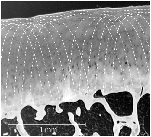
AC plays a vital role in providing a low-friction surface between the bones of articulating joints (Katta et al., Citation2008). Species-specific differences exist with respect to its tissue thickness (Malda et al., Citation2013). It is important to realize that, in major joints, human cartilage is up to 4 mm thick (Sophia Fox et al., Citation2009), which currently restricts full-depth OCT imaging in these joints. The unique properties of AC are largely derived from the ECM surrounding the chondrocytes (Buckwalter et al., Citation2005), providing both structure and function to this tissue (Aigner et al., Citation2006). AC has a characteristic zonal architecture, with a thin surface-near superficial (tangential) zone protecting the deeper layers from shear stresses. This zone, with tightly packed collagens (mainly type II and IX) aligned parallel to the surface, makes up approximately 10% to 20% of the tissue thickness (Sophia Fox et al., Citation2009). The sophisticated collagen network (depicted in ) is providing high tensile strength, while compressive stiffness is attributed to the interactions of osmotically attracted water (i.e. 95%vol of AC in humans) with the negatively charged network of highly sulphated proteoglycan (PG) in the ground substance (Sophia Fox et al., Citation2009).
Figure 1. Role of collagen network in articular cartilage. Artistic impression of Benninghoff’s “arcade model” of collagen fibre organization in human cartilage (Benninghoff, Citation1925), indicated as dashed white lines originating from the calcified zone (bottom), drawn over solarized micrograph. Hyaline cartilage appears greyish, with embedded chondrocytes (black), underlying subchondral bone white and bone marrow dark.
OA is one of the most important chronic health issues in humans and currently affecting about 35% of adults above age 65. During this progressive age-related disease, cartilage is degraded in response to joint biomechanics, long-term low-grade inflammation or a traumatic insult (Goldring & Goldring, Citation2006). OA is projected to cause 3.5 million primary knee replacement procedures by 2030 (Neogi, Citation2013; Neogi & Zhang, Citation2013). Early diagnosis and treatment are, therefore, clinically highly relevant. Despite its devastating impact, the development of effective tools for early diagnosis and disease-modifying therapeutics is hampered by our incomplete understanding of especially the early pathogenesis of OA. Sadly, in humans, currently no therapy can reverse joint damage sustained during OA progression and drug development is largely hampered by our inability to diagnose OA early enough for successful intervention (Chu et al., Citation2012). The gradual destruction of the ECM (Sandell & Aigner, Citation2001) has long been considered the hallmark of OA (Malfait et al., Citation2002; Stanton et al., Citation2005). Once the PGs are lost, mechanical stresses are concentrated on the collagen network, making it more susceptible to enzymatic degradation and turning the process irreversible to initiate a vicious circle progressing into terminal OA (Little & Fosang, Citation2010).
The past decade of OA research has faced two major challenges: (i) identifying and developing quantifiable and measurable diagnostic biomarkers and (ii) developing efficacious disease modifying osteoarthritis drug (DMOAD) to slow down or even halt cartilage degeneration (Dvir-Ginzberg & Reich, Citation2014). Of the characteristic sequence of distinctive morphological and functional changes during the course of OA, the superficial cartilage layer is affected first. Here, PG depletion and alterations in collagen orientation, content and integrity occur (Panula et al., Citation1998; Pritzker et al., Citation2006). Hence, earliest changes include cartilage surface irregularities, erosion and fissuring. With preventive strategies available to modify joint biomechanics by surgical interventions or to improve tissue resilience by pharmaceutical agents, earliest possible detection of degeneration is crucial as the pathology may be reversible at this point (Bay-Jensen et al., Citation2010).
Biomarkers of osteoarthritis
A biomarker is classically defined as a characteristic that is objectively measured and evaluated as an indicator of normal biological processes, pathogenic processes or pharmacological responses to a therapeutic intervention (Biomarkers Definitions Working Group, Citation2001). A recent increase of post-genomic technologies has resulted in a rapid growth and progress in OA biomarker research (Henrotin, Citation2012), mainly focusing on wet biomarker identification that has historically been relatively unsatisfactory – even in combination with proteomics and metabolomics (Zolg, Citation2006). The Osteoarthritis Research Society International (OARSI)/US Food and Drug Administration (FDA) OA biomarkers working group divided biomarkers into (i) so-called soluble or “wet” biomarkers, as measured in body fluids like e.g. blood, serum, plasma, urine or synovial fluid (SF) and (ii) so-called “dry” biomarkers usually consisting of visual analogue scales, performed tasks or imaging (Kraus et al., Citation2010). While the former, currently comprising the majority of biomarkers such as proteins and metabolites, have been comprehensively reviewed (Attur et al., Citation2013; Kraus et al., Citation2015; Lotz et al., Citation2013), we will now review the potential of selected imaging techniques to contribute novel, clinically relevant, dry biomarkers.
Cartilage imaging techniques
AC has been subjected to novel non-contact optical techniques almost since polarized light microscopy (PLM) was developed. Early PLM studies were the main evidential basis for quantitative models of the AC collagen structure by Benninghoff (Citation1925). State-of-the-art optical techniques like quantitative PLM (qPLM), second harmonic generation (SHG) microscopy, Fourier-transform infrared (FTIR) microscopy, Raman or optical hyperspectral reflectance and fluorescence imaging are still providing new insights into AC structure at nano- to mesoscale. A recent review elegantly addresses diverse aspects of modern biophotonic techniques in this perspective (Matcher, Citation2015). Progress in related imaging techniques using either magnetic (i.e. magnetic resonance imaging or MRI) or radiographic (i.e. X-ray micro-tomography or micro-computed tomography or micro-CT) spectra are also beyond the scope of the present review. While MRI holds potential to detect pre-radiographic OA (Sharma et al., 2014), routinely used clinical X-ray or morphological MRI either have low resolution, inter-observer reliability or sensitivity/specificity (Krampla et al., Citation2009) and thus may have limited potential for biomarker discovery. Another interesting emerging technology to characterize AC is diffusion tensor imaging (DTI), which may be used as a biomarker for cartilage composition and structure. DTI is sensitive to PG content and thus holds a lot of diagnostic potential, but its in vivo acquisition in AC is challenging due to the short T2 of this tissue (approx. 40 ms at 3 Tesla) and the high resolution required. However, promising protocols have recently been reviewed (Raya, Citation2015). In this review, we will focus on the clinically applicable optical spectrum and recent developments from trends in 2D and 3D OCT data acquisition to insights from polarization-sensitive OCT (PS-OCT) and their respective diagnostic value.
Imaging osteoarthritic changes
Based on the principle of low coherence interferometry, OCT detects echo time delays and intensities of backscattered near-infrared light. Modern OCT systems can relatively noninvasively image cartilage tissue at micrometre resolution and to millimetre depths, resembling an “optical biopsy” (Tearney et al., Citation1997), similar to low power histology but without requiring biopsy. Similarly to other optical methods, the imaging depth and spatial resolution of OCT are limited by strong light scattering of biological tissues and presence of chromophores absorbing the light (Petrov et al., Citation2012). OCT is a volumetric imaging tool similar conceptually to ultrasound (US) but using light instead of acoustic waves. OCT thus differs in two aspects from clinical US: it is characterized by a higher spatial resolution (i.e. 2–10 μm as compared to about 100–1000 μm) and a smaller imaging depth (i.e. 1–2 mm versus 10–100 mm), respectively. Its penetration depth is thus sufficient to image through to the subchondral bone interface in smaller human joints, while it usually fails to penetrate the thick cartilage of e.g. human femoral condyles in the knee. Interestingly, OCT is apparently able to image cartilage in all relevant animal models of OA where, from mouse to horse, cartilage of the femoral condyles is on average thinner than 1.5 mm (Malda et al., Citation2013). Especially in smaller animal models of OA, OCT imaging was successfully used to directly measure cartilage thinning in response to e.g. chemically induced OA. Patel and co-workers were among the first to image cartilage deterioration and disruption of the bone–cartilage interface in rat knees at 1300 nm (Patel et al., Citation2005).
Herrmann et al. performed the earliest OCT studies with human cartilage: 100 human specimens from different joints of 10 individuals were analysed ex vivo (Herrmann et al., Citation1999). In 1999, their OCT imaging at between 5 and 15 microns axial resolution provided much more information than other non-destructive modalities such as radiography or MRI. Physical penetration into the joint-space, as during visible arthroscopy, was still necessary though, but e.g. fibrosis and surface fibrillations were readily detected. OCT then evolved as an attractive high resolution imaging technology to assess osteoarthritically altered AC microstructures. Interestingly, Brezinski and co-workers already envisioned 15 years ago the ability to integrate OCT technology into small portable arthroscopes that could operate at relatively low costs. Since then, the diagnostic value of OCT-based imaging was demonstrated in vitro and in vivo and in the context of open and arthroscopic knee surgery (Chu et al., Citation2004; Li et al., Citation2005; Xie et al., Citation2006).
OCT yields microscopic cross-sectional images of cartilage in real time and at high, near-histological, resolution. Still, yet, most OCT data are two-dimensional, while 3D-based appreciation of cartilage lesions is clearly desirable. From a clinical point of view, quantitative OCT measurements seem feasible since OCT probes have been miniaturized and applied in patients during arthroscopy or open knee surgery (Saarakkala et al., Citation2009). OCT could furthermore distinguish between native and regenerated equine AC and provided an accurate measurement of cartilage surface roughness ex vivo (Viren et al., Citation2012). Cartilage degeneration is not uniform across the joint and multiple single 2D OCT scans are needed to appreciate the full spectrum of degeneration in a particular joint compartment. Using human cartilage, a diagnostic superiority of real time 3D OCT over conventional 2D OCT was confirmed (Nebelung et al., Citation2015) after showing that OCT was effective in assessing cartilage surface, integrity and homogeneity and to discriminate between unmineralized and mineralized cartilage, respectively (Nebelung et al., Citation2014). Therefore, quantitative OCT holds great potential as a diagnostic tool for more reliable, standardized and objective assessment of cartilage tissue properties.
Imaging structural changes
OCT usually cannot penetrate through the full thickness of cartilage at the clinically important human femoral condyles. Therefore, recent studies focused on the cartilage surface as loss of AC surface integrity is considered the earliest sign of OA, but its reliable detection has not been established by clinical routine diagnostics. Clinical arthroscopic OCT imaging was, however, evaluated as an adjunct tool to improve the detection of surface fibrillation, cracks and fissures on human cadaver knees in situ (Chu et al., Citation2004). Using a rigid arthroscope beam delivery system, generating cross-sectional B-scan images, the authors could image condyles and trochlea in intact joints, but not the tibia plateau. Superiority of OCT over routine arthroscopy was demonstrated by improved detection of subtle surface fibrillation in macroscopically otherwise “normal” areas, as confirmed by histology. OCT-based surface roughness measurements of cartilage was also reported by Saarakkala et al. (Citation2009), showing that the roughness of “healthy” AC ranges within 4–10 μm, while OA samples have a much higher roughness of up to 40 μm. These approaches have in common that determination of surface roughness may be prone to inaccuracy when it comes to the definition of the idealized (i.e. mean) smoothed surface that is used as the reference to determine the roughness. In these studies, the idealized debrided smooth cartilage surface serving as the reference line for subsequent roughness detection was either manually drawn (Chu et al., Citation2004), a semi-automatically determined auxiliary line obtained by manual surface point definition and subsequent interpolation (Cernohorsky et al., Citation2015) or determined by averaged surface positions (Huang et al., Citation2011a; Viren et al., Citation2012). In the light of clinical needs, these approaches seem impractical as they require, potentially variable, user input. Brill et al. therefore used 105 human cartilage samples with variable degrees of degeneration to comprehensively assess the validity of a novel algorithm-based cartilage surface roughness determination approach based on 2D OCT parameters. The authors only included osteoarthritic specimens, but correlated OCT-based roughness parameters to routine clinical Outerbridge (Citation1961) grades. While the majority of their parameters revealed a close-to-linear correlation with cartilage degeneration, cartilage surface integrity should be best assessed by using combined parameters to improve the current accuracy of diagnostics (Brill et al., Citation2015). Such methods may prove especially valuable for OCT-based evaluation of femoral condyles, where AC is characterized by considerable curvature (Terukina et al., Citation2003) which makes surface position averaging challenging. Non-perpendicular optical beam angles are further known to bias the evaluation of cartilage surface degeneration by quantitative OCT (Huang et al., Citation2011b). In a feasibility study, Nebelung et al. performed a comprehensive OCT-based morphometric grading study on human AC representing the full spectrum of arthritic degeneration, ranging from Outerbridge 0 to 4, from macroscopically “normal” to fully eroded cartilage, respectively (Nebelung et al., Citation2014). Although, with appropriate care a good correlation between OCT-based surface assessment and histological grading seems possible (Nebelung et al., Citation2014), standardized OCT-based surface evaluation remains clinically challenging and awaits reliable standards. OCT certainly holds potential as a diagnostic tool for more reliably assessing cartilage tissue properties in a better standardized, objective way (Nebelung et al., Citation2014).
Interestingly, Cernohorsky and colleagues used a 0.9 mm diameter flexible probe, designed for intravascular OCT imaging, to image surface fibrillation and fissuring within a carpometacarpal human cadaver joint to directly determine cartilage thickness (Cernohorsky et al., Citation2012) and demonstrated practical feasibility of a minimal invasive approach. The same probe was also used in equine metacarpophalangeal joints (te Moller et al., Citation2013) in combination with a Dragonfly intravascular OCT catheter (Terashima et al., Citation2012) to reach high-quality images. Overall, OCT allowed a less subjective scoring of cartilage lesions and seems beneficial to select most appropriate treatment and monitoring therapeutic response.
Posttraumatic OA constitutes a major cause of disability in our increasingly elderly population and people at risk have to be identified as soon as possible after a corresponding traumatic insult. By modulating the polarization state of the incident light in correlation to the OCT image, Patel and co-workers concluded that monosodium acetate-induced OA in rats eliminated tissue birefringence, assumingly by destroying directional collagen fibre alignment. Of note, OCT can infer optical properties, such as the near-infrared backscatter and extinction coefficients (Schmitt et al., Citation1993) and this correlates with degeneration and impact loading (Patel et al., Citation2005). Impact injury consistently raised the surface signal intensity and decreased the deep OCT signal intensity in an ex vivo bovine indentation model of tibial cartilage damage (Bear et al., Citation2010). This change in optical properties might have occurred from chondrocyte death and surface collagen matrix disruption as evident from histology. Shyu and colleagues used a more quantitative approach (Shyu et al., Citation2009) by measuring the full OCT image brightness profile versus depth. Comparing this to a theoretical extended Huygens-Fresnel model (Thrane et al., Citation2000), damaged porcine cartilage showed a tendency to produce larger attenuation coefficients than normal cartilage, with a trend towards more isotropic scattering in damaged cartilage (Matcher, Citation2015). While 2D OCT, representing only a snapshot of the tissue, easily misses important structural abnormalities (), 3D OCT gives a good volumetric representation of the tissue integrity. Very recently, the suitability of OCT to evaluate single impact-induced cartilage degeneration was confirmed by de Bont and colleagues in an ex vivo model using 34 macroscopically normal human osteochondral specimen. Standardized single impacts, ranging from 0.25 J to 0.98 J, were evaluated by 3D OCT prior to and directly after impaction as well as 1, 4 and 8 d later. The authors concluded that OCT-based parameterization and quantification is able to reliably detect loss of cartilage surface integrity after high-energy traumatic insults and holds potential to be used for clinical screening of early OA (de Bont et al., Citation2015). Thus, while sensitive screening of the roughness of the cartilage surface by OCT may be employed to evaluate the age-related progression of wear-and-tear erosion of the tissue, OCT may further aid in assessing the patient’s risks to develop posttraumatic OA.
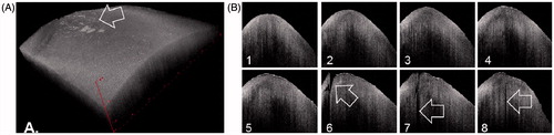
Figure 2. OCT-based articular surface evaluation. Topographical reconstruction of human articular cartilage ex vivo (A). The volumetric dataset to reconstruct this 8 × 8 mm macroscopically only slightly degraded, Outerbridge grade 1, specimen from the medial femoral condyle in 3D consists of 100 adjacent 2D OCT images. Note the essentially smooth articular surface around a focal lesion (arrow). Individual, in silico sliced 2D cross-sectional OCT images from this dataset representing 1 mm intervals (B, front-to-back). Smooth surfaces of early sections (B1 through B5) matching the first half of the in silico reconstructed tissue (A). Large, surfacing clefts (arrows; B6, B7) and a smaller sub-surface cleft (B8) corresponding to (peri-)lesional cartilage damage in A.
Imaging macromolecular deterioration by PS-OCT
Unlike US, light waves are transverse and can carry polarimetric information, which combined with OCT resulted in “polarization-sensitive” OCT (PS-OCT) (de Boer & Milner, Citation2002). Herrmann et al. also early investigated the correlation between changes observed by OCT and the degree of collagen organization in OA cartilage by polarization sensitive OCT (PS-OCT) to assess changes in cartilage collagen organization in vitro. While frequently cartilage appeared “normal” by routine staining, showing cartilage thickness >2 mm and no fibrillations, abnormalities were evident in these specimen by both PS-OCT and Picrosirius red birefringence polarization microscopy (Drexler et al., Citation2001). PS-OCT thus enables detection of areas of altered birefringence in human cartilage (Ugryumova et al., Citation2005). Enhanced birefringence is usually associated with (inappropriate) tissue repair, during which type II collagen of degenerated hyaline cartilage is replaced by type I collagen fibres to result in so-called fibrocartilage. The latter is characterized by thicker and denser collagen fibres, which therefore appear brighter on structural OCT images. Li and co-workers employed polarized light OCT imaging to study human knee cartilage in vivo prior to partial, or total, replacement prior to correlating PS-OCT images to histology (Li et al., Citation2005). While modulation of the incident polarization state of the light due to birefringence from organized, mainly type II, collagen fibres revealed a regular depth-resolved banding pattern with “normal” cartilage, the pattern was lost upon progressive cartilage degeneration. Later, Chu et al. investigated the use of PS-OCT birefringence to grade femoral condyle and trochlea osteoarthritic lesions in human cadaver knees (Chu et al., Citation2007). These authors interpreted the loss of ex vivo birefringence as an early stage of chondrocyte viability loss, based on their acquired (reversible) irresponsiveness to IGF-1. Absence of birefringence might thus be a potential biomarker of early stage cartilage degeneration – at a point where the disease pharmacologically may still be reversible. Excitingly, the Matcher Lab recently introduced two other PS-OCT variants, termed variable-incidence-angle PS-OCT (Kasaragod et al., Citation2012; Ugryumova et al., Citation2006,Citation2009) and conical-scan PS-OCT (Lu et al., Citation2014), respectively, to specifically elucidate the zonal architecture of the collagen fibre network. The apparent birefringence of a specimen is determined not only by the directional organization or abundance of collagen fibres, but also by their axial orientation relative to the direction of the incident light (Ugryumova et al., Citation2005; Xie et al., Citation2006). Birefringence is thus exterminated when both directions are either parallel or anti-parallel, or is maximal at their orthogonal orientation. While both techniques hold tremendous potential for OA biomarker development, handling issues may currently obstruct their direct clinical application.
PS-OCT may, however, not only proof valuable during monitoring OA, but also to improve (osteo-) chondral repair and improve tissue regeneration: some data imply that collagen fibres are “brushed” in a particular direction (Matcher, Citation2015), which is a structural parameter of cartilage that received little attention. In contrast, the split-line direction has been extensively mapped across a variety of species and sites: cartilage has a markedly higher tensile Young’s modulus when loaded in a direction aligned with the split-line versus orthogonal to it (Matcher, Citation2015). Strikingly, Matcher now speculates that the brushing direction might reflect the magnitude and direction of the dominant shear-stress experienced at a particular site during locomotion. It would be interesting to see if matching the brushing direction might provide beneficial osteochondral grafting, like matching the split-line directions of graft and surrounding host tissue at the defect site. To this end, PS-OCT may prove itself a valuable adjunct tool during orthopaedic routine osteochondral autograft transfer.
Earlier studies using cartilage samples from large animals (Ugryumova et al., Citation2005; Xie et al., Citation2006) reported a pronounced variation in apparent birefringence with beam orientation. As the beam must be inclined by a substantial angle to reveal the underlying birefringence, controlled PS-OCT seems challenging during routine clinical applications. Interestingly, in contrast, healthy human knee cartilage appears to have a strong apparent birefringence when the beam is applied at normal incidence (i.e. perpendicular to joint surface) (Chu et al., Citation2007; Li et al., Citation2005), which may point towards species-specific differences for this application. This notion is supported by observations by Rieppo et al., suggesting that the much lower contact pressure in the human knee, as compared to equine or bovine joints, favours a collagen architecture with a physically much thicker superficial zone (Rieppo et al., Citation2009), which would explain this apparent difference. Even within the same species, PS-OCT techniques may thus either detect a loss of birefringence as a result of truly disorganized (i.e. degraded) collagen fibre networks as a result of arthritic tissue deterioration or stress-induced remodelling or debridement (Matcher, Citation2015). This makes controlled PS-OCT currently challenging for routine clinical evaluation of cartilage quality.
Summary and discussion
Most human cartilage available for research is obtained at the time of joint replacement, when OA lesions are end stage and little can be concluded about the factors that played a role in disease development. To overcome this limitation, numerous induced and spontaneous animal models have been utilized to study disease onset and progression, as well as to test novel therapeutic interventions. A recent excellent review by Moon and Beier focuses on our latest insights into OA pathogenesis from mice (Moon & Beier, Citation2015). While only limited conclusions can be drawn from mice, no single “gold standard” animal model of OA exists that accurately reflects all aspects of the human disease – with several key variations in OA pathology among species (McCoy, Citation2015). Most of our knowledge about the onset of OA comes from animal models. Its limited penetration depth makes OCT ideally suited for full depth imaging of cartilage in animal joints, rather than in clinically relevant major human joints. Thus, the challenge now lies in applying the rapidly improving OCT technologies to relevant pre-clinical models of OA induction to evaluate its feasibility and identify potential dry biomarkers of early disease development and/or progression prior to reliably applying this novelty in clinical praxis.
From the currently available data, it appears likely that future diagnostic biomarkers (i.e. markers confirming the existence of a certain degree of pathological alteration) will not only be derived from the “wet” circuit, but will be complemented by “dry” markers derived from imaging technology. The majority of OA biomarkers will probably remain “wet” due to their ability to indicate metabolic responses rather than sole structural changes. A combination of cell- and ECM-derived biomarkers may be beneficial, especially to evaluate OA progression and treatment efficacy (Dvir-Ginzberg & Reich, Citation2014).
Nonetheless, OCT holds potential as future minimal invasive prognostic biomarker for OA (i.e. providing an indication of the likelihood of pathological progression) if especially PS-OCT can be better standardized with respect to its beam angle orientation. Despite some current shortcomings and technical challenges, supplementing conventional arthroscopies with OCT modalities potentially adds further value during assessing hyaline AC and different OCT modalities may thus shortly move from sole characterization to diagnosis. Detection of morphological alterations in collagen architecture could also improve arthroscopically guided interventions such as debridement and monitor the success of regenerative cell-based therapies such as autologous chondrocyte implantation. In animal models, regenerative potential of tissue engineered constructs may be monitored using OCT. While other imaging modalities may allow imaging of the joints even non-invasively, few can easily directly assess cartilage quality. To this end, birefringence may be exploited as a potential anabolic or catabolic biomarker, as detailed above. Accounting for collagen “brushing” and split-line direction may offer a more direct approach to improve current surgical treatments of (osteo-)chondral defects. The latter may prevent or at least delay posttraumatic OA.
OCT is currently facing important technical challenges with respect to normalization strategies and controlling light beam angles. At present, unequivocal image-based differentiation between healthy and early degenerative cartilage by OCT still seems challenging.
Acknowledgements
H.J. is grateful for the support by the Aachen Interdisciplinary Center for Clinical Research (IZKF) and H.J. and S.N. are both indebted to the START-Program of the Faculty of Medicine of the RWTH Aachen.
Declaration of interest
The authors report no declarations of interests. H.J. is a member of D-BOARD; this project has received funding from the European Union’s Seventh Framework Programme for research, technological development and demonstration under grant agreement No. 305815.
References
- Aigner T, Sachse A, Gebhard PM, Roach HI. (2006). Osteoarthritis: pathobiology-targets and ways for therapeutic intervention. Adv Drug Deliv Rev 58:128–49
- Attur M, Krasnokutsky-Samuels S, Samuels J, Abramson SB. (2013). Prognostic biomarkers in osteoarthritis. Curr Opin Rheumatol 25:136–44
- Bay-Jensen AC, Hoegh-Madsen S, Dam E, et al. (2010). Which elements are involved in reversible and irreversible cartilage degradation in osteoarthritis? Rheumatol Int 30:435–42.
- Bear DM, Szczodry M, Kramer S, et al. (2010). Optical coherence tomography detection of subclinical traumatic cartilage injury. J Orthop Trauma 24:577–82
- Benninghoff A. (1925). Der Aufbau des Gelenkknorpels in seinen Beziehungen zur Funktion. Zeitschrift für Zellforschung und mikroskopische Anatomie 2:783–862
- Biomarkers Definitions Working Group. (2001). Biomarkers and surrogate endpoints: preferred definitions and conceptual framework. Clin Pharmacol Ther 69:89–95
- Brill N, Riedel J, Rath B, et al. (2015). Optical coherence tomography-based parameterization and quantification of articular cartilage surface integrity. Biomed Opt Express 6:2398–411
- Buckwalter JA, Mankin HJ, Grodzinsky AJ. (2005). Articular cartilage and osteoarthritis. Instr Course Lect 54:465–80
- Cernohorsky P, de Bruin DM, van Herk M, et al. (2012). In-situ imaging of articular cartilage of the first carpometacarpal joint using co-registered optical coherence tomography and computed tomography. J Biomed Opt 17:060501
- Cernohorsky P, Kok AC, Bruin DM, et al. (2015). Comparison of optical coherence tomography and histopathology in quantitative assessment of goat talus articular cartilage. Acta Orthop 86:257–63
- Chu CR, Izzo NJ, Irrgang JJ, et al. (2007). Clinical diagnosis of potentially treatable early articular cartilage degeneration using optical coherence tomography. J Biomed Opt 12:051703
- Chu CR, Lin D, Geisler JL, et al. (2004). Arthroscopic microscopy of articular cartilage using optical coherence tomography. Am J Sports Med 32:699–709
- Chu CR, Williams AA, Coyle CH, Bowers ME. (2012). Early diagnosis to enable early treatment of pre-osteoarthritis. Arthritis Res Ther 14:212
- de Boer JF, Milner TE. (2002). Review of polarization sensitive optical coherence tomography and Stokes vector determination. J Biomed Opt 7:359–71
- de Bont F, Brill N, Schmitt R, et al. (2015). Evaluation of single-impact-induced cartilage degeneration by optical coherence tomography. Biomed Res Int 2015:486794
- Drexler W, Stamper D, Jesser C, et al. (2001). Correlation of collagen organization with polarization sensitive imaging of in vitro cartilage: implications for osteoarthritis. J Rheumatol 28:1311–18
- Dvir-Ginzberg M, Reich E. (2014). Chopping off the chondrocyte proteome. Biomarkers 1–7. [Epub ahead of print]. DOI:10.3109/1354750X.2014.955884
- Goldring SR, Goldring MB. (2006). Clinical aspects, pathology and pathophysiology of osteoarthritis. J Musculoskelet Neuronal Interact 6:376–8
- Han SK, Chen CW, Wierwille J, et al. (2015). Three dimensional mesoscale analysis of translamellar cross-bridge morphologies in the annulus fibrosus using optical coherence tomography. J Orthop Res 33:304–11
- Henrotin Y. (2012). Osteoarthritis year 2011 in review: biochemical markers of osteoarthritis: an overview of research and initiatives. Osteoarthritis Cartilage 20:215–17
- Herrmann JM, Pitris C, Bouma BE, et al. (1999). High resolution imaging of normal and osteoarthritic cartilage with optical coherence tomography. J Rheumatol 26:627–35
- Huang YP, Saarakkala S, Toyras J, et al. (2011a). Effects of optical beam angle on quantitative optical coherence tomography (OCT) in normal and surface degenerated bovine articular cartilage. Phys Med Biol 56:491–509
- Huang YP, Wang SZ, Saarakkala S, Zheng YP. (2011b). Quantification of stiffness change in degenerated articular cartilage using optical coherence tomography-based air-jet indentation. Connect Tissue Res 52:433–43
- Kasaragod DK, Lu Z, Jacobs J, Matcher SJ. (2012). Experimental validation of an extended Jones matrix calculus model to study the 3D structural orientation of the collagen fibers in articular cartilage using polarization-sensitive optical coherence tomography. Biomed Opt Express 3:378–87
- Katta J, Jin Z, Ingham E, Fisher J. (2008). Biotribology of articular cartilage – a review of the recent advances. Med Eng Phys 30:1349–63
- Kim J, Brown W, Maher JR, et al. (2015). Functional optical coherence tomography: principles and progress. Phys Med Biol 60:R211–37
- Krampla W, Roesel M, Svoboda K, et al. (2009). MRI of the knee: how do field strength and radiologist’s experience influence diagnostic accuracy and interobserver correlation in assessing chondral and meniscal lesions and the integrity of the anterior cruciate ligament? Eur Radiol 19:1519–28.
- Kraus VB, Blanco FJ, Englund M, et al. (2015). OARSI Clinical Trials Recommendations: soluble biomarker assessments in clinical trials in osteoarthritis. Osteoarthritis Cartilage 23:686–97
- Kraus VB, Huebner JL, Degroot J, Bendele A. (2010). The OARSI histopathology initiative – recommendations for histological assessments of osteoarthritis in the guinea pig. Osteoarthritis Cartilage 18:S35–52
- Li X, Martin S, Pitris C, et al. (2005). High-resolution optical coherence tomographic imaging of osteoarthritic cartilage during open knee surgery. Arthritis Res Ther 7:R318–23
- Little CB, Fosang AJ. (2010). Is cartilage matrix breakdown an appropriate therapeutic target in osteoarthritis – insights from studies of aggrecan and collagen proteolysis? Curr Drug Targets 11:561–75.
- Lotz M, Martel-Pelletier J, Christiansen C, et al. (2013). Value of biomarkers in osteoarthritis: current status and perspectives. Ann Rheum Dis 72:1756–63
- Lu Z, Kasaragod D, Matcher SJ. (2014). Conical scan polarization-sensitive optical coherence tomography. Biomed Opt Express 5:752–62
- Malda J, de Grauw JC, Benders KE, et al. (2013). Of mice, men and elephants: the relation between articular cartilage thickness and body mass. PLoS One 8:e57683
- Malfait AM, Liu RQ, Ijiri K, et al. (2002). Inhibition of ADAM-TS4 and ADAM-TS5 prevents aggrecan degradation in osteoarthritic cartilage. J Biol Chem 277:22201–8
- Matcher SJ. (2015). What can biophotonics tell us about the 3D microstructure of articular cartilage? Quant Imaging Med Surg 5:143–58.
- McCoy AM. (2015). Animal models of osteoarthritis: comparisons and key considerations. Vet Pathol 52:803–18
- Moon PM, Beier F. (2015). Novel insights into osteoarthritis joint pathology from studies in mice. Curr Rheumatol Rep 17:524
- Nebelung S, Brill N, Marx U, et al. (2015). Three-dimensional imaging and analysis of human cartilage degeneration using optical coherence tomography. J Orthop Res 33:651–9
- Nebelung S, Marx U, Brill N, et al. (2014). Morphometric grading of osteoarthritis by optical coherence tomography – an ex vivo study. J Orthop Res 32:1381–8
- Neogi T. (2013). The epidemiology and impact of pain in osteoarthritis. Osteoarthritis Cartilage 21:1145–53
- Neogi T, Zhang Y. (2013). Epidemiology of osteoarthritis. Rheum Dis Clin North Am 39:1–19
- Outerbridge RE. (1961). The etiology of chondromalacia patellae. J Bone Joint Surg Br 43-B:752–7
- Panula HE, Hyttinen MM, Arokoski JP, et al. (1998). Articular cartilage superficial zone collagen birefringence reduced and cartilage thickness increased before surface fibrillation in experimental osteoarthritis. Ann Rheum Dis 57:237–45
- Patel NA, Zoeller J, Stamper DL, et al. (2005). Monitoring osteoarthritis in the rat model using optical coherence tomography. IEEE Trans Med Imaging 24:155–9
- Petrov GI, Doronin A, Whelan HT, et al. (2012). Human tissue color as viewed in high dynamic range optical spectral transmission measurements. Biomed Opt Express 3:2154–61
- Pritzker KP, Gay S, Jimenez SA, et al. (2006). Osteoarthritis cartilage histopathology: grading and staging. Osteoarthritis Cartilage 14:13–29
- Raya JG. (2015). Techniques and applications of in vivo diffusion imaging of articular cartilage. J Magn Reson Imaging 41:1487–504
- Rieppo J, Hyttinen MM, Halmesmaki E, et al. (2009). Changes in spatial collagen content and collagen network architecture in porcine articular cartilage during growth and maturation. Osteoarthritis Cartilage 17:448–55
- Saarakkala S, Wang SZ, Huang YP, Zheng YP. (2009). Quantification of the optical surface reflection and surface roughness of articular cartilage using optical coherence tomography. Phys Med Biol 54:6837–52
- Sandell LJ, Aigner T. (2001). Articular cartilage and changes in arthritis. An introduction: cell biology of osteoarthritis. Arthritis Res 3:107–13
- Schmitt JM, Knuttel A, Bonner RF. (1993). Measurement of optical properties of biological tissues by low-coherence reflectometry. Appl Opt 32:6032–42
- Shyu J, Chan CH, Hsiung MW, et al. (2009). Diagnosis of articular cartilage damage by polarization sensitive optical coherence tomography and the extracted optical properties. Prog Electromagn Res 91:365–76
- Sophia Fox AJ, Bedi A, Rodeo SA. (2009). The basic science of articular cartilage: structure, composition, and function. Sports Health 1:461–8
- Stanton H, Rogerson FM, East CJ, et al. (2005). ADAMTS5 is the major aggrecanase in mouse cartilage in vivo and in vitro. Nature 434:648–52
- te Moller NC, Brommer H, Liukkonen J, et al. (2013). Arthroscopic optical coherence tomography provides detailed information on articular cartilage lesions in horses. Vet J 197:589–95
- Tearney GJ, Brezinski ME, Bouma BE, et al. (1997). In vivo endoscopic optical biopsy with optical coherence tomography. Science 276:2037–9
- Terashima M, Kaneda H, Suzuki T. (2012). The role of optical coherence tomography in coronary intervention. Korean J Intern Med 27:1–12
- Terukina M, Fujioka H, Yoshiya S, et al. (2003). Analysis of the thickness and curvature of articular cartilage of the femoral condyle. Arthroscopy 19:969–73
- Thrane L, Yura HT, Andersen PE. (2000). Analysis of optical coherence tomography systems based on the extended Huygens-Fresnel principle. J Opt Soc Am A Opt Image Sci Vis 17:484–90
- Ugryumova N, Attenburrow DP, Winlove CP, Matcher SJ. (2005). The collagen structure of equine articular cartilage, characterized using polarization-sensitive optical coherence tomography. J Phys D: Appl Phys 38:2612–19
- Ugryumova N, Gangnus SV, Matcher SJ. (2006). Three-dimensional optic axis determination using variable-incidence-angle polarization-optical coherence tomography. Opt Lett 31:2305–7
- Ugryumova N, Jacobs J, Bonesi M, Matcher SJ. (2009). Novel optical imaging technique to determine the 3-D orientation of collagen fibers in cartilage: variable-incidence angle polarization-sensitive optical coherence tomography. Osteoarthritis Cartilage 17:33–42
- Viren T, Huang YP, Saarakkala S, et al. (2012). Comparison of ultrasound and optical coherence tomography techniques for evaluation of integrity of spontaneously repaired horse cartilage. J Med Eng Technol 36:185–92
- Xie T, Guo S, Zhang J, et al. (2006). Determination of characteristics of degenerative joint disease using optical coherence tomography and polarization sensitive optical coherence tomography. Lasers Surg Med 38:852–65
- Zolg W. (2006). The proteomic search for diagnostic biomarkers: lost in translation? Mol Cell Proteomics 5:1720–6.