
Abstract
Context: There is insufficient knowledge about the chondrocyte membranome and its molecular composition.
Objective: To develop a Triton X-114 based separation technique using nanoLC-MS/MS combined with shotgun proteomics to identify chondrocyte membrane proteins.
Materials and methods: Articular chondrocytes from equine metacarpophalangeal joints were separated into hydrophobic and hydrophilic fractions; trypsin-digested proteins were analysed by nanoLC-MS/MS.
Results: A total of 315 proteins were identified. The phase extraction method yielded a high proportion of membrane proteins (56%) including CD276, S100-A6 and three VDAC isoforms.
Discussion: Defining the chondrocyte membranome is likely to reveal new biomarker targets for conventional and biological drug discovery.
Introduction
Proteins that are embedded in or associated with biological membranes play critically important roles in a wide range of vital cellular functions including transport, cell–cell communication and signalling processes. As the plasma membrane (PM) acts as the first barrier to the extracellular environment, PM proteins enable cells to sense and respond to external stimuli in a specific manner – they include receptors; cell recognition, cell–cell or cell–matrix adhesion sites; enzymes; as well as channels, pores and transporters for ions, small molecules and nutrients (Cordwell & Thingholm, Citation2010). Based on domain predictions by different methods, membrane proteins comprise approx. 15–30% of the human proteome (Almen et al., Citation2009; Kabbani, Citation2008), highlighting the fundamental importance of membrane-associated physiological processes. PM proteins are also the primary targets of many of the drugs that are currently in our pharmaceutical arsenal; indeed, the majority (over 70%) of currently marketed drugs act on PM proteins (Almen et al., Citation2009; Rabilloud, Citation2003). The qualitative and quantitative composition of the PM proteome is known to be significantly altered during cellular differentiation and disease. Membrane proteins have the potential to be selective and sensitive biomarkers for disease progression and prognosis. Furthermore, membrane proteins that exhibit altered expression in disease states could be suitable candidates for the development of sensitive receptor-targeted imaging agents for non-invasive monitoring of biological and inflammatory processes (Dissoki et al., Citation2015; Samkoe et al., Citation2014; Sega & Low, Citation2008). Therefore, there is a critical need for the development of tools and technologies for identification and characterisation of membrane proteins to complement physiological methods for elucidating their functions. This combined approach will promote the discovery of new and better drugs, and the development of novel treatment strategies of diseases.
Integral membrane proteins have an amphiphilic structure; apart from hydrophilic domains located on the external cytosolic or organellar surfaces, they also contain hydrophobic (membrane-spanning) regions that directly interact with the lipid bilayer of the membranes in which they are embedded. High-resolution and high-throughput proteomic techniques have been widely applied to study the PM proteome of various cell types [for a review please see Cordwell & Thingholm (Citation2010)]. However, there are serious (mainly technical) limitations that currently hinder advances in this field. In addition to their very low relative abundance, their amphiphilic nature and poor solubility makes membrane proteins challenging to purify, identify and characterise on a proteomic scale. The use of non-ionic detergents (e.g. the Triton X series in which the number of hydrophilic oxyethylene units attached to the hydrophobic octylphenyl residue determines the specific physicochemical properties) has enabled the solubilisation and characterisation of these proteins. Their use is based on the principle that water-soluble proteins, unlike amphiphilic membrane proteins, show little or no interaction with these compounds; consequently, only integral membrane proteins form mixed micelles with non-ionic detergents (Bordier, Citation1981). The cloud point, the temperature at which phase separation occurs between the detergent and the aqueous phase, is at approximately 20 °C for Triton X-114, which makes its application particularly convenient in studies aimed at analysing integral membrane proteins (Bordier, Citation1981; English et al., Citation2012; Mathias et al., Citation2011).
In addition to the application of non-ionic detergents, a number of other approaches have been developed over the past decades for the selective enrichment of membrane proteins including precipitation and gradient centrifugation, biotinylation and affinity enrichment or the application of glycoproteomics [reviewed in Cordwell & Thingholm (Citation2010)]. The main technical challenge remaining in the analysis of integral membrane subproteomes, however, is the ability to obtain high purity membrane protein samples without the presence of high abundance contaminating proteins from the cytoplasm or other intracellular organelles. Comprehensive analyses of the membrane protein complement (also known as the membranome) of distinct cell types are relatively scarce; this can at least partially be attributed to the challenges and limitations described above. It is particularly true for chondrocytes, the single cell type in articular cartilage that serves as a specialised load-bearing tissue with unique tribological properties such as a low-friction gliding surface and peculiar rheology in synovial joints. The extracellular matrix (ECM) of hyaline cartilage, in which chondrocytes are embedded, primarily consists of a meshwork of type II collagen fibres and other minor collagens (types VI, IX and XI); large aggregating proteoglycans (e.g. aggrecan) and their constituent glycosaminoglycans (GAGs); as well as high quantities of osmotically bound water (approx. 70% of the net weight of ECM) and counteracting cations attracted by the net negative charge of GAGs (Archer & Francis-West, Citation2003). Because of its avascular nature and the inability of mature chondrocytes to divide in situ, once damaged, articular cartilage seldom regenerates on its own. Therefore, lesions due to either osteoarthritis (OA) or traumatic injuries are associated with progressive degeneration of articular cartilage, pain and disability. OA is still an unresolved clinical problem, and developing novel therapies or drug targets poses a major challenge (Mobasheri, Citation2013).
In order to identify proteins involved in pathological processes affecting the structure and function of articular cartilage such as OA, it is first necessary to characterise the normal protein complement of chondrocytes in healthy tissues. For proteomic studies, cartilage is very challenging as the chondrocyte, its sole cell type, forms only 1–2% of the volume of the tissue (Lambrecht et al., Citation2010). Although the proteome of healthy (Lambrecht et al., Citation2010; Ruiz-Romero et al., Citation2005) and OA-affected chondrocytes (Lambrecht et al., Citation2008; Ruiz-Romero et al., Citation2008; Tsolis et al., Citation2015), as well as the secretory profile (secretome) of a cartilage tissue explant model of OA (Williams et al., Citation2013) has been published, the “hidden” proteome, i.e. low-abundance membrane proteins or other poorly soluble proteins may have remained undiscovered in those studies. Here, we report a technique for profiling integral membrane proteins in primary equine articular chondrocytes using an optimised Triton X-114 phase partitioning technique and LC-MS/MS analysis for protein identification. To the best of our knowledge, this work represents the first and most comprehensive analysis of the integral membrane subproteome in chondrocytes reported. This technique allowed us to establish CD276, S100-A6 (calcyclin) and three VDAC isoforms as key components of the chondrocyte membranome.
Materials and methods
Isolation and culture of primary equine articular chondrocytes
Articular chondrocytes were isolated from equine articular cartilage. The animal used in this study was euthanized in a UK-based abattoir for research-unrelated purposes, and stunned before slaughter in accordance with Welfare of Animals (Slaughter or Killing) Regulations 1995. Ethical approval for the use of abattoir-derived animal tissues was obtained from the Ethics Committee of the School of Veterinary Science and Medicine, University of Nottingham, with input from members of the University of Nottingham Animal Welfare and Ethical Review Body (AWERB). After opening the metacarpophalangeal joint cavity under aseptic conditions and rinsing the articular cartilage surface with sterile physiological saline, articular cartilage shavings were taken from the distal end of the metacarpal bone using a sterile surgical blade and placed in serum-free DMEM (Thermo Fisher Scientific, Inc., Waltham, MA) supplemented with 4% Penicillin/Streptomycin solution (P/S, Sigma-Aldrich, St. Louis, MO) pre-warmed to 37 °C as described previously (Williams et al., Citation2013). The shavings (∼100 μm thick, ∼5 mm in diameter) were taken from the superficial part of macroscopically normal cartilage areas without any visible signs of degeneration, including discolouration, fibrillation and surface irregularities, to avoid the deep (calcified) layers of articular cartilage or the cartilage–bone interface. The surface of articular cartilage did not receive treatment prior to sampling to preserve the lamina splendens (the uppermost surface layer of articular cartilage) (Dunham et al., Citation1988).
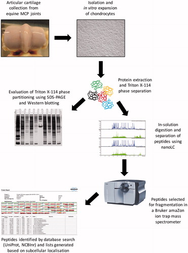
Cartilage shavings were washed three times with sterile PBS containing 10% P/S. Articular chondrocytes were isolated by overnight incubation with 0.1% type II collagenase (from Clostridium histolyticum; Invitrogen, Carlsbad, CA) dissolved in serum-free DMEM containing 4% P/S solution at 37 °C. Following dissociation of cartilage shavings by trituration the solution was filtered through a 70-μm nylon mesh filter to yield a single cell suspension, and centrifuged at 800 ×g for 5 min at room temperature. After washing twice in serum-free DMEM, cells were resuspended in DMEM containing 10% foetal calf serum (FCS; Invitrogen) and 2% P/S solution, seeded into tissue culture flasks (Nunc; Thermo Fisher Scientific), and cultured in a CO2 incubator at 37 °C. Cells were subcultured when they reached approx. 80% confluency. The media was changed on every second day. Cells from the second passage were used for further experiments. A schematic overview of the experimental design is shown in .
Figure 1. Schematic overview of the experimental design used in this study.
Sample preparation, phase partitioning using triton X-114, and methanol/chloroform extraction
Approximately 80% confluent cultures of primary equine articular chondrocytes from passage 2 were washed with PBS, then 2 mL of PBS containing 80 μL of protease inhibitor cocktail (25×, Sigma-Aldrich) was added to the flasks. The flasks were placed on ice, and cells were liberated using a cell scraper (Greiner, Stonehouse, UK). The solution was centrifuged (at 850 ×g for 2 min, room temperature), and the pellet was resuspended in 600 μL of PBS containing 24 μL of 25 × protease inhibitor cocktail. After incubating on ice for 15 min, the suspension was transferred into a glass homogeniser and the cells were lysed.
Following the addition of Triton X-114 (Sigma-Aldrich) at a final concentration of 0.75%, the lysate was incubated on ice for 30 min with vortexing every 5 min. After centrifugation (30 min, 10 000 ×g, 4 °C) the supernatant was retained and incubated at 37 °C for 5 min, and then on ice for 15 min. The sample was centrifuged again (30 min, 10 000 ×g, 4 °C) and the supernatant was incubated at 37 °C for 5 min. Following centrifugation for 3 min (1000 ×g, room temperature), two layers appeared. The upper layer (aqueous phase) contained the hydrophilic proteins, the lower layer (detergent phase) contained the hydrophobic proteins. To maximise the recovery of membrane proteins, the upper layer was extracted further by adding Triton X-114 at a final concentration of 0.75% and the phase partitioning procedure was repeated. Finally, the two lower layers were combined together to constitute the hydrophobic fraction, and the upper layer was treated as the hydrophilic fraction.
To remove Triton X-114 from the samples, four times the sample volume of methanol (Thermo Fisher Scientific) was added to both fractions. After centrifugation at 15 000 ×g for 10 s at room temperature, two times the original sample volume of chloroform (Sigma-Aldrich) was added. The mixture was centrifuged again, and after adding three times the original sample volume of HPLC grade water, the sample was centrifuged for 5 min (15 000 ×g, room temperature). The proteins accumulated at the interface between the two layers formed during the last centrifugation step. Following removal of the upper layer, three times the sample volume of methanol was added, and after spinning for 5 min (15 000 ×g, 4 °C), the pellet containing the proteins was retained and air-dried.
Quantification of proteins
After methanol/chloroform extraction, the pellets were dissolved in sample resuspension buffer containing 4% SDS (Bio-Rad Laboratories, Inc., Hercules, CA), 0.2 M Tris pH 7.4 (Bio-Rad) and 0.15 M NaOH (Thermo Fisher Scientific). Protein concentration in the samples was determined using the Bio-Rad DC Protein Assay Kit according to the manufacturer’s protocol (Bio-Rad). The absorbance of the assayed samples at 655 nm was read using a Bio-Rad Benchmark Microplate Reader.
Polyacrylamide gel electrophoresis (SDS–PAGE)
Loading buffer containing 4 × Laemmli buffer and 3 M dithiothreitol (DTT; Bio-Rad) was added to each sample (typically, 4.8 μL 4 × Laemmli buffer and 1.2 μL 3M DTT was added to 18 μL sample resuspension buffer), and then proteins were fractionated by SDS–PAGE on a 12% polyacrylamide gel. Proteins were initially run at 32 mA constant current, and once the dye front reached the bottom of the stacking gel, the current was increased to 45 mA. Protein bands were visualised by silver staining using a Hoefer Processor Plus automated gel stainer (Amersham, GE Healthcare Life Sciences, UK). The protocol for silver staining was performed as described previously (Yan et al., Citation2000).
Preparation and trypsin digestion of proteins for LC-MS/MS analysis: in-solution digestion
The protein pellets from the methanol/chloroform extraction step were resuspended in a solution of 50 mM ammonium bicarbonate (AMBIC) (Sigma-Aldrich) and 10 mM DTT (Bio-Rad), and incubated at 37 °C for 30 min, vortexing every 10 min. Following the addition of iodoacetamide (IAA, Bio-Rad) at a final concentration of 55 mM, samples were incubated at 37 °C for 45 min in dark. Then, 1.2 mL of –20 °C acetone was added to each sample, and after mixing, the samples were incubated at 4 °C overnight. Protein precipitates were pelleted by centrifugation at 15 000 ×g for 5 min at 4 °C. Pellets were air-dried for 1 min, and then resuspended in 20 μL of trypsin buffer including 50 mM AMBIC and 10 ng/μL Trypsin Gold (Promega, Madison, WA). Samples were vortexed until the pellets were fully dissolved and then incubated at 37 °C for 16 h. Finally, 1 μL of formic acid (1%) was added to each sample to stop the reaction. Samples were stored at –80 °C until analysis.
LC-MS/MS analysis
Samples were injected into a 15 cm C18 Pepmap column using a Bruker Easy-nanoLC UltiMate® (Bruker, Coventry, UK) 3000 RSLCnano chromatography platform with a flow rate of 300 nL/min to separate peptides. Three microlitres of each sample was injected into the HPLC column. After peptide binding and washing processes on the column, the complex peptide mixture was separated and eluted by a gradient of solution A (100% water + 0.1% formic acid) and solution B (100% ACN + 0.1% formic acid) over 115 min, followed by column washing and re-equilibration. The peptides were delivered to a Bruker amaZon ETD ion trap instrument (Bruker, Coventry, UK). The top five most intense ions from each MS scan were selected for fragmentation. The nanoLC-MS/MS analysis was performed three times on the samples (all triplicates).
Peptide and protein identification, data analysis and bioinformatics
Processed data were compiled into *.MGF files and submitted to the Mascot search engine (version: 2.4.1) and compared to mammalian entries in the SwissProt and NCBInr databases. The data search parameters were as follows: two missed trypsin cleavage sites; peptide tolerance, ±0.3 Da; number of C13 = 1; peptide charge, 1+, 2 + and 3 + ions. Carbamidomethyl cysteine was specified as a fixed modification, and oxidised methionine and deamidated asparagine and glutamine residues were specified as variable modifications. Individual ions Mascot scores above 50 indicated identity or extensive homology. Only protein identifications with probability-based protein family Mascot MOWSE scores above the significant threshold of >50 (p < 0.05) were accepted. After mass spectrometric identification, 315 proteins were classified manually using the UniProt (http://www.uniprot.org/) database, considering homologous proteins and further literature information. For many proteins, assigning a definitive cellular compartment and/or function was a difficult task because of the limitations in accurate predictions and lack of experimental evidence. Also, many proteins may actually reside in multiple cellular compartments. To assign identified proteins to specific organelles, the references to subcellular localisations in the UniProt database, as well as gene ontology (GO) annotations were used.
Validation of selected membrane proteins by western blotting
Hydrophobic and hydrophilic protein samples were loaded onto Mini-Protean 3 gels. Approximately 20 μg protein per lane was separated by 7.5% SDS–PAGE gel for immunological detection of selected proteins. Proteins were transferred to PVDF membranes (Immun-Blot™ PVDF Membrane, Bio-Rad). After blocking in 5% non-fat dry milk in PBST, membranes were incubated with the anti-Na+, K+-ATPase primary antibody (diluted 1:100) in blocking solution at 4 °C overnight, with gentle rotation. Membranes were then incubated with the secondary antibody (anti-mouse labelled polymer HRP, DakoCytomation, 1:1000 dilution) in blocking solution at room temperature for 1 h. Membranes were developed by enhanced chemiluminescence reaction (Amersham) according to the instructions of the manufacturer and using auto-radiographic films (Hyperfilm, Amersham). Films were scanned on a calibrated densitometer (Bio-Rad GS800) operated by Quantity One version 4.4.1 software (Bio-Rad). Optical density of bands was determined using ImageJ version 1.47 (ImageJ, Bethesda, MD; http://imagej.nih.gov/ij); data were normalised to the value detectable in the hydrophilic fraction.
Results
Triton X-114 phase separation efficiently enriches membrane proteins from primary chondrocyte cultures
To confirm whether the Triton X-114 phase separation method was able to efficiently extract and enrich lipid-soluble membrane proteins from primary articular chondrocytes cultured in vitro, equal amounts of proteins (25 μg) from the hydrophobic and the hydrophilic fractions were loaded onto polyacrylamide gels. Following SDS–PAGE and silver staining, protein bands with clearly different patterns appeared in the gels with several strong bands present in the hydrophobic fraction only (). To validate the effectiveness of the Triton X-114 extraction method, western blot experiments were performed on both fractions to probe for the presence and relative abundance of a membrane-bound Na+, K+-ATPase. As seen in , the band for this protein in the hydrophobic pool was more than 2.6-fold stronger than that in the hydrophilic pool, demonstrating that lipid-soluble proteins were extracted and enriched in the hydrophobic fraction.
Figure 2. Validation of the efficacy of the Triton X-114 phase separation method. (A) Distribution of protein bands in the hydrophobic (1) and hydrophilic (2) fractions following phase partitioning in total lysates from primary equine articular chondrocyte cultures. After SDS–PAGE, protein bands were visualised using silver staining (M, molecular weight marker). Representative gel image. (B) Western blot experiment performed on both hydrophobic and hydrophilic fractions to probe for the presence and relative abundance of the membrane-bound Na+, K+-ATPase. Numbers below bands represent integrated densities determined by ImageJ freeware. Representative image. (C). The relative distribution of identified proteins following analysis by nanoLC-MS/MS based on their solubility in both hydrophobic and hydrophilic fractions. Numbers outside pie charts represent the actual numbers of proteins identified in each subgroup.
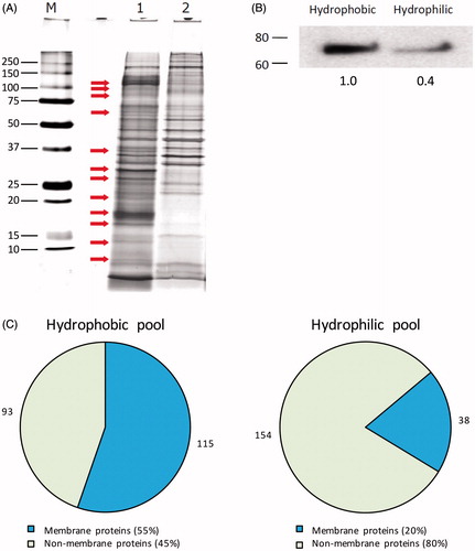
To investigate the protein content of the two fractions, trypsin-digested protein fractions were analysed by nanoLC-MS/MS using Bruker Easy-nanoLC chromatography and a Bruker amaZon ion trap instrument with shotgun proteomics methodologies. A total of 315 unique proteins were reliably (p < 0.05) identified in this study; 208 proteins were detected in the hydrophobic fraction and 192 proteins in the hydrophilic fraction, with 73 (23%) proteins present in both fractions. According to the subcellular localisation data in the UniProt database entries and gene ontology (GO) annotations, in the hydrophobic pool 115 proteins (55%) were membrane proteins and only the remaining 93 proteins (45%) were non-membrane proteins. In contrast, only 38 proteins (20%) were listed as membrane proteins in the hydrophilic fraction, and the other 154 proteins (80%) were non-membrane proteins (). Based on the distribution of membrane versus non-membrane proteins in the two fractions, using the Triton X-114 phase separation method, we successfully extracted and enriched membrane proteins in lysates of primary articular chondrocytes.
Further analysis of the hydrophobic pool reveals various types of membrane proteins
Proteins identified in the hydrophobic fraction were further analysed according to subcellular localisation based on gene ontology (GO) annotation data in the UniProt database entries (). Of the 115 membrane proteins in this pool, PM localisation was indicated for 64 proteins (56%), and the other 51 proteins (44%) were localised in organellar membranes. The PM proteins were further subdivided according to their main functions (). Eighteen proteins (28%) were transporters or involved in membrane/vesicle traffic; 11 and 10 proteins (17 and 16%) were adhesion molecules and proteins with enzyme functions, respectively; 15 proteins (23%) were receptors, and the remaining 10 PM proteins (16%) could not be assigned to any of the previous groups or their function was unknown.
Figure 3. Subcellular distribution of the identified membrane proteins in the hydrophobic fraction. PM proteins were further classified according to their main function based on GO annotations. Numbers outside pie charts represent the actual numbers of proteins identified in each subgroup.
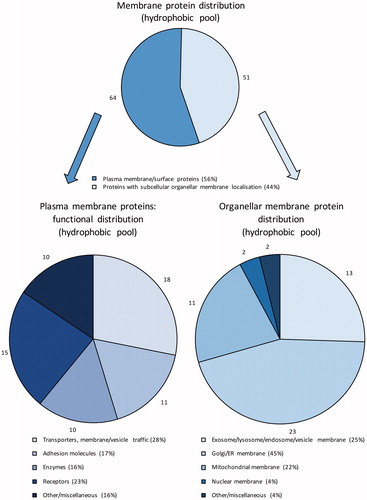
Table 1. Functional classification of PM proteins in the hydrophobic fraction identified in equine articular chondrocytes based on GO annotations.
The membrane proteins with other organellar distributions were also subdivided according to their subcellular localisations (). The majority (23 proteins; 45%) were localised in the membrane of the Golgi complex or the endoplasmic reticulum; 13 proteins (25%) were localised to exosome/lysosome/endosome/other vesicular membranes; another big portion (11 proteins; 22%) were mitochondrial membrane proteins; two proteins (4%) were nuclear membrane proteins; and the remaining two proteins (4%) were ambiguous in terms of specific subcellular localisation.
Table 2. Subcellular distribution of membrane proteins in the hydrophobic pool identified in equine articular chondrocytes based on GO annotations.
The majority of the non-membrane proteins in the hydrophobic pool were cytoplasmic/cytoskeletal proteins (46 proteins; 50%) and secreted (extracellular) proteins (19 entries; 20%). Other subcellular localisations included the lysosome/endosome (4 proteins; 4%), the mitochondrion (1 protein; 1%), the Golgi complex or the endoplasmic reticulum lumen (8 proteins; 9%), the nucleus (10 proteins; 11%), and the remaining five proteins were either contaminants or their subcellular localisation was ambiguous (5%; and ).
Figure 4. Subcellular localisation of the identified non-membrane proteins in the hydrophobic fraction based on GO annotations. Numbers outside pie chart represent the actual numbers of proteins identified in each subgroup.
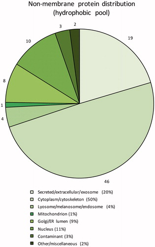
Table 3. Subcellular distribution of non-membrane proteins in the hydrophobic pool identified in equine articular chondrocytes based on GO annotations.
The hydrophilic pool contains proteins with different solubility and subcellular distribution
The Triton X-114 phase separation technique effectively extracted and enriched lipid-soluble membrane proteins in the hydrophobic phase, and left comparably few membrane proteins (only 20%) in the hydrophilic fraction (). As in the case of the hydrophobic fraction, the majority of the 38 lipid-soluble membrane proteins were localised in the PM (25 proteins; 66%), whilst the others were localised in various organellar membranes (Golgi complex/endoplasmic reticulum membrane, 16%; mitochondrial membrane, 8%; nuclear membrane, 2%; and ).
Figure 5. Subcellular localisation of the identified membrane and non-membrane proteins in the hydrophilic fraction based on GO annotations. Numbers outside pie charts represent the actual numbers of proteins identified in each subgroup.
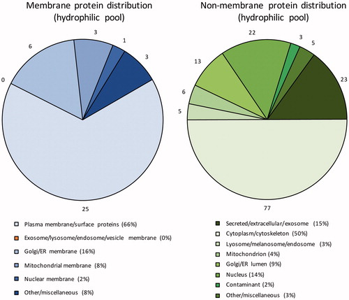
Table 4. Functional classification of PM proteins and other membrane proteins in the hydrophilic pool identified in equine articular chondrocytes based on GO annotations.
Taken together, we have identified 78 unique PM proteins in equine articular chondrocytes in this work. Among them, 32 proteins possessed receptor/adhesion functions; the most important ones are the cluster of differentiation (CD) proteins and integrins. Furthermore, 21 PM proteins with transporter functions were detected in articular chondrocytes ( and ).
The non-membrane protein complement in the hydrophilic fraction comprised 154 proteins, the majority of which (77 proteins; 50%) were cytoplasmic/cytoskeletal proteins. Also in good correlation with the non-membrane protein distribution observed in the hydrophobic samples, the secreted (extracellular) and the nuclear proteins were the second and third largest groups in this fraction (23 proteins, 15%; and 22 proteins, 14%, respectively). Other subcellular localisations included the lysosome/endosome (5 proteins; 3%), the mitochondrion (6 proteins; 4%), the Golgi complex or the endoplasmic reticulum lumen (13 proteins; 9%), and the remaining eight proteins were either contaminants or their subcellular localisation was not determined (5%; and ).
Table 5. Functional classification of non-membrane proteins in the hydrophilic pool identified in equine articular chondrocytes based on GO annotations.
Discussion
The application of mass spectrometry (MS) has recently become an important tool in cartilage biology as it offers numerous advantages over more conventional biochemical approaches such as western blotting. To date, a number of proteomic studies have been performed on cartilage tissue and on chondrocytes, confirming that this analytical tool is particularly suitable for high-throughput and large-scale analysis of the protein complement in health and disease [reviewed in Hsueh et al. (Citation2014) and Williams et al. (Citation2011)]. The first proteomic study carried out on normal human knee articular chondrocyte cultures aimed at creating a two-dimensional gel electrophoresis (2-DE) reference map and generated 93 unique protein identities (Ruiz-Romero et al., Citation2005). A 1-D SDS–PAGE approach combined with MS/MS resulted in the identification of over 100 different proteins from human knee cartilage supernatants (Garcia et al., Citation2006). In the first large-scale MS analysis of human articular cartilage, which was designed to extract both extracellular and intracellular proteins from samples depleted of highly abundant ECM proteins such as collagens and aggrecan to allow the detection of less abundant proteins, a total number of 814 distinct proteins were identified (Wu et al., Citation2007). In a more recent study, the proteome of articular chondrocytes from healthy and OA patients using high resolution label-free MS was analysed, leading to the identification of ∼2400 proteins (Tsolis et al., Citation2015).
Despite these studies and the impressive number of proteins identified, our knowledge about the proteome of cartilage and its resident cell, the chondrocyte, can still be improved. One significant drawback is that at least in some of the studies the identified proteins have not been properly analysed in terms of subcellular locations and/or functions. In addition, the majority of proteins identified in these studies were located in the ECM because of their high abundance relative to cellular proteins in chondrocytes. More specifically, the “hidden” proteome, which comprises low abundance proteins and/or is not accessible by standard methods, is still poorly characterised. In a study that combined extensive pre-fractionation followed by electrospray ionisation mass spectrometry (ESI-MS/MS), 779 unique proteins expressed by cultured chondrocytes were identified, of which 203 were annotated to the membrane (Lambrecht et al., Citation2010). However, the authors did not carry out a detailed analysis with respect to specific subcellular location and/or function of the identified membrane proteins, making further data interpretation attempts challenging.
Therefore, the aim of this study was to extend the current knowledge of the chondrocyte proteome by using the Triton X-114 phase separation technique to discover the membrane subproteome (the membranome). We have chosen to use equine articular chondrocytes in this study, for the horse is widely involved in occupational/sports activities and considered as an excellent animal model for human joint diseases (Aigner et al., Citation2010) and yet, current knowledge is limited and relates to only the protein complement of equine chondrocytes. The unique features of our study are as follows. First, to the best of our knowledge, this is the first application of LC-MS/MS proteomics to study the membrane protein complement of cultured articular chondrocytes. Second, a large proportion (133 proteins; 42%) of the 315 proteins identified in this work consisted of membrane proteins, and for some of these only ambiguous data were available in chondrocytes [e.g. CD276, S100-A6 (calcyclin), VDACs]. The proportion of membrane proteins was even higher in the hydrophobic phase (55%). As far as the proportion of membrane proteins is concerned, our results are in a good agreement with those reported elsewhere (Hansson et al., Citation2010). The aim of that study was to characterise the human pancreatic islet membrane proteome by evaluating five different extraction procedures; while the proportion of membrane proteins in the total extracts was 35%, a considerably higher proportion (61%) of membrane proteins was identified following the use of membrane protein-enriching methods. It is also worth noting that the choice of method for extraction of membrane proteins had a strong influence on the number and identity of proteins detected in that analysis, and the hydrophobic phase of Triton X-114 phase separation was found to be the most efficient extraction method (Hansson et al., Citation2010). These results also underpin that appropriate sample preparation and pre-fractionation methods can increase the amount of identified proteins with specific properties in the MS/MS analysis of proteins in complex biological samples. It was for that reason that we used the Triton X-114 phase separation method in this study.
Although a detailed description of the proteins identified in the chondrocyte membranome is beyond the scope of this article, a few important protein classes merit comment due to their potential involvement in chondrocyte homeostasis. We therefore restrict discussing our results to certain protein groups localised in the PM.
CD antigens and integrins
Cluster of differentiation (CD) proteins are four hydrophobic domain-containing cell surface membrane glycoproteins that mediate a range of cellular processes including development, differentiation, activation, growth and motility. Composed of alpha and beta subunits, integrins are integral PM receptors that mediate attachment between a cell and its surroundings. They transduce information from the ECM to the cell and integrin-mediated signalling pathways influence cell shape, mobility, differentiation and the cell cycle. The integrins identified in this study (integrin beta-1 [CD29], integrin alpha-5 [CD49e], integrin alpha-V [CD51], integrin alpha-3 [CD49c] and integrin beta-3 [CD61]) are in a complete agreement with what has been published earlier (Mobasheri et al., Citation2002a; Shakibaei et al., Citation2008; Woods et al., Citation1994). Our data also confirm reports on CD antigen expression in articular chondrocytes (Diaz-Romero et al., Citation2005). We found that equine articular chondrocytes express tetraspanins (CD9 [tetraspanin-19], CD63 [tetraspanin-30], CD81 [tetraspanin-28] and CD151 [tetraspanin-24]); CD44 (hyaluronan receptor); CD71 (transferrin receptor); CD90 (Thy-1); and CD166 (ALCAM). CD73, an ecto-5′-nucleotidase, which plays a crucial role in extracellular adenosine generation, has been reported to be involved in mechanotransduction pathways following cyclic compressive stimulation (Ode et al., Citation2013). CD107 (LAMP) expression has been recently reported in murine growth plate cartilage and cartilaginous nodules in embryonic limb bud-derived micromass cultures (Hatakeyama et al., Citation2014). CD147 (basigin; also known as extracellular matrix metalloproteinase inducer) is known to be extensively expressed by chondrocytes both in normal and OA cartilage (Orazizadeh & Salter, Citation2008). CD228 (melanotransferrin) has also long been known to facilitate the differentiation of prechondrogenic cells (Suardita et al., Citation2002). While CD276 has not been identified earlier in chondrocytes, it is known to be expressed in undifferentiated mesenchymal stem cells derived from Wharton’s jelly (WJ-MSCs), even after osteogenic, adipogenic and chondrogenic differentiation (La Rocca et al., Citation2013).
S100 proteins
Of particular interest is protein S100-A6 (calcyclin) expression in equine articular chondrocytes. The S100 family of proteins consists of 24 members, which show cell-specific expression patterns and are involved in a wide range of cellular processes including proliferation, differentiation, apoptosis, Ca2+ homeostasis, energy metabolism, inflammation and migration/invasion through interactions with a variety of target proteins ranging from enzymes, cytoskeletal subunits, receptors, transcription factors and nucleic acids [reviewed in Donato et al. (Citation2013)]. In particular, S100-A6 may function as a Ca2+ sensor and modulator, contributing to Ca2+ signalling pathways. It is also implicated in cell proliferation and cytoskeletal dynamics, and known to have a potential role in cell responses to different stressors. Calcyclin was reported to be significantly upregulated in serially passaged adipose tissue-derived MSCs (Capra et al., Citation2012), which may correspond to previous data suggesting that it is frequently upregulated during proliferation and differentiation and it is induced by different growth factors. There are only sporadic data available relating to S100-A6 expression in chondrocytes. Its mRNA transcript has been shown to be downregulated following chondrogenic induction by BMP4 in ATDC5 cells, and it has also been identified in one of the chondrocyte proteome studies discussed earlier (Lambrecht et al., Citation2010).
Other S100 proteins identified in this study include S100-A1 and S100-A11 ( and ). S100-A1 is localised in the cytoplasm where it is associated with cytoskeletal components and mitochondria. It can influence Ca2+ handling in cultured ventricular cardiomyocytes through interaction with the sarcoplasmic reticulum Ca2+-ATPase and RyR2; it also modulates CaV1 channel currents in a PKA-dependent manner. S100-A1 also regulates energy metabolism by stimulating fructose-1,6-biphosphate aldolase and inhibiting phosphoglucomutase and glycogen phosphorylase (Donato et al., Citation2013). Also localised in the cytoplasm, S100-A11 is reported to modulate cell growth via binding either to nucleolin or Rad54B (Donato et al., Citation2013). In particular, S100-A11 can activate the p38 MAPK pathway to accelerate chondrocyte hypertrophy and ECM catabolism that may promote OA progression (Cecil & Terkeltaub, Citation2008). Both S100-A1 and S100-A11 have been reported to be expressed and functional in chondrocytes (Donato et al., Citation2013; Patti et al., Citation1999), and both proteins were identified in a previous MS study (Lambrecht et al., Citation2010).
Transporters
Ion channels and transporters are essential components of chondrocytes that control the movement of ions and other small molecules across the PM. An increasing number of studies have reported the presence of an ever-expanding list of ion channels and transporters in chondrocytes [reviewed in Barrett-Jolley et al. (Citation2010) and Matta et al. (Citation2015)]. Based on GO annotations, 21 proteins with transporter functions were identified in the PM in this study ( and ). Originally described as being localised in the outer mitochondrial membrane (Benz, Citation1994), voltage-dependent anion-selective channels (VDACs), also known as mitochondrial porins, form the pores that allow the transport of small hydrophilic solutes across the membrane. However, accumulating evidence support that VDACs can also be expressed in the PM (De Pinto et al., Citation2010), where they exhibit voltage-gated anion channel activity, and its electrophysiological phenotype is that of a maxi-chloride channel (Lewis et al., Citation2013). Although VDACs have not been unequivocally reported to be expressed and function in chondrocytes, the anion channel identified in some previous studies was the maxi-chloride channel, which is remarkably similar to the maxi-Cl–/VDAC channel (Sugimoto et al., Citation1996; Tsuga et al., Citation2002). Although all three VDAC proteins were identified in chondrocytes in our experiments and also by others (Lambrecht et al., Citation2010), further studies will need to functionally investigate the physiological and pathophysiological roles of these transporters in the chondrocyte PM.
The chloride intracellular channel (CLIC) proteins possess pH-dependent chloride ion channel activity. CLIC1 and CLIC4, in addition to other members of the CLIC family, are often referred to as “p64-related” proteins, and while they may localise to intracellular compartments (e.g. the nucleus), they also appear to be in the PM and could serve a role in secretion (Lewis et al., Citation2013). Once again, although the CLIC1 protein was identified in chondrocytes in this study and by others (Lambrecht et al., Citation2010), its presence and function has not been unambiguously demonstrated earlier.
In addition to anion channels, glucose transporter (GLUT) proteins (facilitative glucose transporter 1 and 3; GLUT 1 and GLUT3) were also identified in our study. Glucose is a key metabolite and a structural precursor for articular cartilage and its transport has significant consequences for cartilage development and functional integrity. Our results are in a good agreement with previously published data (Mobasheri et al., Citation2002b), confirming here by proteomic techniques the expression of these two GLUT isoforms in articular chondrocytes.
Conclusion
In summary, studying the membranome profile of equine articular chondrocytes by LC-MS/MS following enrichment using Triton X-114 pre-fractionation has turned out to be an excellent approach to gain insight into proteins involved in a wide range of membrane-bound processes such as signal transduction, adhesion and transport of ions and other molecules. In spite of the significant enrichment of lipid-soluble membrane proteins in the hydrophobic phase, the proteins that are present in an extremely low abundance in chondrocytes such as the majority of ion channels and other transporter molecules in the PM remained undetectable. Although detergent-based phase partitioning enriches PM proteins relative to total soluble proteins, the membrane proteins in the ER, mitochondria and other organelles are also enriched; and the abundance of proteins in the contaminating organelles can interfere with the ability to detect PM proteins (Zhang & Peck, Citation2011). To mitigate these limitations, a combination of the Triton X-114 phase separation method with other membrane protein enrichment techniques could also be considered.
Our study confirms some previous findings and adds further proteins to the proteomic profile of equine articular chondrocytes. Some of the identified proteins including the CD276 antigen, S100-A6 (calcyclin) or VDACs have not been unambiguously reported before to be components of articular chondrocytes. However, there are certain limitations to this work. First and foremost, protein identifications were somewhat aggravated by the fact that our search results listed the same proteins several times but for different species (primarily human or bovine but no horse entries), suggesting that the protein was present in the sample and that the identification was made by virtue of the horse protein sharing homology with several other species. This is one of the disadvantages when “cross species matching” is used to identify proteins. Another possible disadvantage of using equine articular chondrocytes is that there may be subtle differences between the two species and that the entirety of our results may not be directly applicable to human articular chondrocytes.
A more detailed and comprehensive insight into the chondrocyte membranome is likely to make a significant contribution to the development of novel drugs for arthritic diseases. The development and refinement of proteomics-based techniques will enable a better understanding of regulatory proteins and enhance the search for new drug targets. It may also help to discover novel cartilage disease-specific biomarkers. Thus, our data represent a significant addition to the comprehensive cartilage proteome database that is essential for understanding the molecular mechanisms underlying cartilage function and OA.
Declaration of interest
The authors do not have any commercial relationships that could be construed as biased or inappropriate. The authors report no declarations of interests. C. M. is supported by the European Commission through a Marie Skłodowska-Curie Intra-European Fellowship for career development (project number: 625746; acronym: CHONDRION; FP7-PEOPLE-2013-IEF). A. M. is the co-ordinator of the D-BOARD Consortium funded by European Commission Framework 7 programme (EU FP7; HEALTH.2012.2.4.5-2, project number 305815, Novel Diagnostics and Biomarkers for Early Identification of Chronic Inflammatory Joint Diseases). A. M. has received funding from the Deanship of Scientific Research (DSR), King AbdulAziz University (grant no. 1-141/1434 HiCi). A. M. is also a member of the Arthritis Research UK Centre for Sport, Exercise, and Osteoarthritis, funded by Arthritis Research UK (Grant Reference Number: 20194).
References
- Aigner T, Cook JL, Gerwin N, et al. (2010). Histopathology atlas of animal model systems – overview of guiding principles. Osteoarthritis Cartilage 18 Suppl 3:S2–6
- Almen MS, Nordstrom KJ, Fredriksson R, Schioth HB. (2009). Mapping the human membrane proteome: a majority of the human membrane proteins can be classified according to function and evolutionary origin. BMC Biol 7:50
- Archer CW, Francis-West P. (2003). The chondrocyte. Int J Biochem Cell Biol 35:401–4
- Barrett-Jolley R, Lewis R, Fallman R, Mobasheri A. (2010). The emerging chondrocyte channelome. Front Physiol 1:135
- Benz R. (1994). Permeation of hydrophilic solutes through mitochondrial outer membranes: review on mitochondrial porins. Biochim Biophys Acta 1197:167–96
- Bordier C. (1981). Phase separation of integral membrane proteins in Triton X-114 solution. J Biol Chem 256:1604–7
- Capra E, Beretta R, Parazzi V, et al. (2012). Changes in the proteomic profile of adipose tissue-derived mesenchymal stem cells during passages. Proteome Sci 10:46
- Cecil DL, Terkeltaub R. (2008). Transamidation by transglutaminase 2 transforms S100A11 calgranulin into a procatabolic cytokine for chondrocytes. J Immunol 180:8378–85
- Cordwell SJ, Thingholm TE. (2010). Technologies for plasma membrane proteomics. Proteomics 10:611–27
- De Pinto V, Messina A, Lane DJ, Lawen A. (2010). Voltage-dependent anion-selective channel (VDAC) in the plasma membrane. FEBS Lett 584:1793–9
- Diaz-Romero J, Gaillard JP, Grogan SP, et al. (2005). Immunophenotypic analysis of human articular chondrocytes: changes in surface markers associated with cell expansion in monolayer culture. J Cell Physiol 202:731–42
- Dissoki S, Abourbeh G, Salnikov O, et al. (2015). PET molecular imaging of angiogenesis with a multiple tyrosine kinase receptor-targeted agent in a rat model of myocardial infarction. Mol Imaging Biol 17:222–30
- Donato R, Cannon BR, Sorci G, et al. (2013). Functions of S100 proteins. Curr Mol Med 13:24–57
- Dunham J, Shackleton DR, Billingham MEJ, et al. (1988). A reappraisal of the structure of normal canine articular-cartilage. J Anat 157:89–99
- English JA, Manadas B, Scaife C, et al. (2012). Partitioning the proteome: phase separation for targeted analysis of membrane proteins in human post-mortem brain. PLoS One 7:e39509
- Garcia BA, Platt MD, Born TL, et al. (2006). Protein profile of osteoarthritic human articular cartilage using tandem mass spectrometry. Rapid Commun Mass Spectrom 20:2999–3006
- Hansson SF, Henriksson A, Johansson L, et al. (2010). Membrane protein profiling of human islets of langerhans using several extraction methods. Clin Proteom 6:195–207
- Hatakeyama Y, Hatakeyama J, Oka K, et al. (2014). Immunohistochemical study of amelogenin and lysosome-associate membrane proteins (LAMPs) in cartilage. Int J Morphol 32:618–26
- Hsueh MF, Onnerfjord P, Kraus VB. (2014). Biomarkers and proteomic analysis of osteoarthritis. Matrix Biol 39:56–66
- Kabbani N. (2008). Proteomics of membrane receptors and signaling. Proteomics 8:4146–55
- La Rocca G, Lo Iacono M, Corsello T, et al. (2013). Human Wharton's jelly mesenchymal stem cells maintain the expression of key immunomodulatory molecules when subjected to osteogenic, adipogenic and chondrogenic differentiation in vitro: new perspectives for cellular therapy. Curr Stem Cell Res Ther 8:100–13
- Lambrecht S, Dhaenens M, Almqvist F, et al. (2010). Proteome characterization of human articular chondrocytes leads to novel insights in the function of small heat-shock proteins in chondrocyte homeostasis. Osteoarthr Cartil 18:440–6
- Lambrecht S, Verbruggen G, Verdonk PC, et al. (2008). Differential proteome analysis of normal and osteoarthritic chondrocytes reveals distortion of vimentin network in osteoarthritis. Osteoarthr Cartil 16:163–73
- Lewis R, May H, Mobasheri A, Barrett-Jolley R. (2013). Chondrocyte channel transcriptomics: do microarray data fit with expression and functional data? Channels (Austin) 7:459–67
- Mathias RA, Chen YS, Kapp EA, et al. (2011). Triton X-114 phase separation in the isolation and purification of mouse liver microsomal membrane proteins. Methods 54:396–406
- Matta C, Zakany R, Mobasheri A. (2015). Voltage-dependent calcium channels in chondrocytes: roles in health and disease. Curr Rheumatol Rep 17:43
- Mobasheri A. (2013). The future of osteoarthritis therapeutics: targeted pharmacological therapy. Curr Rheumatol Rep 15:364
- Mobasheri A, Carter SD, Martin-Vasallo P, Shakibaei M. (2002a). Integrins and stretch activated ion channels; putative components of functional cell surface mechanoreceptors in articular chondrocytes. Cell Biol Int 26:1–18
- Mobasheri A, Neama G, Bell S, et al. (2002b). Human articular chondrocytes express three facilitative glucose transporter isoforms: GLUT1, GLUT3 and GLUT9. Cell Biol Int 26:297–300
- Ode A, Schoon J, Kurtz A, et al. (2013). CD73/5'-ecto-nucleotidase acts as a regulatory factor in osteo-/chondrogenic differentiation of mechanically stimulated mesenchymal stromal cells. Eur Cell Mater 25:37–47
- Orazizadeh M, Salter DM. (2008). CD147 (extracellular matrix metalloproteinase inducer-emmprin) expression by human articular chondrocytes. Iran Biomed J 12:153–8
- Patti AM, Gabriele A, Della Rocca C. (1999). Human chondrocyte cell lines from articular cartilage of metatarsal phalangeal joints. Tissue Cell 31:550–4
- Rabilloud T. (2003). Membrane proteins ride shotgun. Nat Biotechnol 21:508–10
- Ruiz-Romero C, Carreira V, Rego I, et al. (2008). Proteomic analysis of human osteoarthritic chondrocytes reveals protein changes in stress and glycolysis. Proteomics 8:495–507
- Ruiz-Romero C, Lopez-Armada MJ, Blanco FJ. (2005). Proteomic characterization of human normal articular chondrocytes: a novel tool for the study of osteoarthritis and other rheumatic diseases. Proteomics 5:3048–59
- Samkoe KS, Tichauer KM, Gunn JR, et al. (2014). Quantitative in vivo immunohistochemistry of epidermal growth factor receptor using a receptor concentration imaging approach. Cancer Res 74:7465–74
- Sega EI, Low PS. (2008). Tumor detection using folate receptor-targeted imaging agents. Cancer Metastasis Rev 27:655–64
- Shakibaei M, Csaki C, Mobasheri A. (2008). Diverse roles of integrin receptors in articular cartilage. Adv Anat Embryol Cell Biol 197:1–60
- Suardita K, Fujimoto K, Oda R, et al. (2002). Effects of overexpression of membrane-bound transferrin-like protein (MTf) on chondrogenic differentiation in vitro. J Biol Chem 277:48579–86
- Sugimoto T, Yoshino M, Nagao M, et al. (1996). Voltage-gated ionic channels in cultured rabbit articular chondrocytes. Comp Biochem Physiol C Pharmacol Toxicol Endocrinol 115:223–32
- Tsolis KC, Bei ES, Papathanasiou I, et al. (2015). Comparative proteomic analysis of hypertrophic chondrocytes in osteoarthritis. Clin Proteomics 12:12
- Tsuga K, Tohse N, Yoshino M, et al. (2002). Chloride conductance determining membrane potential of rabbit articular chondrocytes. J Membr Biol 185:75–81
- Williams A, Smith JR, Allaway D, et al. (2011). Applications of proteomics in cartilage biology and osteoarthritis research. Front Biosci (Landmark Ed) 16:2622–44
- Williams A, Smith JR, Allaway D, et al. (2013). Carprofen inhibits the release of matrix metalloproteinases 1, 3, and 13 in the secretome of an explant model of articular cartilage stimulated with interleukin 1β. Arthritis Res Ther 15:R223
- Woods VL, Jr Schreck PJ, Gesink DS, et al. (1994). Integrin expression by human articular chondrocytes. Arthritis Rheum 37:537–44
- Wu J, Liu W, Bemis A, et al. (2007). Comparative proteomic characterization of articular cartilage tissue from normal donors and patients with osteoarthritis. Arthritis Rheum 56:3675–84
- Yan JX, Wait R, Berkelman T, et al. (2000). A modified silver staining protocol for visualization of proteins compatible with matrix-assisted laser desorption/ionization and electrospray ionization-mass spectrometry. Electrophoresis 21:3666–72
- Zhang ZJ, Peck SC. (2011). Simplified enrichment of plasma membrane proteins for proteomic analyses in Arabidopsis thaliana. Proteomics 11:1780–8