Abstract
Estradiol (E2) is, apart from its role as a reproductive hormone, also important for cardiac function and bone maturation in both genders. It has also been shown to play a role in insulin production, energy expenditure and in inducing lipolysis. The aim of the study was to investigate if low circulating testosterone or E2 levels in combination with variants in the estrogen receptor alpha (ESR1) and estrogen receptor beta (ESR2) genes were of importance for the risk of type-2 diabetes. The single nucleotide polymorphisms rs2207396 and rs1256049, in ESR1 and ESR2, respectively, were analysed by allele specific PCR in 172 elderly men from the population-based Tromsø study. The results were adjusted for age. In individuals with low total (≤11 nmol/L) or free testosterone (≤0.18 nmol/L) being carriers of the variant A-allele in ESR1 was associated with 7.3 and 15.9 times, respectively, increased odds ratio of being diagnosed with diabetes mellitus type 2 (p = 0.025 and p = 0.018, respectively). Lower concentrations of E2 did not seem to increase the risk of being diagnosed with diabetes. In conclusion, in hypogonadal men, the rs2207396 variant in ESR1 predicts the risk of type 2 diabetes.
Introduction
Estrogens are considered as “female sex hormones” but, in both sexes, they have a crucial role in regulating not only the reproductive system but also many other of the functions in the human body, e.g. the cardiovascular function, bone mineral density and the metabolism [Citation1–3].
In males, estradiol (E2) is primarily produced at extragonadal sites by CYP19 controlled aromatization of testosterone [Citation4,Citation5]. In an alternative pathway, androstenedione is converted to estrone, which subsequently is metabolized to E2. Previously, E2 has been shown to inhibit insulin gene receptor transcription, promote insulin resistance and also to up-regulate CYP19 expression in mature adipocytes [Citation6–8]. E2 has also been shown to increase insulin biosynthesis [Citation9].
Low testosterone levels in men has in numerous studies and also in a meta-analysis comprising more than 6000 men been associated with increased risk of type 2 diabetes (referred to as diabetes hereafter) [Citation6]. It has also been shown that testosterone replacement therapy may improve insulin sensitivity [Citation10], whereas androgen deprivation increases insulin resistance in men [Citation11]. However, whether this is attributed to testosterone per se or to the parallel changes in E2 has been discussed. Estrogen action is mediated through estrogen receptor α (ESR1), estrogen receptor β (ESR2) and a G-protein coupled cell surface receptor [Citation12,Citation13].
Total estrogen resistance due to a germ line mutation in the ESR1 gene was in 1994 reported in a 28-year-old man with continued linear growth into adulthood. He also presented with osteoporosis and early atherosclerosis as well as glucose intolerance and hyperinsulinemia [Citation11]. However, testosterone concentration was within normal range and the man had gone through normal puberty, suggesting that lack of estrogen action rather than testosterone deficiency was the cause of his diabetes. Estrogen resistance due to homozygous germ line mutations in the ESR1 gene is however extremely rare, only one kindred described to date. Nevertheless, one cannot exclude inherited differences in estrogen secretion and response, which may have more subtle effects on the metabolic system and over time contribute to the etiology of diabetes. Animal studies are supporting this theory, since ESR1 knockout mice suffer from increased fasting plasma glucose levels and increased insulin insensitivity [Citation14]. In males suffering from mutations in the gene coding for aromatase increased insulin resistance, increased concentrations of insulin and diabetes have been found [Citation15–18]. However, Carani et al. found no changes in metabolic parameters in a patient with a mutation in the gene coding for aromatase [Citation19].
Both the ESR1 and ESR2 genes contain several single nucleotide polymorphisms (SNPs). We have previously reported that G to A substitution in SNP rs2207396, situated in intron 6, of the ESR1 was associated with higher risk of developing azoospermia in childhood cancer survivors [Citation20], pointing to a functional role of this polymorphism. With respect to rs1256049 SNP in ESR2, a synonymous substitution of GTG to GTA in, exon 5 was first shown to be associated with ovulatory dysfunction in women of Chinese origin [Citation21] and later also with male infertility in Caucasian patients [Citation22].
Since subnormal testosterone levels, either due to the male sex hormone action per se or due to parallel decrease in E2, were found to be associated with increased risk of metabolic derangements [Citation23], we wished to investigate whether this association is modified by polymorphisms in the above mentioned SNPs in the ESR1 and ESR2 genes.
Materials and methods
Subjects
The study population and the methods have been presented previously [Citation23]. Shortly, the Tromsø Study is an on-going population-based health survey of the inhabitants from the municipally of Tromsø, mainly focusing on lifestyle related diseases. A subgroup consisting of 68 men with consistent total testosterone ≤11.0 nmol/L and 104 men with consistent total testosterone >11.0 nmol/L were recruited from the fifth Tromsø study. When they were invited to participate in this study in 2005 they were between 60- and 80-years-old.
Blood samples for analysis of levels of testosterone and E2, glucose, insulin and for genetic analyses were drawn 08.00 am. An oral glucose tolerance test with ingestion of 75 g glucose was performed. Diabetes was defined as having either non-fasting glucose >11.1 mmol/L, fasting glucose >7.0 mmol/L or 2 h glucose load >11.1 mmol/L. Height and weight were measured in subjects wearing light clothing without shoes. Body mass index (BMI) was calculated (kg/m2). All participants were genotyped for rs2207396 in ESR1 and rs1256049 in ESR2. Free testosterone was calculated according to Vermeulen’s formula [Citation24]. All participants signed an informed consent form and the study was approved by the Regional Research Ethics Committee, North-Norway.
Sex hormone analyses and biochemistry
Serum total testosterone was analysed by electrochemical luminescence immunoassay using an automated clinical chemistry analyser (Modular E, Roche Diagnostics GmbH, Mannheim, Germany). The total analytical precision expressed as the sum of intra and interassay coefficients of variation (CVa) were 5.6 and 4.7 and the reference values were 10–28 nmol/L and <0.20 nmol/L, respectively. Serum E2 levels were measured using DELFIA® Estradiol-kit (Perkin Elmer, Turku, Finland). The total analytical precision expressed as the sum of interassay CV 20% at 30 pmol/L and 10% at 280 pmol/L. Measuring range was 8–1500 pmol/L. For the E2 analysis, serum samples from five participants were lacking, thus, limiting the study population to 167 males for all calculations involving E2.
Serum SHBG and insulin were analysed by immunometry based on chemiluminescence using an automated clinical chemistry analyzer (Immunulite 2000, Diagnostic Product Corp., Los Angeles, CA). The CVas were 5.2 and 8.8, respectively.
Allele specific PCR
To identify the different SNPs, allele-specific PCR was used and PCR conditions established to generate both a short allele-specific and a longer control band in the presence of the variant, and only the longer control band in its absence. For the ESR1 variant rs2207396, the PCR amplification volume was 25 µl containing 25 ng genomic DNA, 45 mM KCL, 10 mM Tris-HCL, 0.2 mM dNTP (Fermentas GmbH, St. Leon-Rot, Germany), 0.1% Tween 20 (Scharlau Chemie S.A, Barcelona, Spain), 1.5 mM MgCl2, 0.5 µM forward primer (Invitrogen, Edinburgh, Scotland) (), 0.5µM reverse primer, 0.45 µM of either one of the allele specific primers, 0.5 U DNA Polymerase (DyNAzyme II, Finnzymes, Espoo, Finland). Amplifications were carried out for 42 cycles (Eppendorf, Hamburg, Germany); each cycle including denaturation for 1 min at 96 °C, primer annealing at 53.5 °C for 30 s and primer extension at 72 °C for 3 min, with an initial denaturation step for 3 min at 96 °C and a final extension at 72 °C for 5 min.
Table 1. Primers used for amplification of SNPs in ESR1 and ESR2, respectively.
For rs1256049 in ESR2 gene, the PCR amplification volume was 25 µl containing 25 ng genomic DNA, 45 mM KCL, 10 mM Tris-HCL, 0.2 mM dNTP 0.25 µM forward primer, 0.25 µM reverse primer and 0.3 µM of either one of the allele specific primers [Citation22] and 0.5 U DNA Polymerase. Amplifications were carried out for 40 cycles; and primer annealing at 57 °C for 30 s, whereas all other procedures were the same as for ESR1.
To verify the results of the allele-specific PCR, three selected PCR samples representing each genotype, were purified using a JETquick-kit according to the manufacturer’s recommendation (Genomed, Löhne, Germany) and directly sequenced. The sequencing reaction was performed using ABI Prism® BigDye® Terminator v3.1 Cycle Sequencing Kit according to the manufacturer’s recommendation (Applied Biosystems, Austin, TX) and the results were obtained using a 3130x/Genetic Analyzer (Applied Biosystems, Hitatchi, Austin, TX).
Statistics
The subjects were categorised into groups with subnormal and normal testosterone level. For total testosterone the cut off level was set to 11 nmol/L, as has been used for previous studies on these subjects, whereas it was 0.18 nmol/L for the calculated level of free testosterone [Citation25,Citation26].
Using binary regression analysis, the odds ratio (OR) for diabetes in men with low testosterone levels, with the group having normal concentration of this sex hormone as reference, was assessed.
In order to explore whether fluctuations in testosterone levels mirror corresponding changes in E2 concentration, we tested correlation between these two parameters using Spearman’s test.
The association between E2 and the OR for diabetes in the entire study population was assessed using binary regression analysis with median E2 (74.0 pmol/L) as a cut-off point for categorizing E2.
For the combination of total/free testosterone and the genotype four different groups were defined: (a) Normal (total/free) testosterone + GG; (b) Normal (total/free) testosterone +AA/AG; (c) Subnormal (total/free) testosterone + GG and (d) Subnormal (total/free) testosterone + AA/AG. For each genotype we primary performed an interaction analysis (testosterone subnormal/normal * genotype) in order to explore whether there was a statistically significant interaction between sex hormone levels and genotype in relation to risk of developing diabetes.
The interaction analysis was also done for E2*genotype with median E2 levels as cut off for dichotomising the hormone concentrations. Since there are no well-defined subnormal E2 levels for men, we even performed the interaction analysis with E2 as a continuous variable.
Similar strategy was applied for the ESR2 genotypes with the exception that the analysis of differences in ESR2 genotype distribution among males with normal levels of testosterone Fisher’s exact test was used since one of the subgroups contained 0 subjects. Calculations were adjusted for age. Statistical significance was defined as p < 0.05. For statistical analysis, IBM SPSS Statistics 20 (version 20, IBM Corp., Armonk, NY) was used.
Results
Total and free testosterone
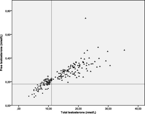
The measured levels of total and free testosterone are shown in . Subnormal levels of total testosterone, irrespective of allele, did not increase the OR of having been diagnosed with diabetes (95% CI: 0.7–6.0; p = 0.196). Subnormal levels of free testosterone, irrespective of allele, increased the OR of having been diagnosed with diabetes (OR = 4.3, 95% CI: 1.2–11.0; p = 0.019,) compared to individuals with normal levels of free testosterone.
Figure 1. Measured levels of total testosterone and free testosterone in men with subnormal levels of both total and free testosterone (×), subnormal levels of total testosterone (○) and men with normal levels of both total and free testosterone (Δ). The cut-off values (11 and 0.18 nmol/L) are indicated by the vertical and horizontal lines, respectively.
Estradiol
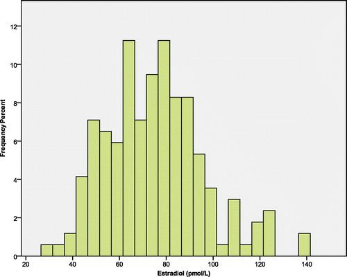
The distribution of E2 in the study population is shown in . There was a statistically significant although weak positive correlation between testosterone and E2 (p < 0.0005, β: 0.327). The OR of having diabetes in those with E2 below median did not differ from those with E2 above this value (OR = 1.4; 95% CI: 0.460–4.184; p = 0.571).
Figure 2. The distribution of E2 in the study population (n = 167).
ESR1
The frequency of the variants of rs2207396 SNP in ESR1 was GG (n = 98, 57.0%), AG (n = 61, 35.5%) and AA (n = 13, 7.6%), which is in Hardy Weinberg equilibrium. In the cohort, in total 15% of the men were diagnosed with diabetes. Of these, 53% had the AA or the AG-variant. No statistical significant difference in the OR between the different allele groups was observed (OR = 1.6; 95% CI: 0.569–4.901; p = 0.350).
Interaction between ESR1 and testosterone
There was a statistically significant interaction between both total and free testosterone groups (subnormal and normal) and allele groups in relation to the OR of having been diagnosed with diabetes (p = 0.026 and p = 0.013, respectively).
In carriers of an A-allele and subnormal levels of total testosterone, the OR of having diabetes was 7.3 (95% CI: 1.3–41.7; p = 0.025) as compared to men homozygous for the G-allele (). For those with low free testosterone, men with the A-variant had 15.9 times increased OR (95% CI: 1.6–166.7; p = 0.018) for diabetes compared to counterparts with the G-variant (). In men with normal levels of total or free testosterone, carriers of an A-allele, as compared to men homozygous for the G-allele did not differ significantly with respect to the diabetes risk (p = 0.354 and p = 0.323, respectively) ().
Table 2. Risk of having developed diabetes for carriers of ESR1 variants with normal or subnormal total testosterone concentrations, adjusted for age.
Table 3. Risk of having developed diabetes for carriers of ESR1 variants with normal or subnormal free testosterone concentrations, adjusted for age.
Interaction between ESR1 and E2
No statistically significant interaction between E2, as dichotomized or continuous variable and ESR1 genotype in relation to the OR of having been diagnosed with diabetes (p = 0.425 and p = 0.477, respectively) was observed.
ESR2
In ESR2, the frequency of the variants of rs1256049 was GG (n = 159, 92.1%), AG (n = 12, 7.3%) and AA (n = 1, 0.6%) which is in Hardy Weinberg equilibrium. No statistical significant difference in the OR between the different allele groups was observed (OR = 1.1; 95% CI: 0.124–8.929; p = 0.962).
Interaction between ESR2 and testosterone
There was no interaction between total, nor free, testosterone (subnormal or normal) and genotype in relation to the OR of having been diagnosed with diabetes (p = 0.999 and p = 0.678, respectively). Neither for subnormal levels of total nor free testosterone was there any statistically significant change in the OR of having been diagnosed with diabetes when carriers of an A-allele were compared with those homozygous for the G-allele ( and ).
Table 4. Risk of having developed diabetes for carriers of ESR2 variants with normal or subnormal total testosterone concentrations, adjusted for age.
Table 5. Risk of having developed diabetes for carriers of ESR2 variants with normal or subnormal free testosterone concentrations, adjusted for age.
Interaction between ESR2 and E2
There was no interaction between E2, as dichotomized or continuous variable, and genotype in relation to the OR of having been diagnosed with diabetes (p = 0.668 and p = 0.163, respectively).
Discussion
The main finding of current study was significant interaction between the ESR1 genotype of the SNP rs2207396 and testosterone levels in relation to the risk of having been diagnosed with diabetes. In men with subnormal testosterone levels, carriers of the less frequent A variant presented with increased OR for diabetes as compared to those homozygous for G. However, in men with normal testosterone levels, the genotype did not matter. This effect does not seem to be mediated through the conversion of testosterone to E2, since there was only a weak correlation between the serum levels of this two hormones and E2 concentration did not affect the OR for having diabetes, regardless of the ESR1 or ESR2 genotype.
The finding that men with low androgen concentrations were at higher risk for diabetes has been reported before and is consistent with a recent meta- analysis comprising more than 6000 men [Citation6]. Earlier studies have also found an association between variants in ESR1 (rs3020314 and rs2234693, which are not in linkage disequilibrium with rs2207396), and increased risk of developing diabetes [Citation27,Citation28]. However, the interaction between low androgen levels and variants of the ESR1 gene in relation to diabetes risk is a novel finding. Moreover, in current study, E2 was not associated with diabetes risk, neither as an independent factor nor in combination with any genotype tested. However, care should be taken when interpreting this result due to the 20% CV for the method used for analysis of E2.
The G to A change in rs2207396 does not lead to a change of amino acid in the protein but might be in linkage disequilibrium with other genetic variations that could affect gene expression or function. However, another option is that polymorphisms in the SNP rs2207396, changes the secondary structure of the ESR1 mRNA, possibly leading to changes in mRNA synthesis, splicing, maturation, transport, translation or degradation [Citation29]. The importance of this SNP is stressed by the fact that same variation in the ESR1 was previously reported to determine the risk of azoospermia in men who underwent cancer treatment during childhood [Citation20].
The most obvious limitation to this study is the relatively small population size. However, since there were no obvious reasons to believe any selection bias related to the SNPs, we believe that our findings mirror a true biological association. The strength of the study is the genetic homogeneity of the subjects, which minimized noise due to ethnic origin.
In this study, we have not corrected for BMI. The reason for this was that as testosterone is inversely correlated to BMI [Citation30]. Thus, inclusion of BMI in the analysis would lead to an over-adjustment of the effect of low testosterone levels. Furthermore, the genetic variant had most impact in men with low free testosterone, which is regarded as a parameter that is rather robust to the variation in BMI.
Our finding has biological and clinical implications. Further investigation of the role of ESR1 in the pathogenesis of diabetes may help in understanding the biology of this frequent disease and in this context genetic and endocrine risk markers can be used in defining high risk subjects and in prevention trials.
In summary, this study shows that older men with low free testosterone levels have markedly increased risk of diabetes if they in addition are carriers of the rs2207396 A allele in the ESR1 gene. A larger study on this topic is warranted.
Declaration of interest
None of the authors have financial and/or personal relationships with people or organizations that could inappropriately influence (bias) their work. The corresponding author had full access to all data in the study and had the final responsibility for the decision to submit the manuscript for publication.
This work was supported by The Swedish Cancer Society (grants CAN 2011/572 and 5148-B11-05PDF), The Research Fund and Cancer Research Fund of Malmö University Hospital and The Gunnar Nilsson Cancer Foundation.
References
- Weihua Z, Lathe R, Warner M, Gustafsson JA. An endocrine pathway in the prostate, ERbeta, AR, 5alpha-androstane-3beta,17beta-diol, and CYP7B1, regulates prostate growth. Proc Natl Acad Sci USA 2002;99:13589–94
- Clapauch R, Mattos TM, Silva P, et al. Total estradiol, rather than testosterone levels, predicts osteoporosis in aging men. Arq Bras Endocrinol Metabol 2009;53:1020–5
- Hill JW, Elmquist JK, Elias CF. Hypothalamic pathways linking energy balance and reproduction. Am J Physiol Endocrinol Metab 2008;294:E827–32
- Vermeulen A, Kaufman JM, Goemaere S, van Pottelberg I. Estradiol in elderly men. Aging Male 2002;5:98–102
- Harada N, Ogawa H, Shozu M, et al. Biochemical and molecular genetic analyses on placental aromatase (P-450AROM) deficiency. J Biol Chem 1992;267:4781–5
- Ding EL, Song Y, Malik VS, Liu S. Sex differences of endogenous sex hormones and risk of type 2 diabetes: a systematic review and meta-analysis. JAMA 2006;295:1288–99
- Garcia-Arencibia M, Molero S, Davila N, et al. 17beta-Estradiol transcriptionally represses human insulin receptor gene expression causing cellular insulin resistance. Leuk Res 2005;29:79–87
- McTernan PG, Anwar A, Eggo MC, et al. Gender differences in the regulation of P450 aromatase expression and activity in human adipose tissue. Int J Obes Relat Metab Disord 2000;24:875–81
- Nadal A, Ropero AB, Laribi O, et al. Nongenomic actions of estrogens and xenoestrogens by binding at a plasma membrane receptor unrelated to estrogen receptor alpha and estrogen receptor beta. Proc Natl Acad Sci USA 2000;97:11603–8
- Kapoor D, Goodwin E, Channer KS, Jones TH. Testosterone replacement therapy improves insulin resistance, glycaemic control, visceral adiposity and hypercholesterolaemia in hypogonadal men with type 2 diabetes. Eur J Endocrinol 2006;154:899–906
- Smith EP, Boyd J, Frank GR, et al. Estrogen resistance caused by a mutation in the estrogen-receptor gene in a man. N Engl J Med 1994;331:1056–61
- Nilsson S, Makela S, Treuter E, et al. Mechanisms of estrogen action. Physiol Rev 2001;81:1535–65
- Revankar CM, Cimino DF, Sklar LA, et al. A transmembrane intracellular estrogen receptor mediates rapid cell signaling. Science 2005;307:1625–30
- Ribas V, Nguyen MT, Henstridge DC, et al. Impaired oxidative metabolism and inflammation are associated with insulin resistance in ERalpha-deficient mice. Am J Physiol Endocrinol Metab 2010;298:E304–19
- Maffei L, Murata Y, Rochira V, et al. Dysmetabolic syndrome in a man with a novel mutation of the aromatase gene: effects of testosterone, alendronate, and estradiol treatment. J Clin Endocrinol Metab 2004;89:61–70
- Morishima A, Grumbach MM, Simpson ER, et al. Aromatase deficiency in male and female siblings caused by a novel mutation and the physiological role of estrogens. J Clin Endocrinol Metab 1995;80:3689–98
- Bilezikian JP, Morishima A, Bell J, Grumbach MM. Increased bone mass as a result of estrogen therapy in a man with aromatase deficiency. N Engl J Med 1998;339:599–603
- Herrmann BL, Saller B, Janssen OE, et al. Impact of estrogen replacement therapy in a male with congenital aromatase deficiency caused by a novel mutation in the CYP19 gene. J Clin Endocrinol Metab 2002;87:5476–84
- Carani C, Qin K, Simoni M, et al. Effect of testosterone and estradiol in a man with aromatase deficiency. N Engl J Med 1997;337:91–5
- Romerius P, Giwercman A, Moell C, et al. Estrogen receptor alpha single nucleotide polymorphism modifies the risk of azoospermia in childhood cancer survivors. Pharmacogenet Genomics 2011;21:263–9
- Sundarrajan C, Liao WX, Roy AC, Ng SC. Association between estrogen receptor-beta gene polymorphisms and ovulatory dysfunctions in patients with menstrual disorders. J Clin Endocrinol Metab 2001;86:135–9
- Aschim EL, Giwercman A, Stahl O, et al. The RsaI polymorphism in the estrogen receptor-beta gene is associated with male infertility. J Clin Endocrinol Metab 2005;90:5343–8
- Svartberg J, Agledahl I, Figenschau Y, et al. Testosterone treatment in elderly men with subnormal testosterone levels improves body composition and BMD in the hip. Int J Impot Res 2008;20:378–87
- Vermeulen A, Verdonck L, Kaufman JM. A critical evaluation of simple methods for the estimation of free testosterone in serum. J Clin Endocrinol Metab 1999;84:3666–72
- Kaufman JM, Vermeulen A. Declining gonadal function in elderly men. Baillieres Clin Endocrinol Metab 1997;11:289–309
- Agledahl I, Brodin E, Svartberg J, Hansen JB. Impact of long-term testosterone treatment on plasma levels of free TFPI and TF-induced thrombin generation ex vivo in elderly men with low testosterone levels. Thromb Haemost 2009;102:945–50
- Dahlman I, Vaxillaire M, Nilsson M, et al. Estrogen receptor alpha gene variants associate with type 2 diabetes and fasting plasma glucose. Pharmacogenet Genomics 2008;18:967–75
- Meshkani R, Saberi H, Mohammadtaghvaei N, Tabatabaiefar MA. Estrogen receptor alpha gene polymorphisms are associated with type 2 diabetes and fasting glucose in male subjects. Mol Cell Biochem 2012;359:225–33
- Hunt R, Sauna ZE, Ambudkar SV, et al. Silent (synonymous) SNPs: should we care about them? Methods Mol Biol 2009;578:23–39
- Wu FC, von Eckardstein A. Androgens and coronary artery disease. Endocr Rev 2003;24:183–217