
Abstract
Objective: The role of insulin-like growth factor-1 (IGF-1) in bone health in men is debatable. This study aimed to determine whether IGF-1 is a mediator in age-related decline of bone health status measured by calcaneal speed of sound (SOS) in Malaysian men.
Methods: The study recruited 279 Chinese and Malay men. Their demographic data, weight, height, calcaneal SOS were taken and fasting blood was collected for total testosterone, sex-hormone binding globulin and IGF-1 assays. The associations between the studied variables were assessed using multiple linear regression (MLR) analysis. Mediator analysis was performed using Sobel test.
Results: There was a significant and parallel decrease of IGF-1 and SOS with age (p < 0.05). Serum IGF-1 was significantly and positively associated with SOS (p < 0.05) but after further adjustment for age, the significance was lost (p > 0.05). The strength of the association between age and SOS decreased after adjusting for IGF-1 level but it remained significant (p < 0.05). Sobel test revealed that IGF-1 was a significant partial mediator in the relationship between age and SOS (z = −4.3).
Conclusion: Serum IGF-1 is a partial mediator in the age-related decline of bone health in men as determined by calcaneal ultrasound. A prospective study should be performed to validate this relationship.
Introduction
Insulin-like growth factor 1 (IGF-1) is an anabolic hormone secreted mainly by liver and various other tissues in regulating growth, and it plays a vital role in the regulation of growth hormone (GH) [Citation1]. Growth hormone/IGF-1 axis has been shown to play an important part in the maintenance of bone health. Due to the intricate relationship between GH and IGF-1, it is difficult to differentiate the individual effects between these two hormones on bone. Hence, various mouse models of IGF-1 deficiency have been generated and a reduction in IGF-1 level has been shown to translate to significant reductions in bone formation, mineralization rate and bone mineral density in partial knock-out mice [Citation2,Citation3]. In humans, there is a parallel decrease of IGF-1 and bone density with age [Citation4,Citation5]. Cross-sectional studies have found that IGF-1 is significantly associated with bone density in the post-menopausal population, even after adjustment for age [Citation4–6]. The relationship between IGF-1 and bone density in men is debatable. While some studies agree that IGF-1 is associated with BMD in men [Citation7–9], the others have found that the association is not significant [Citation4,Citation10], suggesting a gender difference in the regulation of IGF-1 on bone.
Bone mineral density (BMD) measurement using dual-energy X-ray absorptiometry (DEXA) is the recommended bone health assessment technique by the World Health Organization [Citation11]. However, it is not accessible to all populations as an osteoporosis screening tool, especially in developing countries [Citation12]. Quantitative ultrasound offers a suitable alternative to DEXA in the screening of osteoporosis [Citation13]. It has been shown previously that factors affecting bone mineral density, such as body mass index (BMI), physical activity level and sex hormone levels, also influence indices of QUS [Citation14,Citation15]. To the best of our current knowledge, no study evaluating the association between serum IGF-1 level and QUS parameters has been conducted.
This study aimed to determine the association between calcaneal speed of sound (SOS) and serum IGF-1 level in a group of Malaysian men, as part of the Malaysian Aging Male study. Quantitative ultrasound was done at the calcaneus as it is the recommended site by the International Society of Clinical Densitometry [Citation16]. Previously we have shown that there is an age-related decline of SOS in Malaysian men [Citation17,Citation18]. In this study, we hypothesized that IGF-1 was a significant mediator that contributed to the age-related decline of SOS in men. A simplified mediation analysis was used to illustrate this association.
Materials and methods
The recruitment for this cross-sectional study was conducted from September 2009 to September 2011. Subjects recruited were Chinese and Malay men aged 20 years and above living in Klang Valley, Malaysia. They were not previously diagnosed with osteoporosis, osteomalacia, Paget's disease or other bone deformities. Subjects with mobility problems (need walking aids), had suffered from a fracture or had undergone a major surgery within 6 months prior to the screening date were excluded. Medical history taking and basic physical examination were performed by qualified physicians to ensure that subjects were not suffering from the above conditions and/or taking any medications affecting bone metabolism. Solicitation was conducted via advertisements on major newspapers, radio broadcasts and public announcements in community centers and religious places. The inclusion and exclusion criteria were stated clearly in the advertisements. Subjects participated in this study on their own volition. They were briefed on the details of this study and gave written consent before enrolling in this study. The subjects answered a questionnaire on demographic characteristics. Their age was assessed using records on their identification card and their ethnicity was self-declared.
The weight of the subjects with light clothing but without shoes was determined using a weighing scale (Tanita Corp., Tokyo, Japan) and was recorded to the nearest 0.1 kg. Their height without shoes was determined using a portable stadiometer (Seca, Hamburg, Germany) and was recorded to the nearest 0.1 cm. Body mass index (BMI) was determined using the conventional formula: BMI (kg/m2) = body weight in kg/square of height in m. The calcaneal SOS of the subjects was determined using a CM-200 calcaneal sonometer (Furuno, Noshinimiya City, Japan) in a sitting position. The right calcaneus of the subject was in contact with transducers positioned at both its lateral sides. The speed of the ultrasound waves passing from one transducer to the other transducer through the calcaneus was measured and interpreted by the device. Measurement was repeated thrice for each subject and the average value was taken. Calibration was performed prior to each screening session. A trained technician handled all the QUS measurements. The short-term coefficient of variation (CV) of the device was 0.19%.
Blood collection was performed between 8:30 a.m. and 10:30 a.m. at the same day of the screening by phlebotomists or physicians. Subjects were required to fast overnight (for at least 8 h) prior to blood collection. Serum was extracted and stored at −70 °C until analyzed for total testosterone (TT), sex hormone-binding globulin (SHBG) and IGF-1 levels. The evaluation of SHBG and IGF-1 levels were performed at Pharmacology Department, Universiti Kebangsaan Malaysia, while TT level was evaluated by an accredited external laboratory. Total testosterone level was measured using the ADVIA Centaur automated analyzer (Siemens Healthcare Diagnostic, Deerfield, IL) based on immuno-chemiluminescence technique. Enzyme-linked immunosorbent assay kits were used to determine the level of SHBG (IBL International, Hamburg, Germany) and IGF-1 (Abnova, Taipei City, Taiwan). All assays were run in batches and in duplicates. The intra-assays coefficient of variations (CVs) for TT, SHBG and IGF-1 were 2.3–6.2, 5.3–9.0 and ≤10%. The inter-assays CVs for TT, SHBG and IGF-1 were 1.4–4.4, 3.1–8.0 and ≤15%.
The study protocol was reviewed and approved by the Ethics Committee of Universiti Kebangsaan Malaysia Medical Centre (code: UKM-AP-TKP-09-2009).
Statistical analysis
Normality of the data was determined using the Kolmogorov–Smirnov test with the aid of histograms. Skewed data (SHBG and IGF-1) was logarithm-transformed to improve the distribution, and was used for the following analysis. Comparison of the basic characteristics between the Chinese and the Malay subjects were performed using independent t test. Spearman's correlation analysis was performed to evaluate the associations between the studied variables. Multiple linear regression analysis was used to study the association among age, IGF-1 and SOS with adjustment for confounders (BMI, TT and SHBG). The standardized and unstandardized regression coefficients with their respective standard errors (SEs) for the associations between age and SOS, age and IGF-1, and IGF-1 and SOS were computed. These values were used in the Sobel equation to determine whether IGF-1 was a significant mediator in the relationship between age and SOS. Briefly, the mediator analysis required that the associations between age and SOS, age and IGF-1, and IGF-1 and SOS to be significant (), and the result of Sobel test (z) to be less than−1.96 or greater than+1.96 to justify a significant mediation [Citation19,Citation20]. All significance was defined as p < 0.05 unless mentioned otherwise. Statistical evaluation was performed using Statistical Package for Social Sciences version 16 (SPSS Inc., Chicago, IL). The formula for Sobel test was as follows:
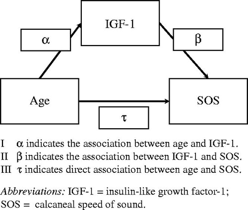
Where α = the regression coefficient of the association a (age and IGF-1); β = the regression coefficient of the association b (IGF-1 and SOS); SEα = standard error of the regression coefficient α; SEβ = standard error of the regression coefficient β.
Figure 1. The proposed scheme of mediation.
Results
The subjects recruited consisted of 140 Chinese and 139 Malay men. Their age ranged from 20 to 80 years old, with a mean age of 44.9 ± 15.3 years. The Chinese subjects were significantly taller, lower in weight and BMI and their serum SHBG level was significantly higher compared to Malay subjects (p < 0.05). There were no significant differences in age, calcaneal SOS, serum TT and IGF-1 levels between the two ethnic groups (p > 0.05; ). The Chinese and Malay subjects were grouped in the following analysis since there were no major differences in the variables of interest (age, IGF-1 and SOS).
Table 1. Characteristics of the subjects.
Spearman's correlation analysis showed that age correlated negatively and significantly with IGF-1 (r = −0.783; p < 0.05) and SOS (r = −0.318; p < 0.05). Serum IGF-1 correlated positively and significantly with SOS (r = 0.231; p < 0.05). It also revealed significant associations between the studied variables (age, IGF-1 and SOS) with potential confounders (BMI, TT and SHBG; p < 0.05). Hence, for the following regression analysis, BMI, TT and SHBG were adjusted ().
Table 2. Correlation analysis among the studied variables.
Multiple regression analysis revealed that there was a parallel and significant decrease of SOS and IGF-1 with increasing age (p < 0.05; Model 1 and 4, ). There was a significant positive association between IGF-1 and SOS after adjustments for BMI, SHBG and TT levels (p < 0.05; Model 2, ). However, after adjustment for age, this relationship reverted to non-significance (p > 0.05; Model 3, ).
Table 3. Multiple regression analysis on the associations among age, insulin-like growth factor-1 and calcaneal speed of sound.
The strength of the association between age and SOS, as assessed by standardized regression value was reduced after IGF-1 level was adjusted, but the relationship remained significant (p < 0.05; Model 5, ). Sobel test revealed that the mediation of IGF-1 was significant, as indicated by a significant z-value (z = −4.3, which was less than the critical value of −1.96). The mediation was partial because the association between age and SOS remained significant after IGF-1 level was controlled.
Discussion
Age-related decline of bone health status in men is mediated by a multitude of factors, such as changes in the bioavailability and quantity of testosterone, sex hormone-binding globulin and parathyroid hormone [Citation21]. This study showed that serum IGF-1 level was a mediator that explained age-related decline of bone health status in men, as measured using the QUS technique, via a simplified mediator analysis. In agreement with previous studies employing BMD as the bone health determinant [Citation4,Citation10], this study showed that adjustment for age reduced the variation of SOS explained by serum IGF-1 and the association became not significant. However, this study further explored that relationship between IGF-1 and bone health by mediator analysis and found a significant mediation effect of IGF-1 in the relationship between age and SOS. These results indicated that although serum IGF-1 might not explain more variation in SOS than age alone, it was still a component of the complex network of factors that explained age-related decline in bone health status in men.
A number of studies found that the association between IGF-1 and bone health was significant and independent of age in women but not in men. In the Framingham Heart Study involving 425 women and 257 men aged 72–84 years, serum IGF-1 was significantly and positively associated with BMD at several sites in elderly women but not in elderly men after age-adjustment [Citation4]. The same findings were obtained in the Rancho Bernardo Study involving 483 men and 455 post-menopausal women aged 50 years and above, whereby total spine and hip BMD was significantly related to serum IGF-1 in women but not in men [Citation10]. These findings were confirmed by a prospective study conducted by Seck et al., involving 107 women and 173 men aged 51–82 years, with a mean follow-up period of 3.3 years. They discovered a significant association between serum IGF-1 level with the loss of femoral neck BMD in women but not in men [Citation22]. The explanation for this gender discrepancy has not been found. None of these studies attempted a mediator analysis similar in this study. There was no study done that had assessed bone health status using QUS technique, so we did not have similar studies to be compared to.
Nevertheless, previous studies reporting age-independent association between IGF-1 and BMD were also found. Krassas et al. found that IGF-1 was significantly associated with BMD at the femoral neck and trochanter in a group of healthy Greek men. They found that the associations between IGF-1 and BMDs at the respective regions were stronger than age itself [Citation8]. It was noteworthy that 11% of their subjects were osteoporotic. This might contribute to a greater variance in bone health compared to this study, whereby the subjects screened were not osteoporotic. Gillberg et al. [Citation7] reported that serum IGF-1 was associated with femoral neck BMD in a relatively small sample size (n = 55) of Swedish men after adjustment for age and weight. Using partial correlation analysis, Szulc et al. demonstrated that after adjusting for age; total hip BMD, bone mineral content (BMC) and bone mineral apparent density, whole body BMD and BMC, femoral cortical thickness were significantly correlated with IGF-1. Their sample size was more than twice as large (721 subjects) as compared to this study and the power of the study was greater [Citation9].
Several experimental studies using knock-out mice illustrated the importance of IGF-1 on skeletal health. Heterozygote IGF-1 deficient (HET) mice had lower mineralization rate and bone formation rate compared to wild type [Citation2]. They also showed significant reductions in femoral length, bone mineral content, BMD, trabecular volume and cortical bone parameters [Citation3,Citation23,Citation24]. Similarly, IGF-1 receptor disruption in osteoblasts in mice caused a reduction in bone structural, static and dynamic histomorphometric parameters [Citation25]. In vitro studies showed that proliferation and differentiation of osteoblasts of HET mice were lower compared to osteoblasts of wild-type mice [Citation23]. Besides that, IGF-1 signaling was also shown to induce osteoblast migration [Citation26], matrix synthesis [Citation25] and mineralization [Citation27].
This study adopted the calcaneal SOS as a determinant of bone health status. Calcaneal SOS was shown to reflect BMD and microarchitecture of the bone [Citation28,Citation29]. The calcaneus consists of more than 95% trabecular bone, which is highly porous and provides a high bone surface per volume ratio for the bone exposure to humorous factors like IGF-1 [Citation30]. Hence, this site might be relatively responsive to the variation of these factors in men.
The association between IGF-1 and bone health status suggests that it may be a potential therapeutic target in osteoporosis. Several clinical studies had indicated that serum IGF-1 level was lower in male idiopathic osteoporotic patients [Citation31,Citation32]. The use of recombinant IGF-1 was shown to improve bone health status in IGF-1 deficient mice [Citation2] and in ovariectomized rats (in combination with other therapies) [Citation33]. However, one clinical trial on post-menopausal women treated with recombinant IGF-1 for 1 year concluded that there was no improvement in BMD [Citation34]. No similar study has been conducted in osteoporotic men. Hence, the potential of IGF-1 as an anti-osteoporotic agent is still in question and warrants further research.
This study possessed several limitations. It employed a non-randomized sampling technique due to logistic difficulties so the generalization of the results of this study should be conducted with caution. However, the sample in this study consisted of an equal proportion of Chinese and Malay subjects, which was similar to the previously reported population composition of Klang Valley, Malaysia [Citation35]. IGF-1 is bound by several types of binding proteins (IGFBPs) and some studies indicated that these IGFBPs were also related to bone health status in humans [Citation7,Citation8]. However, IGFBP levels were not measured in this study. The QUS device, CM-200 generated SOS as the sole bone health determinant, but not broadband ultrasound attenuation, another common QUS index. However, previous studies showed SOS was more related to BMD and elasticity [Citation28,Citation29]. Due to the cross-sectional nature of this study, the causal relationship between IGF-1 and SOS could not be determined. A prospective study should be performed for this purpose. This study was novel in that it was the first to determine the association between serum IGF-1 and QUS indices, and the first to use mediator analysis to evaluate the role of IGF-1 in the association between age and bone health status. Potential confounders for SOS measurement, such as BMI, testosterone and SHBG were considered and adjusted in the analysis.
Conclusion
In conclusion, serum IGF-1 is a partial mediator in age-related decline of bone health status in men, using calcaneal SOS as the determinant. A prospective study should be conducted to validate this relationship.
Declaration of interest
The authors reported no conflict of interest. This work was supported by Arus Perdana Grant (UKM-AP-TKP-09-2009), Postgraduate Research Grant (FF-376-2010) and Impak Perdana Grant (DIP-2012-07).
References
- Laron Z. Insulin-like growth factor 1 (IGF-1): a growth hormone. Mol Pathol 2001;54:311–6
- Bikle D, Majumdar S, Laib A, et al. The skeletal structure of insulin-like growth factor I-deficient mice. J Bone Miner Res 2001;16:2320–9
- Mohan S, Baylink DJ. Impaired skeletal growth in mice with haploinsufficiency of IGF-I: genetic evidence that differences in IGF-I expression could contribute to peak bone mineral density differences. J Endocrinol 2005;185:415–20
- Langlois JA, Rosen CJ, Visser M, et al. Association between insulin-like growth factor I and bone mineral density in older women and men: the Framingham heart study. J Clin Endocrinol Metab 1998;83:4257–62
- Yamaguchi T, Kanatani M, Yamauchi M, et al. Serum levels of insulin-like growth factor (IGF); IGF-binding proteins-3, -4, and -5; and their relationships to bone mineral density and the risk of vertebral fractures in postmenopausal women. Calcif Tissue Int 2006;78:18–24
- Kim JG, Shin CS, Choi YM, et al. The relationship among circulating insulin-like growth factor components, biochemical markers of bone turnover and bone mineral density in postmenopausal women under the age of 60. Clin Endocrinol (Oxf) 1999;51:301–7
- Gillberg P, Olofsson H, Mallmin H, et al. Bone mineral density in femoral neck is positively correlated to circulating insulin-like growth factor (IGF)-I and IGF-binding protein (IGFBP)-3 in Swedish men. Calcif Tissue Int 2002;70:22–9
- Krassas G, Papadopoulou P, Koliakos G, et al. Growth hormone, insulin growth factor-1, and igf binding protein-3 axis relationship with bone mineral density among healthy men. Arch Androl 2003;49:191–9
- Szulc P, Joly-Pharaboz MO, Marchand F, Delmas PD. Insulin-like growth factor I Is a determinant of hip bone mineral density in men less than 60 years of age: MINOS study. Calcif Tissue Int 2004;74:322–9
- Barrett-Connor E, Goodman-Gruen D. Gender differences in insulin-like growth factor and bone mineral density association in old age: the Rancho Bernardo Study. J Bone Miner Res 1998;13:1343–9
- World Health Organization. Assessment of fracture risk and its application to screening for postmenopausal osteoporosis: report of a World Health Organization Study Group. In WHO Technical Report Series No 843. Geneva: World Health Organization; 1994
- Handa R, Ali Kalla A, Maalouf G. Osteoporosis in developing countries. Best Pract Res Clin Rheumatol 2008;22:693–708
- Laugier P. An overview of bone sonometry. Int Congr Ser 2004;1274:23–32
- Chin KY, Soelaiman IN, Mohamed IN, et al. The effects of age, physical activity level, and body anthropometry on calcaneal speed of sound value in men. Arch Osteoporos 2012;7:135–45
- Chin KY, Soelaiman IN, Mohamed IN, Ngah WZ. Serum testosterone, sex hormone-binding globulin and total calcium levels predict the calcaneal speed of sound in men. Clinics 2012;67:911–6
- Krieg M-A, Barkmann R, Gonnelli S, et al. Quantitative ultrasound in the management of osteoporosis: the 2007 ISDN official positions. J Clin Densitom 2008;11:163–87
- Chin K-Y, Soelaiman I-N, Mohamed IN, et al. Discrepancy between the quantitative ultrasound value of Malaysian men and the manufacturer's reference and the impact on classification of bone health status. J Clin Densitom 2013;16:189–95
- Chin K-Y, Soelaiman I-N, Naina Mohamed I, et al. Testosterone is associated with age-related changes in bone health status, muscle strength and body composition in men. Aging Male 2012;15:240–5
- Sobel M. Asymptotic confidence intervals for indirect effects in structural equation models. Sociol Methodol 1982;13:290–321
- Baron RM, Kenny DA. The moderator-mediator variable distinction in social psychological research – conceptual, strategic, and statistical considerations. J Pers Soc Psychol 1986;51:1173–82
- Riggs BL, Khosla S, Melton LJ. Sex steroids and the construction and conservation of the adult skeleton. Endocr Rev 2002;23:279–302
- Seck T, Scheidt-Nave C, Leidig-Bruckner G, et al. Low serum concentrations of insulin-like growth factor I are associated with femoral bone loss in a population-based sample of postmenopausal women. Clin Endocrinol (Oxf) 2001;55:101–6
- He J, Rosen CJ, Adams DJ, Kream BE. Postnatal growth and bone mass in mice with IGF-I haploinsufficiency. Bone 2006;38:826–35
- Yakar S, Rosen CJ, Beamer WG, et al. Circulating levels of IGF-1 directly regulate bone growth and density. J Clin Invest 2002;110:771–81
- Zhang M, Xuan S, Bouxsein ML, et al. Osteoblast-specific knockout of the insulin-like growth factor (IGF) receptor gene reveals an essential role of IGF signaling in bone matrix mineralization. J Biol Chem 2002;277:44005–12
- Nakasaki M, Yoshioka K, Miyamoto Y, et al. IGF-I secreted by osteoblasts acts as a potent chemotactic factor for osteoblasts. Bone 2008;43:869–79
- Tiago DM, Cancela ML, Laize V. Proliferative and mineralogenic effects of insulin, IGF-1, and vanadate in fish osteoblast-like cells. J Bone Miner Metab 2011;29:377–82
- Guglielmi G, de Terlizzi F. Quantitative ultrasound in the assessment of osteoporosis. Eur J Radiol 2009;71:425–31
- Chin K-Y, Ima-Nirwana S. Calcaneal quantitative ultrasound as a determinant of bone health status: what properties of bone does it reflect? Int J Med Sci 2013;10:1778–83
- Chin K, Ima-Nirwana S, Mohamed I, et al. Thyroid-stimulating hormone is significantly associated with bone health status in men. Int J Med Sci 2013;10:857–63
- Kurland ES, Rosen CJ, Cosman F, et al. Insulin-like growth factor-I in men with idiopathic osteoporosis. J Clin Endocrinol Metab 1997;82:2799–805
- Ljunghall S, Johansson AG, Burman P, et al. Low plasma levels of insulin-like growth factor 1 (IGF-1) in male patients with idiopathic osteoporosis. J Intern Med 1992;232:59–64
- Ammann P, Rizzoli R, Muller K, et al. IGF-I and pamidronate increase bone mineral density in ovariectomized adult rats. Am J Physiol Endocrinol Metab 1993;265:E770–6
- Friedlander AL, Butterfield GE, Moynihan S, et al. One year of insulin-like growth factor I treatment does not affect bone density, body composition, or psychological measures in postmenopausal women. J Clin Endocrinol Metab 2001;86:1496–503
- Statistics Department of Malaysia. Population distribution and basic demographic characteristics. Putrajaya: Statistics Department of Malaysia; 2011