Abstract
Antioxidative, skin protective activities, and cytotoxicity of three extracts (water, methanol, and hexane) from the fruit hull of mangosteen (Garcinia mangostana Linn. (Guttiferae)) and their phenolic constituents such as α-mangostin, epicatechin, and tannin, were evaluated. The amounts of α-mangostin, total flavonoid, and total tannin were different among the three extracts, except those of total tannin in methanol and hexane extracts. For the 1,1-diphenyl-2-picrylhydrazyl (DPPH) free-radical scavenging, hydroxyl radical-scavenging, and inhibition of lipid peroxidation experiment, the water extract showed higher activity than the methanol extract and hexane extract. α-Mangostin, epicatechin, and tannin also revealed these antioxidant and free radical-scavenging activities. When added simultaneously with H2O2 (200 μM) to keratinocyte cells, the water extract (50 μg/mL), epicatechin (200 μM), and tannin (200 μM) effectively protected cells from oxidative damage, but the methanol extract, hexane extract, and α-mangostin did not. The methanol extract and hexane extract exhibited moderate cytotoxicity, whereas α-mangostin showed strong cytotoxicity. The present study provides the evidence that Garcinia mangostana extracts, especially the G. mangostana water extract, act as antioxidants and cytoprotective agents against oxidative damage, which is at least partly due to its phenolic compounds in mangosteen.
Introduction
Recently, reactive oxygen species (ROS) have been of interest in free radical research due to their effects on human health (CitationMatés & Sánchez-Jiménez, 2000). In dermatology it is generally agreed that ROS are one of the major and important factors contributing to skin aging, skin disorders and skin diseases (CitationHarman, 1992; CitationRieger & Pains, 1993). Oxidative damage occurs due to the imbalance between antioxidant defense and free radicals derived from endogenous and exogenous sources (CitationParejo et al., 2003). Therefore, many efforts in studying and searching the substances that prevent or inhibit the activity of free radical-induced skin degenerative diseases have been investigated (CitationWeisburger, 1999).
Garcinia mangostana Linn. (Guttiferae) has a long history in folk medicine. The fruit rinds of G. mangostana have been used for the treatment of diarrhea, skin infections, wounds, and chronic ulcers. Several studies have revealed that G. mangostana exhibits antimicrobial, antiproliferative, antioxidant, and anti- inflammatory properties (CitationGopalakrishnan et al., 1980; CitationWilliams et al., 1995). Phytochemical studies have revealed that the pericarp of G. mangostana is a source of xanthones, tannins, flavonoids, vitamin C, and other bioactive substances (CitationFarnsworth & Bunyapraphatsara, 1992). These compounds are potent antioxidants and free radical scavengers. Therefore, we speculated that G. mangostana extract and its constituents might have skin protective activities, at least partly due to its antioxidant activity.
Extraction is one of the important steps in obtaining active constituents from medicinal plants. The biological quality of G. mangostana-derived constituents is based on the contents of the active compounds. Different solvents can extract different amounts of active compounds due to distinct affinities for compounds. Methanol extract of G. mangostana has been widely used in the literature for evaluating the antioxidant activity (CitationYoshikawa et al., 1994; CitationMoongkarndi et al., 2004; CitationChomnawang et al., 2007). However, little data are available for the comparison of this activity among different solvent extracts of G. mangostana. In this study we investigated the antioxidant and skin protective actions of three solvent extracts of G. mangostana as well as its xanthone (α-mangostin), flavonoid (epicatechin), and tannin.
Materials and methods
Chemicals
Normal human epidermal keratinocytes of adult skin (male, age 47 years, NHEK125) and Keratinocyte Growth Medium® (KGM II) were purchased from Clonetics (San Diego, CA). 1,1-Diphenyl-2-picryl-hydrazyl (DPPH), epicatechin, tannin, 3-(4,5-dimethylthiazol-2-yl)-2,5-diphenyl tetrazolium bromide (MTT), malondialdehyde (MDA), and vitamin E were purchased from Sigma (St Louis, MO). α-Mangostin was purchased from Chroma Dex (Santa Ana, CA). Dulbecco’s modified phosphate-buffered saline (PBS) was obtained from Gibco (Grand Island, NY). All other reagents and solvents were commercially available and of analytical grade.
Plant material and extraction
Garcinia mangostana plants collected from Chantaburi Province (Thailand) in May 2006 were identified by C. Sittisombut, Botanist, Faculty of Pharmacy, Silpakorn University, Nakhon Pathom, Thailand. A voucher specimen was deposited in the Herbarium of the Faculty of Pharmacy, Silpakorn University, Nakhon Pathom, Thailand. The milled fruit hulls of G. mangostana were macerated at room temperature for 7 days with distilled water, methanol, or hexane. The mixture was shaken occasionally. The mixture was filtered through Whatman No. 1 filter paper. The crude extract was then evaporated in a boiling water bath until a constant weight was obtained.
Determination of α-mangostin content
The HPLC method for analysis of α-mangostin was followed (CitationWalker, 2007) with slight modification. The HPLC system consisted of a pump (LC-6AD, Shimadzu, Kyoto), a Spherisorb ODS 5-C18, 4.6 mm × 250 mm column (YMC, Kyoto), an auto-injector, a variable UV detector, and an integrator. A mobile phase consisted of 0.1% (v/v) of formic acid (A) and methanol (B). The optimal separation was achieved using a gradient of 65% to 90% B over 0-30 min. The injection volume was 20 μl. The flow rate of the mobile phase was 1 mL/min. The detection wavelength was set at 254 nm.
Determination of total flavonoid content
The total flavonoid content was determined using the method of CitationMeda et al. (2005). The extract (500 μg/mL) was mixed with the same volume of 2% aluminum trichloride in methanol. The mixture was mixed together and allowed to stand at room temperature for 10 min. The absorbance of the mixture was measured by spectrophotometer (U-2000, Hitachi, Japan) at 415 nm against a blank sample without aluminum trichloride. Epicatechin was used as a standard. The total flavonoid content was expressed as epicatechin equivalents (EE) in g/100 g of extract.
Determination of total tannin content
The tannin content was determined using the method of CitationHangerman and Butler (1978) with slight modification. Bovine serum albumin (2 mL) was mixed with sample solution (1 mL). The mixture was kept for 20 min, and then centrifuged at 3,500 rpm for 15 min, and the supernatant was rinsed off. The pellet was dissolved in 4 mL sodium dodecyl sulfate and triethanolamine. Ferric chloride (1 mL) was added and the mixture was shaken vigorously. The absorbance of the mixture was measured with a spectrophotometer at 510 nm. Tannic acid was used as a standard. The total tannin content was expressed as tannic acid equivalents (TAE) in g/100 g of extract.
DPPH free radical scavenging activity
The DPPH radical scavenging activity was measured by the methodology of CitationBlois (1958). Briefly, stock solutions of the test compounds were prepared by dissolving 0.1 g dry extract in 50 mL methanol. The stock solution was diluted with methanol to obtain sample solutions at various concentrations. The sample solutions were thoroughly mixed with freshly prepared 0.01% DPPH methanol solution at the ratio of 1:1 then kept in the dark for 30 min. The amount of the reaction mixture was determined by spectrophotometer at 517 nm against a blank sample. The measurement was performed in three independent experiments. The antioxidant activity of the test compound was calculated as percentage inhibition from the following equation:
where ODblank is the absorbance of the control and ODsample is the absorbance of the test sample. The radical scavenging activity of DPPH was expressed as IC50. This value represents the concentration of a test compound required to inhibit 50% of the initial DPPH free radical.
Scavenging of hydroxyl radicals activity
The formation of hydroxyl radicals (O•H) from Fenton reagents was quantified using 2-deoxyribose oxidative degradation according to the method of CitationElizabeth and Rao (1990). The reaction mixture contained deoxyribose (2.8 mM); FeCl3 (100 μM); KH2PO4-KOH buffer (20 mM, pH 7.4); EDTA (100 μM); H2O2 (1 mM); ascorbic acid (100 μM); and various concentrations of the test compounds in a final volume of 1 mL. The reaction mixture was incubated at 37°C for 60 min. After incubation, 1 mL 2.8% TCA was added to 0.5 mL of sample. After the samples were centrifuged at 3,000 g for 15 min, the supernatant was incubated with 1 mL 0.6% TBA aqueous solution at 100°C for 15 min. After a cooling period, the absorbance of thiobarbituric acid-reactive substance (TBARS) was measured with a spectrophotometer at 532 nm against a blank sample. Mannitol, a classical O•H scavenger, was utilized as a positive control. Decreased absorbance of the reaction mixture indicates increased hydroxyl radical scavenging activity. The hydroxyl radical-scavenging activity was calculated using the equation described above for DPPH. The hydroxyl radical-scavenging activity was expressed as IC50. This value represents the concentration of a test compound required to inhibit 50% of the initial hydroxyl radical.
Inhibition of lipid peroxidation
Eight-week-old male BALB/c mice (National Animal Center, Mahidol University, Bangkok, Thailand) were housed under standard experimental conditions. Experiments were performed in accordance with the Guideline for Animal Experimentation (CitationOECD, 2000). Under pentobarbital anesthesia the abdominal skin of the mice was shaved and freshly excised before the experiments; the mice were then sacrificed by cervical dislocation. Nine volumes of cold phosphate-buffered saline (pH 7.4) were added to the isolated skins and then homogenized with a tissue homogenizer. The protein content of the homogenate was determined by the Lowry method using BSA as a reference standard. The inhibition of lipid peroxidation activity was determined by quantification of TBARS using the method of Buege and Aust with a slight modification (1978). The skin homogenate (0.5 mL) was mixed with phosphate buffer (20 mM, pH 7.4) 0.9 mL 0.01 mM FeSO4 mixed with 0.5 mL ascorbic acid, and 0.1 mL of test compounds or vehicles. The reaction mixtures were incubated at 37°C for 30 min and the reaction was stopped on ice by adding 0.5 mL of 35% perchloric acid. After centrifugation at 3,000 g for 15 min, the supernatant was incubated with 0.5 mL of 0.6% TBA at 100°C for 15 min. After cooling period, the absorbance of TBARS was measured with a spectrophotometer at 532 nm. MDA was used as a standard. The lipid peroxidation-inhibition activity was calculated using the equation described above for DPPH. The lipid peroxidation-inhibition activity was expressed as IC50. This value represents the concentration of a test compound required to inhibit 50% of the initial TBARS.
Cell cultures
Normal human epidermal keratinocytes were stored as cryopreserved cells. The culture medium for human keratinocytes was Keratinocyte Growth Medium® (KGF). After thawing a cryovial of keratinocytes, the cells were cultured in 25 cm2 flask with 5 mL complete medium. Keratinocytes were maintained at 37°C in a 5% CO2 incubator.
MTT assay for cell viability
MTT solution (1 mg/mL in medium, 100 μL) was added to each well of 96-well plates and then incubated for 3 h at 37°C. The medium was removed and 100 μL DMSO was then added to each well and vigorously mixed to dissolve the formazan crystals. The absorbance of eluted sample was measured directly in a microplate reader at 550 nm (Microplate Manager, Bio-Rad Laboratories, Hercules, CA). The percentage cell viability was calculated from the following equation:
Scavenging activity of extracts in H2O2- induced oxidative cell damage
Keratinocyte cells were plated at a density of 2.5 × 10³ cells/well in 96-well plates and incubated at 37°C with 5% CO2 for 48 h. After incubation, the cells were treated for the test experiments. The test compound was first dissolved in DMSO and later mixed with the culture medium to give a final concentration of 0.5% v/v of DMSO. In our preliminary results, DMSO at this concentration had no effect on cell viability and did not show any protective effect against H2O2-induced oxidative damage in keratinocyte cells (data not shown). DMSO was also present at this level in control cells (those not treated with test compounds). Keratinocyte cells were simultaneously treated with various concentrations of test compound and H2O2 (200 μM). The controls were the cells that were not treated with extracts or an oxidant agent and cells exposed to H2O2-induced oxidative damage in the absence of test compounds. Cells were incubated at 37°C with 5% CO2 for 3 h, washed twice with PBS, and fresh media were added to the wells. Cell viability was then determined using the MTT assay.
Cytotoxicity assay
Cells were incubated with various concentrations of test compound at 37°C with 5% CO2 for 24 h. After 24 h incubation, cell viability was determined using the MTT assay. The cytotoxicity of test compounds was expressed as IC50. This value represents the concentration of the test compounds required to inhibit 50% of the initial cell viability of control.
Statistical analysis
All results were expressed as mean ± SD. Data were analyzed by one-way analysis of variance (ANOVA) followed by LSD post hoc test. Differences of p < 0.05 were considered statistically significant.
Results
Contents of α-mangostin, total flavonoid compounds, and total tannin in G. mangostana extracts
shows the content of α-mangostin, total flavonoid and total tannin in G. mangostana water, methanol, and hexane extracts. The content of α-mangostin could not be detected in the water extract, whereas higher α-mangostin content was observed in the hexane extract (28.7% w/w) than the methanol extract (15.5% w/w). Among the various extracts, the water extract had the highest total flavonoid content (12.1 g EE/100 g extract) and total tannin content (59.6 g TAE/100 g extract). The total flavonoid content in the methanol extract (8.7 g EE/100 g extract) was slightly higher than in the hexane extract (6.8 g EE/100 g extract), whereas the total tannin content of these extracts was not significantly different (36.4–37.7 g TAE/100 g extract).
Table 1. Contents of alpha-mangostin, total phenolic compounds, total tannin in various G. mangostana extractsa.
DPPH radical-scavenging activity
The DPPH radical scavenging activity, hydroxyl radical scavenging activity and inhibition of lipid peroxidation in skin homogenates of test compounds are shown in . The DPPH radical scavenging activity of the water extract (IC50 11 μg/mL) and methanol extract (IC50 14.7 μg/mL) were more efficient than that of the hexane extract (IC50 41.2 μg/mL). Vitamin E, a reference standard, exhibited scavenging activity with an IC50 26.2 μM. α-Mangostin, epicatechin, and tannin also revealed scavenging activity on DPPH radicals with IC50 of 89.2, 13.5, and 12.3 μM, respectively.
Table 2. IC50 of DPPH radical-scavenging activity, hydroxyl radical-scavenging, and inhibition of lipid peroxidation activity in various G.mangostana extracts and test compoundsa.
Scavenging of hydroxyl radicals activity
Mannitol exhibited deoxyribose degradation with an IC50 value of 4 mM. G. mangostana extracts also suppressed deoxyribose degradation in the following order: water extract (1.14 mg/mL) > methanol extract (1.38 mg/mL) > hexane extract (1.53 mg/mL). α-Mangostin, epicatechin, and tannin had inhibitory effects on deoxyribose degradation. Their respective IC50 values were 2.61, 2.65, and 1.42 mM.
Inhibition of lipid peroxidation
Vitamin E had an IC50 value of 140.5 μM. The suppressive effect of the G. mangostana water extract (IC50 85.1 μg/mL) on the lipid peroxidation was the most potent, followed by the methanol extract (IC50 172.1 μg/mL), and the hexane extract (IC50 203.5 μg/mL). α-Mangostin, epicatechin, and tannin also showed a significant suppression of TBARS formation in skin tissue homogenates. Their respective IC50 values were 402.7, 64.2, and 104.4 μM.
H2O2- induced oxidative cell damage in keratinocytes
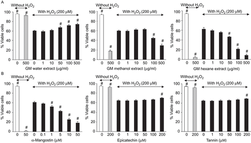
Human keratinocyte cells were treated with various concentrations of H2O2 (100, 200, 300, 400, or 500 μM) for 3 h, then cell viability was determined. The results showed that H2O2 significantly reduced cell viability in a concentration-dependent manner with an IC50 value of 200 μM (data unpublished). At this concentration cell viability was about 60% of the control. The concentration of H2O2 at 200 μM was therefore used as the control for H2O2-induced oxidative cell damage. When G. mangostana water extract (50-500 μg/mL), epicatechin (200 μM), or tannin (200 μM) were added together with H2O2, they significantly increased the viability of keratinocyte cells in a concentration- dependent manner compared with H2O2 treatment alone (). There was no significant increase in cell viability when the methanol extract, hexane extract, or α-mangostin was added simultaneously with H2O2. However, when the higher concentrations of methanol extract (50-500 μg/mL), hexane extract (10-500 μg/mL), and α-mangostin (1-50 μM) were added simultaneously with H2O2 to human keratinocyte cells, the cell viability significantly decreased.
Figure 1. Effect of test compounds on H2O2-induced cell damage in keratinocyte cells when added with H2O2. Keratinocyte cells were treated with H2O2 (200 μM) with various test compounds: (A) water extract, methanol extract, and hexane extract (B) phenolic compounds contained in G. mangostana extracts (α-mangostin, epicatechin, and tannin). After 3 h of incubation, cell viability was measured using MTT assay. Data are expressed as mean ± SD (n = 3). #p < 0.05 compared with the H2O2-treated group.

Cytotoxicity in human keratinocyte cells
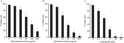
The toxicity of test compounds in human keratinocyte cells was examined. The water extract, epicatechin, and tannin lacked detectable cytotoxicity under tested concentrations (). The methanol extract (IC50 72 μg/mL) and hexane extract (IC50 30 μg/mL) showed moderate cytotoxicity against keratinocyte, and α-mangostin (IC50 2.5 μM; 0.94 μg/mL) exhibited strong cytotoxicity ().
Figure 2. Effect of test compounds on toxicity in keratinocyte cells. Keratinocyte cells were treated with various test compounds: (A) methanol extract, (B) hexane extract, (C) α-mangostin. After 24 h of incubation, cell viability was measured using MTT assay. Data are expressed as mean ± SD (n = 3). #p < 0.05 compared with the control (without test compounds).

Discussion
In this study we investigated the possible antioxidant properties and skin protective activities of G. mangostana extracts prepared by different solvents (water, methanol, and hexane) and their phenolic constituents (α-mangostin, epicatechin, and tannin). The examination of the defense systems against skin oxidative damage by antioxidants may contribute to the understanding of skin aging and the mechanisms involved in the various skin pathology processes (CitationRieger & Pains, 1993).
The phenolic compounds such as α-mangostin, flavonoid and tannin are the composition in the pericarp of G. mangostana (CitationFarnsworth & Bunyapraphatsara, 1992; CitationYu et al., 2007). Variation in the contents of α-mangostin, total flavonoid, and total tannin in different G. mangostana extracts was observed. α-Mangostin, a naturally occurring xanthone, is insoluble in water, and its difference in solubility in variety of non-polar solvents has been reported (CitationBudavari, 1989). The variation in the amounts of these constituents in different extracts might be due to the distinct solubility of the compounds in the particular solvents used for extraction, and the abilities of the solvents to penetrate the sample during extraction (CitationWalker, 2007).
The free radical scavenging activity of G. mangostana extracts and their phenolic constituents was evaluated based on their ability to quench the synthetic radical DPPH. The bleaching of DPPH absorption is representative of the capacity of test compounds to scavenge free radicals. Our results demonstrate that all G. mangostana extracts and their phenolic constituents are effective in DPPH free radical scavenging.
The hydroxyl radical is a highly potent oxidant that reacts with almost all biomolecules found in living cells. The deoxyribose assay allows the determination of the rate constant of reaction with OH•. In this model, deoxyribose is degraded by OH• generated by Fenton systems and results in a series of reactions during which MDA is formed and may be detected by its ability to react with TBA to form a pink chromogen. Therefore, the ability to diminish the amount of color formation has been adapted as one measurement of antioxidative properties. The G. mangostana extracts and their phenolic constituents exhibit an inhibitory effect on hydroxyl radical-induced deoxyribose degradation.
The inhibition of lipid peroxidation has been used as a model to elucidate antioxidant activity. Superoxide indirectly initiates lipid peroxidations because it is a precursor of singlet oxygen and OH• (CitationGao et al., 2000). Hydroxyl radicals eliminate hydrogen atoms in the membrane lipids, which results in lipid peroxidation. The inhibition of lipid peroxidation by G. mangostana extracts and its phenolic constituents may be due to their free radical scavenging activity.
The skin protective effect of G. mangostana extracts and their phenolic constituents on H2O2-induced cell damage of human keratinocyte cells was evaluated. Keratinocyte cells are normally used as a skin model in physiology and pharmacology resewarch. In this study they were chosen as a skin model for studying skin protective effects because they are reported to be more sensitive to oxidative damage than fibroblasts (CitationPhan et al., 2001). Oxidative stress is believed to be an important mediator of cell death; therefore, H2O2 was chosen to induce in oxidative damage in the cell culture. H2O2 was reported to cause fatal injury to keratinocytes, to block cell signaling by inhibiting of epidermal growth factor (EGF) receptor internalization, and to inhibit keratinocyte migration (CitationSimon et al., 1981).
The G. mangostana water extract exhibited protective effects against H2O2-induced decreases in cell viability. This extract lacked detectable cytotoxicity of keratinocyte cells under the concentrations investigated. Topical application of compounds with free radical scavenging properties to patients or animals enhances the protection of tissues from oxidative damage (CitationFoschi et al., 2002). Therefore, it is possible that the skin protective effect may at least partly result from antioxidant and free radical scavenging activities.
The ethanol extract of G. mangostana shows cytotoxicity against Caco-2 cells and blood mononuclear cells with the IC50 values of 32 and 4.9 μg/mL, respectively (CitationOkonogi et al., 2007). α-Mangostin is the major xanthone found in non-polar extracts of G. mangostana (CitationJung et al., 2006). It exhibited a strong cytotoxicity in keratinocyte cells. It is therefore possible to suggest that the presence of α-mangostin in the methanol and hexane extract may play some role in the decrease in cell viability in cytotoxicity assay. The decrease in skin protective activity of the G. mangostana methanol extract, hexane extract, and α-mangostin at high concentrations of these test compounds may be due to their toxicity to keratinocyte cells.
G. mangostana methanol, G. mangostana hexane, and their components: epicatechin, tannin, and α-mangostin have hydroxyl radical scavenging and anti-lipid peroxidation activity; therefore, G. mangostana methanol and G. mangostana hexane extract might interact with H2O2 or ROS derived from H2O2 leading to reduction of oxidative damage from H2O2. The difference in total content of epicatechin, tannin, and α-mangostin in methanol and hexane extracts at similar extract concentration might cause variation in cell viability in these different oxidative stress conditions.
The antioxidant activities of G. mangostana extract might be partly from their phenolic constituents. Polyphenolic compounds are widely distributed in plants. Many phenolic compounds have been shown to have biological actions, including antioxidant activity (CitationIshige et al., 2001). Epicatechin and tannin exhibit antioxidant, free-radical scavenging, and skin protective activity. α-Mangostin shows antioxidative activity in cell-free assay, however, it has a strong toxicity to keratinocyte cells. Among the various extracts, the G. mangostana water extract had the highest total flavonoid and total tannin content, but had no α-mangostin. Our study provides the evidence that the G. mangostana water extract has antioxidant and cytoprotective properties, which is partly due to some phenolic compounds such as epicatechin and tannin.
In the present study our results clearly demonstrate that G. mangostana extracted by different solvents exhibits a variety of antioxidant, free-radical scavenging activity and cytotoxicity due to the distinction in the contents of the compounds among extracts. The G. mangostana water extract would have been expected to be superior skin protection effects compared to the methanol extract and the hexane extract, since α-mangostin, which is not included in G. mangostana water extract, is strongly involved in cytotoxicity. The antioxidant and cytoprotective agent of G. mangostana extracts against oxidative damage might be partly due to phenolic compounds. However, unknown factors may participate in the cytotoxic restraining action of the water extract. The antioxidant and skin protective activity, especially in the water extract, may explain the effects of traditional treatment with G. mangostana in wound healing and chronic ulcer.
Declaration of interest: This work was supported by the National Research Council of Thailand, the Thailand Research Fund and the Commission on Higher Education, Ministry of Education, Thailand (RSA 4880001). T. Ngawhirunpat acknowledges a fellowship for researchers from the National Research Council of Thailand and the Japanese Society for the Promotion of Science (NRCT-JSPS) under Core University Program. The authors alone are responsible for the content and writing of the paper.
References
- Blois MS (1958): Antioxidant determination by the use of a stable free radical. Nature 181: 1199–1200.
- Budavari S (1989): Merck Index: An Enclyclopedia of Chemicals, Drugs, and Biologicals. NJ, White House Station, New Jersey, pp. 123–124.
- Buege JA, Aust SD (1978): Microsomal lipid peroxidation. Meth Enzymol 52: 302–310.
- Chomnawang MT, Surassmo S, Nukoolkarn VS, Gritsanapan W (2007): Effect of Garcinia mangostana on inflammation caused by Propiobacterium acnes. Fitoterapia 78: 401–408.
- Elizabeth K, Rao MNA (1990): Oxygen radical scavenging activity of curcumin. Int J Pharm 58: 237–240.
- Farnsworth NR, Bunyapraphatsara N (1992): Mangosteen, in: Farnsworth NR, Bunyapraphatsara N, eds, Thai Medicinal Plant: Recommended for Primary Health Care System. Bangkok, Prachachon Company, pp. 25–50.
- Foschi D, Trabucchi E, Musazzi M, Castoldi L, Dimattia D, Radaelli E, Marazzi M, Franzini P, Berlusconi A (2002): Prostaglandin cytoprotection in man: Effectiveness of rosaprostol. Int J Tiss Reac 10: 373–379.
- Gao J, Igarashi K, Nukina M (2000): Three new phenylethanoid glycosides from Caryopteris incana and their antioxidative activity. Chem Pharm Bull 48: 1075–1078.
- Gopalakrishnan C, Shankaranarayanan D, Kameswara L, Nazimudern SK (1980): Effect of mangostin, a xanthone from Garcinia mangostana in immunopathological and inflammatory reactions. Ind J Exp Biol 18: 843–846.
- Hangerman AE, Butler LG (1978): Protein precipitation method for quantitative determination of tannins. J Agric Food Chem 26: 809–812.
- Harman D (1992): Role of free radicals in aging and disease. Ann NY Acad Sci 673: 126–141.
- Ishige K, Schubert D, Sagara Y (2001): Flavonoids protect neuronal cells from oxidative stress by three distinct mechanisms. Free Radic Biol Med 30: 433–446.
- Jung HA, Su BN, Keller WJ, Mehta RG, Kinghorn AD (2006): Antioxidant xanthones from the pericarp of Garcinia mangostana (Mangosteen). J Agric Food Chem 54: 2077–2082.
- Matés JM, Sánchez-Jiménez FM (2000): Role of reactive oxygen species in apoptosis: Implications for cancer therapy. Int J Biochem Cell Biol 32: 157–170.
- Meda A, Lamien CE, Romita M, Millogo J, Nacoulma OG (2005): Determination of total phenolic, flavonoid and praline contents in Burkina Fasan honey, as well as their radical scavenging activity. Food Chem 91: 571–577.
- Moongkarndi P, Kosem N, Kaslungka S, Luanratana O, Pongpan N, Neungton N (2004): Antiproliferation, antioxidation and induction of apoptosis by Garcinia mangostana (mangosteen) on SKBR3 human breast cancer cell line. J Ethnopharmacol 90: 161–166.
- Organisation for Economic Co-operation and Development (OECD) (2000): OECD New Guideline Proposal on in vitro Percutaneous Absorption of Chemicals. Paris, OECD, pp. 1–40.
- Okonogi S, Duangrat C, Anuchpreeda S, Tachakittirungrod S, Chowwanapoonpohn S (2007): Comparison of antioxidant capacities and cytotoxicities of certain fruit peels. Food Chem 103: 839–836.
- Parejo I, Viladomat F, Bastida J, Rosas-Romero A, Saavedra G, Murcia MA, Jimenez AM, Codina C, Jimenez AM, Codina C (2003): Investigation of Bolivian plant extracts for their radical scavenging activity and antioxidant activity. Life Sci 73: 1667–1681.
- Phan TT, See T, Lee ST, Chan SY (2001): Anti-oxidant effects of the extracts from the leaves of Chromolaena odorata on human dermal fibroblasts and epidermal keratinocytes against hydrogen peroxide and hypoxanthine-xanthine oxidase induced damage. Burns 27: 319–327.
- Rieger MM, Pains M (1993): Oxidative reactions in and on skin: mechanism and prevention. Cosmetic Toiletries 108: 43–56.
- Simon RH, Scoggin CH, Patterson D (1981): Hydrogen peroxide causes the fatal injury to human fibroblasts exposed to oxygen radicals. J Biol Chem 256: 7181–7186.
- Walker EB (2007): HPLC analysis of selected xanthones in mangosteen fruit. J Sep Sci 30: 1229–1234.
- Weisburger JH (1999): Mechanisms of action of antioxidants as exemplified in vegetables, tomatoes, and tea. Food Chem Toxicol 37: 943–948.
- Williams P, Ongsakul M, Proudfoot J, Croft K, Bellin L (1995): Mangostin inhibits the oxidative modification of human low density lipoprotein. Free Radic Res 23: 175–184.
- Yoshikawa M, Harada E, Miki A, Tsukamoto K, Si QL, Yamahara J, Murakami N (1994): Antioxidant activity of the extract from mangosteen (Garcinia mangostana). Yakugaku Zasshi 114: 129–133.
- Yu L, Zhao M, Yang B, Zhao Q, Jiang Y (2007): Phenolics from hull of Garcinia mangostana fruit and their antioxidant activities. Food Chem 104: 176–181.