Abstract
Matrine, one of the main active components extracted from dry roots of Sophora flavescens Ait (Leguminosae), has been reported to have anticancer effects on a number of cancer cell lines, but the anticancer mechanism of matrine remains elusive. This study shows that matrine also displays anticancer activity on human hepatocellular carcinoma (HepG2) cells. In this work, the optimal cultivation condition for HepG2 cells was determined using the combinatorial orthogonal test design [L18 (21 × 37)]. Exposure of HepG2 cells to matrine resulted in inhibition of proliferation in both a time- and dose-dependent manner, as measured by morphology observation, hematoxylin and eosin (H&E) staining, and MTT assay (p<0.05). Further immunohistochemical analyses revealed that the expression of alpha fetal protein (AFP), proliferating cell nuclear antigen (PCNA), C-myc and Bcl-2 was down-regulated significantly, but the expression of Bax was up-regulated higher than untreated cells. The results demonstrated that matrine inhibited HepG2 cells proliferation primarily via up-regulating or down-regulating expression of the tumor relevant proteins.
Introduction
Hepatocellular carcinoma (HCC), ranked fifth in cancer frequency, is the third most common cause of cancer-related death (CitationMerle, 2005). It usually occurs among people between the third and fifth decades of life with the estimation 0.5 to 1 million new cases and 662,000 deaths per year in the world. By the year 2010, HCC will have exceeded lung cancer as the foremost cause of cancer mortality (CitationClark et al., 2005). The current treatments are unfortunately ineffective in curing HCC, due to the drug-resistance and radio-resistance of HCC cells and the difficulty of achieving complete tumor resection. There is therefore an urgent need to seek novel therapeutic drugs to combat HCC.
Matrine, one of the main components extracted from a traditional Chinese herb, Sophora flavescens Ait (Leguminosae), with a molecular formula of C15H24N2O (CitationLi et al., 2007), was widely studied because of its pharmacological and clinical application. Matrine has displayed a wide range of pharmacological effects such as anti-Hepatitis B Virus (anti-HBV) for treatment of chronic hepatitis B (CitationLiu et al., 2003; CitationLong et al., 2004), anti-inflammation by inhibition of the expression of substance P receptor and regulation of inflammation cytokines production (CitationHu et al., 1996), antihepatocyte injury of sinusoidal endothelial cells in rat orthotropic liver transplantation (CitationLiang et al., 1999), anti-liver fibrosis induced by CCl4 in rats (CitationZhang et al., 2001), and promotes apoptosis in leukemia U937, JM, K562 cells by inhibition of NF-kB activation and multiple receptor tyrosine kinase activation in vitro (CitationLuo et al., 2007). Matrine has been used for the treatment of skin diseases, viral hepatitis, arrhythmia and liver cancer in China (CitationTao & Wang, 1992).
Recently, it has been reported that matrine exhibited antitumor effects by inhibiting proliferation and inducing apoptosis of cancer cells (CitationLi Y et al., 2007; CitationJiang et al., 2007). The anticancer activity of matrine may rely on its effects on the proliferation- and apoptosis-related proteins including alpha fetal protein (AFP), proliferating cell nuclear antigen (PCNA), N-ras, p53, C-myc, E2F-1, Apaf-1, Rb, Bcl-2 family, and caspases (CitationZhang et al., 2001, Citation2007; CitationLuo et al., 2007). However, the mechanism underlying the inhibition process by matrine remains elusive. This study investigated the mechanism of matrine-induced inhibition of the proliferation and expression of tumor relevant proteins of HepG2 cells, a cell line derived from liver tissue with a well differentiated hepatocellular carcinoma.
Materials and methods
Reagents
HepG2 cell line was obtained from Nanjing Keygen Biotech (Nanjing, China). Matrine (product standard 200510-19 99% purity) were purchased from Jilin Yuhuang Pharma (Changchun, China). Mouse anti-human monoclonal antibodies SABC kit (AFP, PCNA, C-myc, Bcl-2, Bax) and DBA kit were purchased from Wuhan Boster (Wuhan, China). RPMI-1640, DMEM, and trypsin were purchased from Gibco (Carlsbad, CA). Ethylenediamine tetra-acetic acid (EDTA) and fetal bovine serum (FBS) were purchased from Shanghai Sunway Biotech (Shanghai, China). Bovine serum albumin (BSA), dimethylsulfoxide (DMSO), and [3-(4,5-dimethylthiazol-2-yl)-2,5-diphenyl tetrazolium bromide] (MTT) were purchased from Sigma-Aldrich (St. Louis, MO). Other analytical reagents were purchased from Shenyang Chemical Reagents Factory (Shengyang, China).
Optimization of cultivation condition
Prior to treating cells with matrine, it was essential to determine the optimal cultivation condition of HepG2 cells. In the following, eight impact factors including culture medium, cell density, serum content, double antibiotics concentration (penicillin and streptomycin), pH, washing times (Hanks’ solution), trypsin concentration and digestion time were selected according to the combinatorial orthogonal test design [L18 (21×37)], as illustrated in . HepG2 cell was put in 75 mL tissue culture flasks sterilized using sterile techniques in a CO2 incubator (Thermo HEPA Class 100, Massachusetts, USA) and grown in a humidified atmosphere containing 5% CO2 at 37°C. Cell morphology was viewed under invert light microscope once every 4 h to assess cell growth condition. The number of cell nucleoli was confirmed using Wright’s staining. The following cell auxodrome and the divisional index of the HepG2 cells were obtained. Average stage of HepG2 growth was scored from 0 to 10 corresponding to every parallel treatment. Group 5 reached the highest score, 9.7, and was determined as the optimal cultivation condition.
Table 1. L18 (21×37) scheme of the combinatorial orthogonal test.
Preparation of cell monolayer
HepG2 cells (4 × 10⁵/mL × 4mL) were seeded in DMEM (Dulbecco’s modified Eagle’s medium) supplemented with 10% FBS (fetal bovine serum), penicillin (50 IU/mL) and streptomycin (50 IU/mL) in 75 mL tissue culture flasks at 37°C in humidified air containing 5% CO2 in CO2 incubator. Then the cells reached 90% confluence on slides, and the medium was replaced with fresh DMEM to continue the growth of cell monolayer.
Groups design
Matrine was dissolved in cell culture medium at a stock concentration of 10 mg/mL, and stored at 4°C. Fresh dilutions in medium were made for each experiment. Matrine-treated groups and control group (pH 7.2, 10 mM PBS) were set up as shown in , respectively.
Table 2. Groups and concentration of matrine.
Morphology and H&E staining
HepG2 cell monolayer was prepared using the above methods. After the supernatant was removed, the fresh DMEM or same medium containing 0.3, 0.5, 0.8, 1, 1.5, 2, 2.5, 3, 3.5 mg/mL of matrine were added and the cells were incubated with matrine for 24 h, followed by morphology viewing under an invert light microscope (37XB, Shanghai). After being cultured for a further 96 h, cells were stained by H&E for 10 min at room temperature in the dark and viewed under a multifunctional microscope (Leica DM2500, German).
MTT assay
MTT assay described by Mosmann was used for determining the proliferation of HepG2 cells (CitationMosmann, 1983). Briefly, 4 × 10³ (0.1 mL × 4 × 104/mL) HepG2 cells were seeded in 100 μL of DMEM into 96-well plates at 37°C in the CO2 incubator. Matrine was added to each well with the final drug concentration shown in respectively, and the total medium volume of each well was 200 μL. Every dosage was repeated six times in the same plate. After the further incubation for 24, 48, 72, 96 h, 20 μL of MTT (5 mg/mL in PBS (pH 7.4, 10 mM)) was added to each well followed by incubation for an additional 4 h. The medium was discarded and 150 μL DMSO was added into each well to dissolve the purple-blue formazan precipitate, and incubated for 20 min. The optical density (OD570nm) was measured with a microplate reader (550 Bio-rad, California, USA) and the proliferation inhibition rate (%) was calculated according to the formula: (1-experimental OD value/control OD value) × 100.
Immunohistochemistry
Immunohistochemical analysis was carried out following the manufacturer’s kit protocol (Wuhan Boster). Serial antibody dilutions were screened on HepG2 cells to determine the optimal dilution. The assays were carried out on slides and every dosage was performed with six replicates. Briefly, HepG2 cells were washed with PBS (pH 7.4, 10 mM), fixed with 4% paraformaldehyde and coated with bovine serum albumin (BSA) at room temperature for 30 min, followed by the addition of primary monoclonal antibody, second monoclonal antibody and DBA coloration. The stained cells were viewed under multifunctional microscope to compare the expression of tumor relevant proteins.
Statistical analysis
All measurements were performed with six replicates. The analysis of variance (one-way ANOVA) was performed for evaluating statistical significance. A value less than 0.05 (P<0.05) was used for statistical significance.
Results
Matrine changed cell morphology of HepG2 cells
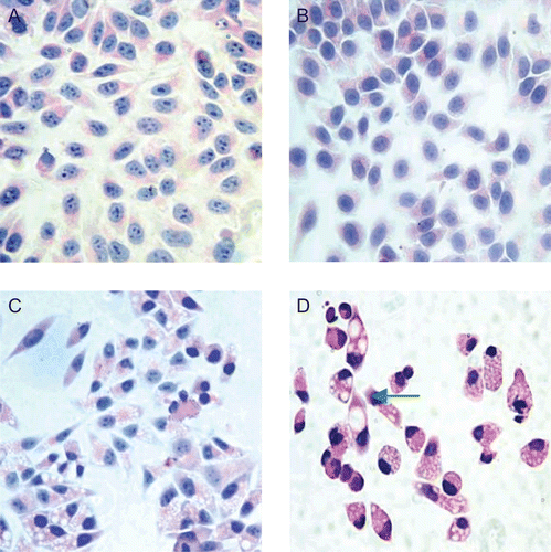
The morphology effect of matrine on HepG2 cells was viewed under a light microscope, as shown in . Below the dosage of 0.3 mg/mL, morphological characteristics of HepG2 cells were similar to normal cells. After HepG2 cells were treated with matrine above the dosage of 0.3 mg/mL for 24 h, the number of scattered cells increased sharply. Prolongation of incubation time to 48 or 72 h had more significant effects on HepG2 cells, compared to the incubation for 24 h. With the final matrine concentration above 1.5 mg/mL, most of the HepG2 cells went plasmatorrhexis and death ultimately.
Figure 1. Matrine changed the morphology of HepG2 and induced part cells to death. HepG2 cell incubated with different concentration of matrine for 4 days were harvested for H&E staining (×400magnification) to view cell morphology under multifunctional microscope. Untreated cells served as control (A), and treated cells were shown in 0.2 mg/mL (B); 1.5 mg/mL (C); 3.0 mg/mL (D) and the green arrow indicated the plasmatorrhexis.

Matrine inhibited proliferation of HepG2 cells in a dose-dependent manner
To confirm the relationship between dosage and effect, HepG2 cells were cultured in the presence of increasing doses of matrine for 4 days, the inhibition effects of matrine on HepG2 cells was determined with MTT assay. The concentration of 0.2 mg/mL in (left) could be discriminated as inflexion. After HepG2 cells were treated with matrine for 24 h, negative-inhibition effects were found at the concentration of matrine below 0.2 mg/mL. However, positive-inhibition effects above 0.3 mg/mL were confirmed and prolongation of treated time to 96 h had more effects on the inhibition of proliferation of HepG2. The results showed the exposure of HepG2 cells to matrine caused the significant inhibition of proliferation in a dose-dependent manner.
Figure 2. Matrine inhibited the proliferation of HepG2 cells in vitro compared with the control. HepG2 cells were incubated in presence of matrine at different concentrations (from 0.1 to 0.5 mg/mL, and from0.3 to 3.5 mg/mL) for 4 days, and cell proliferation was determined by MTT assay to calculate the proliferation inhibition rate (%). Negative-inhibition effects were found at the concentration of matrine below 0.2 mg/mL as shown in left section, whereas positive dose-dependent inhibition of HepG2 cell growth by matrine as described in right section.

Matrine down-regulated the expression of AFP, PCNA
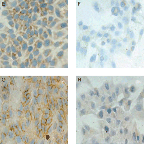
The results of immunohistochemical analyses revealing the expressions of AFP and PCNA protein were shown in . Weaker immunostaining of AFP occurred diffusely in the cytoplasm of matrine-treated HepG2 cells in which the mean grade of staining intensity was lower. PCNA was mainly located around the cell nucleolus and karyotheca in matrine-treated HepG2 cells with weaker staining intensity compared to control cells. The decrease achieved significance at 0.8 mg/mL and demonstrated the down-regulation of protein expression.
Figure 3. Matrine down-regulated the expression of AFP and PCNA proteins. HepG2 cells incubated with absence (E, G) or presence of 0.8 mg/mL matrine for 2 days were determined with immunohistochemistry analysis. HepG2 cells were viewed under multifunctional microscope (×400magnification). Immunostaining intensity of AFP (F) and PCNA (H) were weaker than control. The decrease demonstrated the down-regulation of protein expression.

Matrine down-regulated the expression of C-myc and Bcl-2; up-regulated Bax
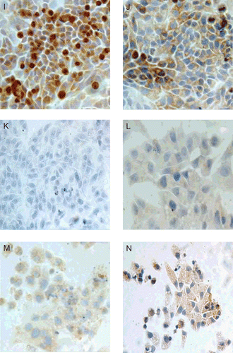
The results of immunohistochemical examination are shown in . Expression of C-myc and Bcl-2 were seen in the cell nucleolus and cytoplasm of matrine-treated cells. Significantly specific staining was detected and the staining intensity was weaker than that of untreated cells. In contrast, the expression of Bax in cell nucleoli and the cytoplasm of matrine-treated cells were stronger than that of untreated cells. The mean grade of staining intensity was higher compared with control cells. With the increasing of matrine dosage, the tendency toward increased grades in matrine-treated cells appeared to be slightly higher.
Figure 4. Matrine down-regulated the expression of C-myc, Bcl-2, and up-regulated the expression of Bax. HepG2 cells cultured in DMEM (upper row, I, J, K) or DMEM containing 0.8 mg/mL matrine (lower row, L, M, N) for 2 days were detected with immunostaining. HepG2 cells were viewed under multifunctional microscope (×400magnification). Expression of C-myc (first column) and Bcl-2 (second column) were seen in the cell nucleolus and cytoplasm of matrine-treated cells and the staining intensity was weaker than that of untreated cells. Bax (third column) expressed stronger than untreated cells. The mean grade of staining intensity was higher compared with control cells.

Discussion
Our present study has demonstrated that matrine could inhibit the proliferation of HepG2 cells in vitro, mainly through changing expression of tumor relevant proteins. Similarly, matrine was shown to change the expression of Bcl-2, Bax, WP53, etc. (CitationSi et al., 2001). The inhibition rate showed both a time- and dose-dependent manner in accordance with the previous report (CitationLuo et al., 2007).
HepG2 cells treated with matrine grew more slowly and decreased the malignant phenotype (CitationHang et al., 1996; CitationSi et al., 2000). Matrine at 0.25 and 0.50 mg/mL dosages had a notable inhibition effect on MKN45 cells (CitationLuo et al., 2007) than lower dosage. In our study, matrine below 0.2 mg/mL dosage had little influence on the HepG2 cells. However, matrine above 0.3 mg/mL dosage had a prominent inhibition effect than the lower dosages. Round and transparent granules were viewed in matrine-treated cells, which made cells to be vacuolar denaturalization. The lipid granules were observed in HepG2 cells treated with matrine from 0.3 to 1 mg/mL dosage. The reason was probably that matrine could interfere with the lipid protein metabolism, or destroy the cell structure and induce the lipid protein hydrolysis further to form lipid granules (CitationSi et al., 2001). Cell chromatin concentrated when treated with 1.5 mg/mL matrine dosage, which were presumed apoptosis in HepG2 cells. Cell membranes were destroyed as treated with 2.5 mg/mL matrine. The above results further supported the evidence that matrine had the ability to change HepG2 cells malignant phenotype. Moreover, along with the increasing of dosage of matrine and prolongation of exposure time, HepG2 cells grew dispersing and shelling, which resulted in death of cells. The results of MTT assay indicated that the inhibition rate of matrine exhibited two tendencies. Below the 0.2 mg/mL doses it showed a promoting tendency, while an inhibited tendency occurred above 0.3 mg/mL doses. It seemed that 0.25 mg/mL was a watershed of matrine effect. Positive-inhibition effect above 0.3 mg/mL was confirmed in accordance with morphology. However, it was noteworthy that a negative-inhibition effect was found below 0.2 mg/mL dosage. Similar results were observed in Luo’s study (CitationLuo et al., 2007). The reason was probably related to the change of ambient conditions of HepG2 cell, followed by the change of ectocellular matrix, which resulted in promoting cell proliferation. On the other hand, a signal transduction pathway probably existed between the ectocelluar matrix and integrin stimulated cell proliferation.
AFP, carcinoembryonic protein, is the most established tumor marker in HCC and the gold standard by which early liver cancer is judged (CitationZhang et al., 1998). AFP protein does not exist in natural hepatocyte and exactly represents the malignant degree of the hepatoma. This study showed that the expression of AFP clearly decreased in HepG2 cells treated with matrine. It suggested the malignant proliferation of HepG2 cell has been inhibited. PCNA participated in a variety of essential cellular processes including DNA replication, DNA repair, and cell-cycle control by interacting with proteins involved in these processes (CitationHe et al., 2001). The PCNA protein expresses little in resting phase and more is synthesized at the end of G1 phase, reaching climax in S phase, whereas PCNA decreases obviously in G2 and M phase. The seasonal variation is similar to the process of cell proliferation, and its expression represents the ability of cell proliferation. The study herein resulted in the obvious decrease of PCNA protein in matrine-treated cells. It may indicate that matrine could prevent HepG2 into S stage to fulfill duplication of DNA molecule and result in the failure of cell division.
The transcription factors of E2F family control the G1/S transition in eukaryotic cells. E2F has the ability to induce both cell cycle progression and apoptosis, making it possible for this transcription factor to have both tumor-promoting as well as tumor-suppressive effects. During most of G1 phase, the unphosphorylated Rb is coupled with the E2F protein. The E2F-Rb complex bind with regulatory sites in the promoter regions of numerous genes involved in cell-cycle progression, acting as a repressor that blocks gene expression. It might be important for c-myc oncogene in regulating cell proliferation and differentiation. If E2F-Rb complex was destroyed and the apoptosis was inhibited, c-myc gene would express continually in hepatoma, resulting in cell proliferation (CitationLeone et al., 1997; CitationJiang et al., 2007). This study indicates that the expression of C-myc was reduced along with increasing the concentration of matrine, which suggests that matrine could reduce the expression of C-myc directly or indirectly, further inhibiting the proliferation of HepG2 cells. Similarly, matrine has been shown to inhibit proliferation of C6 cells and down-regulated expression of proto-oncogene c-myc (CitationDeng et al., 2004).
Bcl-2 protein is an apoptosis-related protein and plays an important role in regulating apoptosis (CitationKarczmarek et al., 2003; CitationFarczadi et al., 2003). Apoptosis of cancer cells in response to cytotoxic agents requires an intact mitochondrial apoptosis pathway in most cell types. The Bcl-2 family proteins are central regulators of this process. The balance between the pro-apoptotic and pro-survival of the Bcl-2 family member determines whether the cell survives or undergoes apoptosis. Bcl-2 protein is mainly distributed in mitochondria ectoblast or endoplasmic reticulum, and regulates the permeability of membrane. Bcl-2 could inhibit the permeability of mitochondria membrane and prevent the releasing of cytochrome C to maintain the stability. The expression of Bcl-2 protein and releasing of cytochrome C prevented apoptosis. Cytochrome C binds to Apaf-1 and pro-caspase-9, further active caspase-9 in turn directly activates pro-caspase-3, initiating a cascade of additional caspase activation (CitationChou et al., 1999; CitationSatio et al., 2000; CitationKuwana & Newmeyer, 2003). In this paper, the amount of Bcl-2 was reduced along with increasing the concentration gradient of matrine, which suggests that matrine could have influence to the permeability of mitochondria membrane.
Bax is a homologous protein of Bcl-2, in the form of homopolymer (Bax/Bax) or isodipolymer (Bcl-2/Bax) and distributed extensively. A previous study demonstrated that the ratio of Bax to Bcl-2 protein influences the apoptotic rate of cells (CitationNakamura et al., 2003). The expression of bax gene was much stronger in apoptosis cells than normal cells. Its biological function mainly lay in antagonist of Bcl-2 and resulted in forming double polymer. The level of Bax directly regulated cell apoptosis. The increasing of matrine following the increasing of Bax, suggests that the apoptosis of HepG2 cells was coupled with up-regulation of bax gene. The effect mechanism is probably similar to the activation of p53 gene. It has also been shown that matrine triggered apoptosis via up-regulating cell cycle protein E2F-1 in K562 cells (CitationJiang et al., 2007), or pro-apoptotic molecules of Bcl-2 family in MKN45 cells (CitationLuo et al., 2007), or reducing Bcl-2/Bax ratio in cardiac fibroblasts (CitationLi et al., 2007).
In conclusion, these results indicate that matrine could inhibit HepG2 cell proliferation by down-regulating the expression of AFP, PCNA C-myc, Bal-2, and up-regulating the expression of Bax in vitro. However, the mechanisms accounting for cell-specific proliferative or apoptotic responses to matrine remain unclear. Further investigations are definitely required to clarify the specific molecules affected by matrine in molecular pathways at protein levels, and the answers to these questions would improve our understanding of the molecular mechanisms of matrine on inhibition of HepG2 cells.
Acknowledgements
We are sincerely grateful to Ji-Yun Yin, Si-Si Liu, Xu Xin who have contributed in many ways toward the completion of this work and Yang Jun (National University of Singapore) and Li-Jun Pu for their reviews of the manuscript.
Declaration of interest
The authors report no conflict of interests. The study was supported by the Technology Research Foundation of Education Department of HeiLongJiang Province, China (11511247).
References
- Chou JJ, Li H, Salvesen GS, Yan J, Wagner G (1999): Solution structure of BID: An intracellular amplifier of apoptotic signaling. Cell 96: 615–624.
- Clark HP, Carson WF, Kavanagh PV, Ho CP, Shen P, Zagoria RJ (2005): Staging and current treatment of hepatocellular carcinoma. Radiographics 25: S3–S23.
- Deng H, Luo H, Huang F, Li X, Gao Q (2004): Inhibition of proliferation and influence of proto-oncogene expression by matrine in C6 cell. Zhong Yao Cai 27: 416–419.
- Farczadi E, Kaszas I, Baki M, Szende B (2003): Changes in apoptotic and mitotic index, bcl2, and p53 expression in rectum carcinomas after short-term cytostatic treatment. Ann N Y Acad Sci 1010: 780–783.
- Hang Z, Wei Y, Zhao X (1996): Electron microscopic observation of hepatocellular carcinoma in 15 cases: An ultrastructural comparison with human embryonic liver. J West Chin Univ Med Sci 27: 332–336.
- He H, Taneat K, Downey M, Antero GS (2001): A tumor necrosis factor α- and interleukin 6-inducible protein that interacts with the small subunit of DNA polymerase δ and proliferating cell nuclear antigen. Proc Natl Acad Sci USA 98: 11979–11984.
- Hu ZL, Zhang JP, Qian DH, Lin W, Xie WF, Zhang XR, Chen WZ (1996): Effects of matrine on mouse splenocyte proliferation and release of interleukin-1 and -6 from peritoneal macrophages in vitro. Acta Pharmacol Sin 17: 259–261.
- Jiang H, Hou C, Zhang S, Xie H, Zhou W, Jin Q, Cheng X, Qian R, Zhang X (2007): Matrine upregulates the cell cycle protein E2F-1 and triggers apoptosis via the mitochondrial pathway in K562 cells. Eur J Pharmacol 559: 98–108.
- Karczmarek-Borowska B, Filip A, Zdunek M, Korobowicz E, Wojcierowski J, Korszen-Pilecka I, Furmanik F (2003): The preliminary estimation of bcl-2, bcl-X (L) and p53 genes expression in locally advanced non-small cell lung cancer. Ann Univ Mariae Curie Sklodowska 58: 124–131.
- Kuwana T, Newmeyer DD (2003): Bcl-2-family proteins and the role of mitochondria in apoptosis. Curr Opin Cell Biol 15: 691–699.
- Leone G, Gregori JD, Sears R, Jakoi L, Nevins JR (1997): Myc and Ras collaborate in inducing accumulation of active cyclin E/cdk2 and E2F. Nature 387: 422–426.
- Li Y, Wang B, Zhou C, Bi Y (2007): Matrine induces apoptosis in angiotensin II-stimulated hyperplasia of cardiac fibroblasts: Effects on Bcl-2/Bax expression and caspase-3 activation. Basic Clin Pharmacol Toxicol 101: 1–8.
- Liang P, Bo AH, Xue GP, Han R, Li HF, Xu YL (1999): Study on the mechanism of matrine on immune liver injury in rats. World Chi J Digestol 7: 104–108.
- Liu J, Zhu M, Shi R, Yang M (2003): Radix Sophora flavescentis for chronic hepatitis B: A systematic review of randomized trials. Am J Chin Med 31: 337–354.
- Long Y, Lin XT, Zeng KL, Zhang L (2004): Efficacy of intramuscular matrine in the treatment of chronic hepatitis B. Hepatobiliary Pancreat Dis Int 3: 69–72.
- Luo C, Zhu YL, Jiang TJ, Lu XY, Zhang W, Jing QF, Li J, Pang LR, Chen KJ, Qiu FM, Yu XY, Yang JH, Huang J (2007): Matrine induced gastric cancer MKN45 cells apoptosis via increasing pro-apoptotic molecules of Bcl-2 family. Toxicology 229: 245–252.
- Merle P (2005): Epidemiology natural history and pathogenesis of hepatocellular carcinoma. Cancer Radiother 9: 452–457.
- Mosmann T (1983): Rapid colorimetric assay for cellular growth and survival: Application to proliferation and cytotoxicity assays. J Immunol Meth 65: 55–63.
- Nakamura H, Kumei Y, Morita S, Shimokawa H, Ohya K, Shinomiya K (2003): Antagonism between apoptotic (Bax/Bcl-2) and anti- apoptotic (IAP) signals in human osteoblastic cells under vector-averaged gravity condition. Ann N Y Acad Sci 1010: 143–147.
- Satio M, Korsmeyer SJ, Schlesinger PH (2000): BAX-dependent transport of cytochrome c reconstituted in pure liposomes. Nat Cell Biol 2: 553–555.
- Si WK, Chen A, Li P, Liu B, Gao LH, Yao J (2001): Study on apoptosis of human hepatoma cell line HepG2 induced by matrine. Acta Acad Med Mil Tert 23: 816–820.
- Si WK, Xiao TY, Kang GF (2000): Effects of matrine on cell morphology and related factors of hepatocellular carcinoma cell HepG2 in vitro. Acta Acad Med Mil Tert 22: 553–556.
- Tao SC, Wang JZ (1992): Pharmacological effects of alkaloids from Sophora alopecuroides. Chin Pharm J 27: 201–204.
- Zhang JP, Zhang M, Zhou JP, Liu FT, Zhou B, Xie WF, Guo C (2001): Antifibrotic effects of matrine on in vitro and in vivo models of liver fibrosis in rats. Acta Pharmacol Sin 22: 183–186.
- Zhang L, Li SN, Wang XN (1998): CEA and AFP expression in human hepatoma cells transfected with antisense IGF-I gene. World J Gastroenterol 4: 30–32.
- Zhang LJ, Wang TT, Wen XM, Wei Y, Peng XC, Li H, Wei L (2007): Effect of matrine on HeLa cell adhesion and migration. Eur J Pharmacol 563: 69–76.
- Zhang LP, Jiang JK, Tam JWO, Zhang Y, Liu XS, Xu XY, Liu BZ (2001): Effects of matrine on proliferation and differentiation in K-562 cells. Leuk Res 25: 793–800.